

Introduction
One of the most important concerns of infertility practice is chemotherapy-induced diminished ovarian reserve [1]. Hence, protection of the ovarian follicle loss in women with different types of cancer has gained increasing attention [2, 3]. Use of gonadotropin releasing hormone analogs, cryopreservation of oocytes, and cryopreservation of ovarian cortical tissues are commonly considered strategies for preservation of ovarian reserve in young cancer patients undergoing chemotherapy. Due to limited data about the possible mechanism of action of anti-cancer drugs regarding ovarian follicle survival there had been limited improvement in the fertility preservation methods [2, 3]. This limitation led us to think that use of protective adjuvants with anti-cancer properties may prevent the loss of follicle depletion during chemotherapy and they would be appropriate for young female cancer patients.
Toxic effects of anti-cancer drugs on reproductive tissues are a major problem during treatment of young women with cancer. Chemotherapeutics not only target the cancer cells but also attack non-cancerous dividing cells including oocytes [4, 5]. Toxic insult due to anti-cancer drugs may lead to undesired adverse effects on the growing follicle that may cause premature ovarian failure. Unfortunately, most of the drugs or surgical interventions developed for minimizing follicle depletion show low efficacy resulting in premature ovarian aging [4, 5]. In order to prevent chemotherapy-induced follicle loss, many plant-derived polyphenols have been used due to their chemopreventive features [6–8]. In this purpose we investigated the impact of three well-known natural molecules, resveratrol (3,4ʹ,5-trihydroxy-transstilbene), caffeic acid phenethyl ester (CAPE), and coenzyme Q10 (CoQ10) on chemotherapy-induced follicle loss. Resveratrol (RSV) is a polyphenol found in grapes and peanuts that has demonstrated antioxidant, anti-inflammatory, cell protecting and anti-cancer properties that could facilitate its therapeutic application in many clinical conditions including ovarian aging [9–11]. It is a plant produced in a range of species as a defense mechanism against environmental stressors such as pathogens and UV radiation [12]. Administration of RSV has beneficial effects in the prevention of ischemia-reperfusion injury of the ovaries [13]. The oocyte preserving effects of resveratrol, if they really exist, could be derived from its metabolites, which have demonstrated more potent activity than resveratrol [13, 14]. Caffeic acid phenethyl ester is a phenolic compound produced from propolis. It has antimitogenic, antioxidant and anti-cancer activities that inhibit the production of free oxygen radicals [15]. In addition to its nuclear factor kappa B inhibitory activity, CAPE has been reported to show anti-inflammatory and immunomodulatory effects [16]. Celik et al. showed that CAPE administration ameliorates ischemia-reperfusion injury in rat ovary by decreasing xanthine and malondialdehyde and by increasing glutathione levels [15].
Coenzyme Q10, a fat-soluble component of nearly all cell membranes, is located in the inner mitochondrial membrane. Coenzyme Q10 contributes energy production in mitochondrial electron transport chain and has antioxidant properties [17]. It has been reported that CoQ10 supplementation causes a significant increase of CoQ10 levels in ovaries [18]. Ozcan et al. [19] reported that ovarian reserve was preserved with 150 mg/kg/day CoQ10 application in a chemotherapy-induced ovarian injury model. In connection with this, by preventing cell membrane peroxidation and free radical oxidation CoQ10 may be involved in inhibition of follicle depletion due to chemotherapy. However, there are no clear data on whether these three molecules have a follicle protective effect on the POI model. In this study, RSV, CAPE and CoQ10, which have been scientifically proven to have strong antioxidant, antiapoptotic and cell survival effects [1–3], were used to investigate the possible protective role of these molecules on ovarian follicle depletion in animals with cisplatin-induced premature ovarian insufficiency (POI). The aim of this study was to investigate the possible follicle-protective effect of RSV, CAPE, or CoQ10 on an experimentally induced POI model.
Material and methods
Grouping of animals
In the current study, the care of the rats and all surgical procedures to be performed were planned in accordance with Guide for the Care and use of Laboratory Animals. The study was carried out in Altinbas University Faculty of Medicine. Local ethical approval of this study was obtained from Altinbas University Hospital. Twenty-eight female Wistar Albino strains weighing 200–350 g were kept under standard environmental, heat, housing and feeding conditions. They were equally divided into four groups with 7 rats in each group: the groups consisted of a vehicle-treated control group (group 1), or chemotherapy-treated rats received resveratrol injection (group 2), or chemotherapy-treated rats received oral CoQ10 (group 3), or chemotherapy-treated rats received CAPE injection (group 4). A sham group (placebo surgery) consisting of five animals was created to measure baseline ovarian weights. On the first day of cisplatin injection to other groups, animals in the sham group underwent laparotomy and their ovaries were surgically removed to calculate the ovarian weights. Unlike other groups, animals in the sham group were not exposed to cisplatin, so the POI model was not applied in animals in this group. In addition, the animals in this group underwent laparotomy without any follicle-sparing treatment and the weights of their ovaries were measured.
Creating a rat model of premature ovarian insufficiency with cisplatin
Rats were chosen to create the POI model because it is easily accessible and their ovarian physiology is similar to human ovarian physiology. We also chose to use cisplatin as the use of this drug is preferred in most POI models, and the harmful effects of cisplatin on follicles have been shown in a previous study [20]. The rat model of POI was established by 10 consecutive days of intraperitoneal injection of cisplatin that was administered once daily at doses of 2.0 mg/kg. Rats in groups 2, 3, and 4 were given either RSV (100 mg/kg, i.p.), CoQ10 (20 mg/kg, oral), or CAPE (10 mg/kg, i.p.) respectively. Treatment with RSV, CoQ10, or CAPE was started at the 1st day of chemotherapy and continued for 10 days during chemotherapy. Since CoQ10, as with all water-insoluble compounds, has been reported to be better absorbed in the intestines, we chose to give the CoQ10 orally [21]. Unlike CoQ10, the oral bioavailability of RSV or CAPE is very low due to its incomplete intestinal absorption and intense catabolism, which limits their oral application [22]. For this reason, we chose to deliver these two molecules by the intraperitoneal route.
Control animals in group 1 received injections of phosphate buffered saline solution. The rats in each group were observed daily for clinical signs including weight loss, food intake and motor function. In addition serum levels of anti-Mullerian hormone (AMH) and estrous cycle characteristics of animals were also monitored. The body weights of each rat were measured on the day of the first injection of cisplatin and on the day of ovariectomy. The animals were sacrificed 2 weeks after the first injection of cisplatin, and their ovaries were excised for histopathological analysis. Ovarian weights of animals were also recorded. The initial ovarian weights of the sham group were compared with the ovarian weights of the animals in the treatment groups 15 days later. Histological sections were evaluated with the H-score method. This is a semi-quantitative method consisting of the percentages of positively stained ovarian cortical and medullar cells multiplied by a weighted intensity of staining: H-score = Ʃ Pi (i + 1), where Pi is the percentage of stained ovarian cells in each intensity category (0–100%), and i is the intensity indicating weak (i = 1), moderate (i = 2) or strong staining (i = 3) [23].
Anti-Mullerian hormone assay
After venous blood collection, serum for assay of AMH was separated and frozen. All samples were analyzed using an ultra-sensitive AMH Gen II ELISA kit (Beckman-Coulter, Inc., Webster, USA). The lower limit of AMH detection was 0.16 µg/l. Inter-assay variation was 10% at 0.27 µg/l. All values are expressed in ng/ml.
Counting follicles
The ovaries of each rats were fixed in paraformaldehyde embedded in paraffin, serially sectioned, and stained with hematoxylin for histology and differential follicle counting. An average of 8 sections were obtained 5 µm in thickness. Follicle counts were performed on representative sections from seven rats from each group, and the mean count per section was calculated. In each section, primordial, primary, antral and Graafian follicle counts were noted. A primordial follicle was defined as an oocyte surrounded by a single layer of granulosa cells. A primary follicle was defined as an oocyte surrounded by a single layer of cubic epithelium. Antral follicles were defined as follicles with an antral space between the multilayered granulosa cells. A Graafian follicle was defined as a follicle with a large antrum [24].
Statistical analysis
All data analysis was applied using the software package SPSS 18.0 for Windows (SPSS, Inc., Chicago, IL, USA). The normality of individual group parameters was assessed with the one-sample Kolmogorov-Smirnov Z test. Variables with normal distributions were compared between groups using independent samples tests. Because they were found to be abnormally distributed in both groups the Mann-Whitney U and ANOVA tests were used for comparisons of results. Data were given as mean ± standard deviation. P-values < 0.05 were considered to indicate statistical significance.
Results
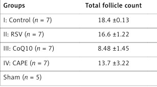
Rats given CAPE or RSV showed vaginal smears with superficial cells showing an estrogenic effect. H-score evaluation of the ovaries demonstrated a higher mean follicle count in group 2 and group 4 compared to group 3. The ovarian cross-sectional area was not significantly different between groups 1, 2, and 4 in terms of the number of individual follicle types. The total follicle number of animals in the control group was approximately 2.5 times that of animals in group 3 (18.4 ±0.13 vs. 8.48 ±1.45, p < 0.001). When the control group and the RSV group were compared, the total follicle numbers were similar (18.4 ±0.13 vs. 16.6 ±1.22, p < 0.41). The total number of follicles in the control group was higher than CAPE, but the difference was not significant (18.4 ±0.13 vs. 13.7 ±3.22, p < 0.57). However, rats in group 3 had a lower total number of ovarian follicles than those in groups 2 (8.48 ±1.45 vs. 16.6 ±1.22, p < 0.05) and 4 (8.48 ±1.45 vs. 13.7 ±3.22, p < 0.05).
Groups 2 (4.96 ±1.41 vs. 3.52 ±1.44 ng/ml, p < 0.05) and 4 (5.10 ±2.70 vs. 3.52 ±1.44 ng/ml, p < 0.05) had significantly higher serum AMH levels than in group 3. Compared to control group rats the RSV or CAPE group rats showed an insignificant decrease in serum levels of AMH. Compared to the control group, AMH level was significantly lower in rats given CoQ10 (5.35 ±6.06 ng/ml vs. 3.52 ±1.44 ng/ml, p < 0.03). Increases in total body weight and ovarian weight were observed at the end of the second week in group 1 animals. The weight of the animals in the control group at the end of the 2nd week was higher than that of the animals in the other three groups. However, only the difference between animals in the CoQ10 group and controls was significant (294.2 ±13.2 vs. 220.1 ±9.01, p < 0.02). The weight differences of animals given CAPE or RSV and animals in the control group did not reach statistical significance. Compared to other groups, significant weight loss was detected in animals given CoQ10. Animals in the RSV or CAPE groups showed insignificant weight loss. While two rats in the CoQ10 group, died none of the rats in the RSV or CAPE group died (Table I).
Table I
Comparison of the groups according to measured parameters
| Groups | Total follicle count | AMH [ng/ml] | Ovarian* weight [g] | Body weight [g] |
|---|---|---|---|---|
| I: Control (n = 7) | 18.4 ±0.13 | 5.35 ±6.06 | 86.1 ±0.29 | 294.2 ±13.2 |
| II: RSV (n = 7) | 16.6 ±1.22 | 4.96 ±1.41 | 78.1 ±9.18 | 286.4 ±1.02 |
| III: CoQ10 (n = 7) | 8.48 ±1.45 | 3.52 ±1.44 | 67.1 ±50.9 | 220.1 ±9.01 |
| IV: CAPE (n = 7) | 13.7 ±3.22 | 5.10 ±2.70 | 77.1 ±43.1 | 278.5 ±6.45 |
| Sham (n = 5) | 84.5 ±12.2 |
Ovarian weights of sham and control groups were found to be similar on the 15th day. Ovarian weights of the animals in the CoQ10 group were significantly lower compared to both sham and control groups. The ovarian weights of the rats in the RSV and CAPE groups were lower compared to the control and sham groups, but the difference was not significant. Ovarian weights of the animals in the RSV and CAPE groups were higher than in the CoQ10 group, but the differences did not reach statistical significance. The ovaries of the rats in the control group showed follicles in all stages of development. The ovaries of the rats in the CoQ10 group showed increased numbers of primary follicles at day 15. Other stages of follicle significantly decreased in rats that were given CoQ10. While the ovaries of the rats in the RSV or CAPE group showed significantly decreased numbers of primordial follicles they showed increased numbers of early growing follicles at day 15. They showed increased numbers of primary and antral follicles. While the number of somatic cells decreased in the ovarian tissue in rats given CoQ10, there was a significant increase in inflammatory cells. Conversely, both somatic and inflammatory cell characteristics of animals given CAPE or RSV were similar to the control group.
Discussion
Chemotherapeutic agents damage the developing follicles in the ovaries in different ways. Oocyte and granulosa cells are vulnerable to damage caused by cisplatin. Some chemotherapeutics cause follicle death in the ovaries, while others block cell division steps. The ovaries activate some natural protective mechanisms to protect against the toxic effects of drugs. The regulation of apoptosis comes first among these mechanisms. A study conducted on xenograft models clearly demonstrated that chemotherapy-induced follicle death in the human ovary occurs via an apoptotic mechanism [25]. Chemotherapeutic agents reduce the ovarian reserve by damaging the DNA of developing follicles. Follicle cells try to prevent this damage by using their DNA repair enzymes [26]. In addition, chemotherapeutics reduce blood flow by damaging vascular and stromal cells in the ovary and preventing oxygenation, leading to the death of follicles. Insufficient oxygenation can also increase the production of reactive oxygen species (ROS), leading to follicle death. The ovarian follicles make efforts to prevent the harmful effect of chemotherapeutics by increasing neovascularization. A study by Soleimani et al. showed an inverse association between ovarian vascular structures and primordial follicle apoptosis [27]. Oocytes with sufficient DNA repair ability and antioxidant capacity may survive this gonadotoxic stress while others with a less efficient repair mechanism and antioxidant enzymes may be lost as a result of severe DNA damage triggering apoptotic death pathways. To offset these risks, women can be offered several options for fertility preservation, including cryopreservation of oocyte, embryo or ovarian tissue. If the patient does not accept fertility preservation methods, it may be necessary to use some medications or supplements to prevent follicle loss due to chemotherapy. Resveratrol and CAPE are two different antioxidant supplements that have recently been used to prevent chemo-induced ovarian damage. Although the oocyte protective mechanism of action of these two molecules is not known exactly, some mechanisms are explained in experimental models. In this study, we do not know precisely the oocyte-protecting mechanism of action of RSV or CAPE since we only evaluate ovarian reserve over follicle count and AMH values. For this reason, our comments may be somewhat speculative.
In this preliminary and experimental study we found that CAPE therapy reduced chemotherapy-induced follicle loss. We do not clearly know how CAPE prevents chemotherapy-induced ovarian damage. Our comments on the chemopreventive effect of CAPE are somewhat speculative, but are based on our laboratory and clinical observations on animals. The findings that support the positive effects of CAPE are that the ovaries of the animals in the CAPE group contain sufficient early growing follicles, the decrease in serum AMH levels is insignificant, and the weight lost by the animals is similar to the control group. Apoptosis induction is one of the mechanisms in which chemotherapeutics show their toxic effects on ovaries. Preserved follicle count and AMH levels in rats that were given CAPE may be due to antiapoptotic or cell cycle regulatory actions of CAPE or its metabolites. Although we did not examine apoptotic changes in the ovary in our study, it is a known fact that CAPE stimulates cell cycle arrest in cancer cells [28, 29]. Chemotherapeutics can lead to the emergence of free oxygen radicals and exert their toxic effects on the oocytes. Since CAPE is one of the strongest molecules with an antioxidant effect, it can prevent follicle loss by reducing oxidant damage due to chemotherapy [30]. More comprehensive studies are needed to clarify all these mechanisms that we believe to be related to the use of CAPE.
The reason for the high number of follicles detected in animals receiving RSV or CAPE may be due to the positive effects of these two molecules on cell life. It has been reported that RSV or CAPE interacts with a variety of molecular targets associated with cell cycle survival pathways [31–33]. It has been reported that RSV inhibits pro-inflammatory enzyme expression, and reduces nuclear factor-κB activation and cytokine release. Treatment with resveratrol activates multiple intracellular molecules involved in cell survival and programmed cell death [34]. RSV protects the patient’s metabolic status by preventing insulin resistance and contributes to the individual’s fight against the harmful effects of chemotherapeutics [35]. The follicle count and AMH levels of animals on CoQ10 were found to be decreased. Despite its antioxidant effect, CoQ10 did not prevent follicle loss due to chemotherapy. This may be due to the fact that CoQ10 cannot be easily diffused into the oocyte. Because chemotherapy disturbs the mitochondrial function of the oocyte CoQ10 may be ineffective for restoration of the follicle. As CoQ10 is fat soluble and is found in tissues with high energy demands [36] restoration of dying cells due to chemotherapy may not be possible [37]. In addition to the increase in follicle loss in animals given CoQ10, weight loss was also greater than in other groups. The reason for these negative effects may be related to the dose of CoQ10 used or the administration route. The dose of 20 mg/kg/day used in this study may not be sufficient for a follicle protective effect.
In accordance with this opinion, a previous study reported that when given at a dose of 150 mg/kg, CoQ10 alleviates ovarian reserve reduction due to chemotherapy [19]. However, since the best absorption form of CoQ10 is provided orally, we think that the route of use does not play a role in these negative effects. For all these reasons, studies applying CoQ10 in different doses and in different ways are required.
Another possible cause of follicle preserving effects of RSV or CAPE may be due to regulatory function of these molecules in ROS generation during chemotherapy [38]. Both molecules can also exert their cell protecting function at the level of the oocyte or granulosa cell mitochondrial membrane. A supportive, apoptotic pathway located in the mitochondria has been reported to be responsible for cancer cell death [39, 40]. By inhibiting apoptosis, either RSV or CAPE might protect the oocyte or granulosa against cell death in animals with cisplatin-induced POI. The oocyte-protecting effect of RSV could also be derived from its sulfated metabolites, which have demonstrated more potent activity than RSV [14]. The generation of ROS during chemotherapy plays a pivotal role during apoptotic cell death. By decreasing glutathione and superoxide dismutase levels ROS lead to apoptotic cell loss. Therefore, antioxidant action of RSV can inhibit ROS-mediated cell death and may allow survival of the oocyte [41, 42]. Furthermore, stimulation of glutathione S-transferase (GST) with RSV may be considered a potential strategy for POI prevention in POI subjects due to chemotherapy. Glutathione S-transferase may protects against exposure to chemotherapy-induced oxidative stress [43–45]. Supporting this, both the expression and catalytic activity of GST were restored with low dose RSV supplementation [40]. Given the relation between increased GST activity and reduced cancer risk [41], stimulation of this enzyme could be a significant mechanism by which resveratrol protects the oocyte from harmful effects of anti-cancer drugs.
This experimental study has its strengths and weaknesses. Successful creation of the POI model in rats makes the study privileged, because many experimental POI models do not turn out as desired, and most animals die. On the other hand, since we evaluated both the number of follicles and AMH levels in our study, we were able to make stronger comments about the effect of these three molecules on the ovarian reserve. Moreover, the combination of three powerful antioxidant molecules enabled us to obtain strong data on the prevention of chemotherapy-induced ovarian damage. In addition to all these positive effects, our study also has limitations. The low number of animals in the experimental groups is the basic handicap. Also, giving RSV, CAPE and CoQ10 as a single dose in the experimental groups weakens the study. The use of these three substances in different doses and in different ways of application could make our results more objective.
In conclusion, while RSV or CAPE administration does not prevent primordial follicle loss, they preserve both primary and antral follicles in a cisplatin-induced POI model. Both molecules may be used as an effective oocyte preservation method particularly among young cancer patients undergoing chemotherapy. Although the mechanisms of action are unclear, their antioxidant and antiapoptotic effects may ameliorate harmful effects of chemotherapeutic agents and preserve ovarian reserve. Taken together, both CAPE and RSV supplementation might be considered as an oocyte preserving strategy in young cancer patients desiring to preserve their fertility potential. However, in order to establish a clear conclusion, more research is still necessary in this area.

