
Orbital-sinonasal interface tumors (OSITs) are mass-forming pathological processes that continuously involve both the orbit and the sinonasal compartment, including the nasal cavity and/or paranasal sinuses [1]. They may extend across the orbital walls or through natural anatomical pathways, such as the lamina papyracea, orbital floor, nasolacrimal pathway, or adjacent foramina [2]. For the purposes of this clinical-anatomical classification, the term OSITs encompasses benign and malignant neoplasms as well as inflammatory tumor-like lesions that radiologically and clinically present as orbital-sinonasal masses [3]. When pathological categories are discussed, these entities are distinguished as benign tumors, malignant tumors, and inflammatory lesions. Patients may present with eyelid swelling, proptosis, visual impairment, restricted ocular motility, nasal obstruction, rhinorrhea, facial paresthesia, or other symptoms depending on the site and direction of extension.
The orbit’s close anatomical relationship with the paranasal sinuses and critical neurovascular pathways makes the diagnosis and surgical management of tumors in this region particularly complex. Advanced imaging techniques such as computed tomography (CT) scanning and magnetic resonance imaging (MRI) are indispensable for accurately assessing tumor extent, planning surgical strategies and predicting patient prognosis [4, 5]; however, clinical management remains challenging due to a lack of standard classification systems that enable surgeons to make informed decisions [6]. More than 20 surgical techniques for orbital and sinus tumors have been described in the past [5], resulting in significant variations in approach between specialties. For example, otolaryngologists favor combined craniofacial approaches for sino-orbital tumors [3], whereas neurosurgeons prefer transcranial routes to shorten operative time and enhance safety – especially for large lesions [1]. Endoscopic transnasal techniques broaden access to deep/medial tumors with lower morbidity [7]. Ophthalmologists perform tailored orbitotomies to preserve vision. Despite these advancements, no consensus exists on the optimal strategy for lesions spanning the nose and orbit. Recent ophthalmic series have shown that sinonasal masses may initially present with proptosis, orbital pain, diplopia, visual impairment, and ocular motility disturbance, emphasizing the importance of coordinated orbital and sinonasal assessment [8, 9].
Current classification systems mainly rely on histopathology or sinonasal staging [10], but they provide limited spatial guidance for tumors crossing the orbital-sinonasal interface [11]. Histopathological subtypes [12] and TNM staging [13] remain important, yet they do not adequately guide surgical route selection when lesions involve both compartments. Therefore, this study retrospectively analyzed 31 cases of OSITs treated at Union Hospital, Huazhong University of Science and Technology and proposes a five-type anatomical classification based on the anatomical origin and direction of extension of the mass. We evaluated the classification using imaging-defined anatomical origin and extension, histopathology, ocular and nasal manifestations, surgical approach, completeness of resection, and postoperative complications.
Methods
Study design and patients
This single-center retrospective observational study was conducted at the Department of Ophthalmology, Affiliated Union Hospital of Huazhong University of Science and Technology, China. Medical records of patients diagnosed with OSITs between January 2021 and April 2025 were reviewed. Eligible patients were 18–75 years old, had CT or MRI evidence of continuous orbital-sinonasal involvement, and had histopathological confirmation by biopsy or excision. Patients with inadequate imaging or records, prior orbital surgery or radiotherapy, known secondary orbital/sinonasal involvement from systemic malignancy, or extensive intracranial extension precluding standard treatment were excluded. Thirty-one consecutive patients were included. The study was approved by the institutional ethics committee and adhered to the Declaration of Helsinki.
Imaging, classification, and pathological assessment
All patients underwent high-resolution orbital CT and contrast-enhanced MRI. Imaging findings were independently reviewed by two radiologists who were blinded to pathological diagnosis. Based on imaging and intraoperative findings, OSITs were classified into five anatomical types: ethmoid, frontal, maxillary, sphenoid, and mixed. The classification was derived from this cohort and was intended to describe the anatomical origin and direction of extension of each mass for surgical planning. Resected or biopsied specimens were examined using hematoxylin and eosin staining, with immunohistochemistry performed when required. Final pathological diagnoses were categorized as benign tumors, malignant tumors, or inflammatory lesions. Neoplastic diagnoses were classified with reference to the 2022 WHO Classification of Head and Neck Tumours.
Treatment and outcome measures
Treatment strategy was individualized according to tumor location, invasion pattern, pathology, and surgical accessibility. Surgical approaches included anterior orbitotomy, medial orbitotomy, transconjunctival access, endoscopic transnasal surgery, combined approaches, and orbital exenteration when clinically indicated. The primary outcomes were treatment intent, completeness of resection, and major treatment-related complications. Clinical outcomes included changes in visual symptoms, ocular motility, proptosis, nasal symptoms, and postoperative ocular, nasal, facial, or systemic complications. Follow-up duration was recorded when available to assess recurrence and delayed complications.
Statistical analysis
Continuous variables were summarized as mean ± standard deviation or median with range/interquartile range, as appropriate. Categorical variables were presented as frequencies and percentages. Because of the small sample size and sparse cells, Fisher’s exact test was used for categorical comparisons. Analyses focused primarily on resection status and major complications; symptom-level comparisons were considered exploratory and hypothesis-generating. Statistical significance was set at p < 0.05.
Results
Patient demographics and clinical characteristics
A total of 31 patients with OSITs were included, comprising 20 (64.5%) males and 11 (35.5%) females, with a mean age of 52.26 ±19.29 years. The median disease duration was 0.5 years (interquartile range: 0.2–7.0 years; range: 0.1–40.0 years). Among them, 5 (16.1%) patients reported a history of facial trauma (Table I).
Table I
Clinical characteristics
Anatomical classification and histopathological categories
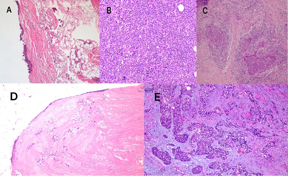
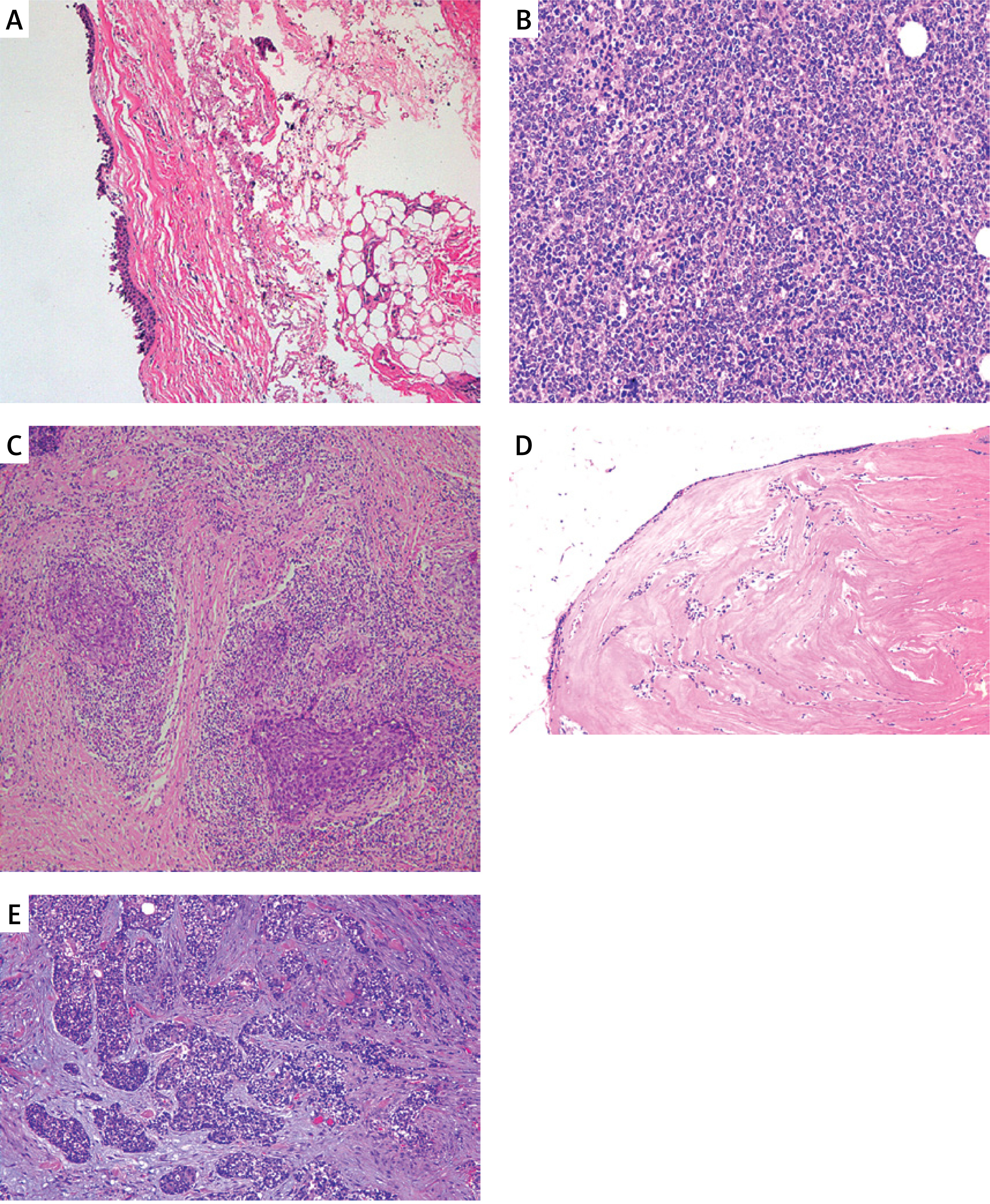
Histopathology showed substantial diagnostic heterogeneity: 16 lesions were benign, 7 malignant, and 8 inflammatory. Representative entities included angiofibroma, dermoid cyst, lymphoma, NUT carcinoma, idiopathic orbital inflammation, and Langerhans cell histiocytosis. Representative imaging and histopathological findings are shown in Figure 1.
Figure 1
Representative histopathological and radiological features of orbital-sinonasal interface tumors (OSITs), including benign tumors, malignant tumors, and inflammatory tumor-like lesions. A – Benign vascular (capillary hemangioma); B – diffuse large B-cell lymphoma (DLBCL); C – idiopathic orbital inflammation (IOI); D – benign epithelial (epidermal cyst); E – malignant epithelial (NUT carcinoma)
Symptomatology and ocular findings
Common symptoms were vision loss (13, 41.9%), exophthalmos (11, 35.5%), ocular pain (10, 32.3%), and external masses (8, 25.8%); less frequent were headache (8, 25.8%), tearing/nasal obstruction (6 patients each, 19.4%), diplopia (3, 9.7%), and facial paresthesia (3, 9.7%). Signs included conjunctival congestion/edema (14, 45.2%), restricted motility (11, 35.5%), ocular misalignment (7, 22.6%), and optic disc edema (3, 9.7%). Ptosis was more frequent in inflammatory tumors (p = 0.004), and optic disc edema occurred only in inflammatory lesions (p = 0.017) (Table II).
Table II
Clinical symptoms and ocular signs according to pathological category
| Variable | Benign N = 16 | Malignant N = 7 | Inflammatory N = 8 | Total N = 31 | P-value |
|---|---|---|---|---|---|
| Symptoms | |||||
| Eye swelling | 1 (6.3%) | 1 (14.3%) | 3 (37.5%) | 5 (16.1%) | 0.144 |
| Exophthalmos | 7 (43.8%) | 1 (14.3%) | 3 (37.5%) | 11 (35.5%) | 0.393 |
| External masses | 2 (12.5%) | 5 (71.4%) | 1 (12.5%) | 8 (25.8%) | 0.007* |
| Vision loss | 6 (37.5%) | 2 (28.6%) | 5 (62.5%) | 13 (41.9%) | 0.362 |
| Tearing | 2 (12.5%) | 2 (28.6%) | 2 (25%) | 6 (19.4%) | 0.599 |
| Ocular pain | 5 (31.3%) | 1 (14.3%) | 4 (50%) | 10 (32.3%) | 0.334 |
| Headache | 5 (31.3%) | 0 | 3 (37.5%) | 8 (25.8%) | 0.197 |
| Diplopia | 1 (6.3%) | 1 (14.3%) | 1 (12.5%) | 3 (9.7%) | 0.795 |
| Facial paresthesia | 1 (6.3%) | 2 (28.6%) | 0 | 3 (9.7%) | 0.140 |
| Nasal symptoms | 2 (12.5%) | 3 (42.9%) | 1 (12.5%) | 6 (19.4%) | 0.202 |
| Signs | |||||
| Ptosis | 3 (18.8%) | 2 (28.6%) | 7 (87.5%) | 12 (38.7%) | 0.004* |
| Conjunctival congestion and edema | 4 (25%) | 5 (71.4%) | 5 (62.5%) | 14 (45.2%) | 0.062 |
| Corneal edema | 3 (18.8%) | 0 | 0 | 3 (9.7%) | 0.211 |
| Optic disk edema | 0 | 0 | 3 (37.5%) | 3 (9.7%) | 0.017* |
| Restricted motility | 3 (18.8%) | 2 (28.6%) | 6 (75%) | 11 (35.5%) | 0.023* |
| Ocular misalignment | 3 (18.8%) | 0 | 4 (50%) | 7 (22.6%) | 0.060 |
External masses were more common in malignant tumors (71.4%, p = 0.007), while ptosis (87.5%, p = 0.004), optic disc edema (p = 0.017), and ocular motility restriction (75%, p = 0.023) were significantly associated with inflammatory lesions.
Tumor location and invasion pattern, treatment modalities, and surgical outcomes. Tumor invasion site and treatment protocol are presented in Supplementary Tables SI and SII. Surgery aimed to eradicate the lesion (malignant or benign) while preserving ocular/neurovascular structures, restoring anatomy, and preventing recurrence/complications. All patients had preoperative CT/MRI, and approaches were chosen by tumor location, depth, and histopathology. Techniques: anterior orbitotomy (7, 22.6%), medial orbitotomy (5, 16.1%), transconjunctival (3, 9.7%), and orbital exenteration (5, 16.1%); no lateral orbitotomies were performed (Table III).
Table III
Surgical approach
Postoperative clinical improvement and complications
Overall, most patients demonstrated symptomatic improvement after definitive treatment (surgery or medical therapy), particularly in pain, proptosis, and nasal symptoms. Among the 27 surgically treated patients, there was no perioperative mortality or major neurological event; however, postoperative complications occurred in 14 (51.9%) patients, representing 45.2% of the total cohort, including post-treatment vision loss in 2 cases, and outcomes varied by pathology and treatment extent. Complications occurred in 14 patients of the total cohort (45.2%): ocular (6, 19.4%), nasal (4, 12.9%), facial numbness (3, 9.7%), systemic (1, 3.2%). Ocular complications were more frequent in malignant lesions (p = 0.018); nasal complications were more frequent in the malignant and inflammatory groups (p = 0.012); facial numbness was also associated with malignancy (p = 0.008) (Table IV).
Table IV
Postoperative complications according to pathological category
| Parameter | Benign N = 16 | Malignant N = 7 | Inflammatory N = 8 | Total N = 31 | P-value |
|---|---|---|---|---|---|
| Ocular complications | 1 (6.25%) | 4 (57.1%) | 1 (12.5%) | 6 (19.4%) | 0.018* |
| Vision loss | 0 | 1 (14.3%) | 1 (12.5%) | 2 (6.5%) | 0.226 |
| Ptosis | 0 | 1 (14.3%) | 0 | 1 (3.2%) | 0.17 |
| Ocular motility disorders | 1 (6.25%) | 1 (14.3%) | 0 | 2 (6.5%) | 0.467 |
| Tearfulness | 0 | 1 (14.3%) | 0 | 1 (3.2%) | 0.226 |
| Nasal complications | 0 | 3 (42.9%) | 1 (12.5%) | 4 (12.9%) | 0.012* |
| Rhinorrhea | 0 | 2 (28.6%) | 1 (12.5%) | 3 (9.7%) | 0.058 |
| Oral nasal fistula | 0 | 1 (14.3%) | 0 | 1 (3.2%) | 0.226 |
| Facial numbness | 0 | 3 (42.9%) | 0 | 3 (9.7%) | 0.008* |
| Systemic | 0 | 0 | 1 (12.5%) | 1 (3.2%) | 0.484 |
| Infection | 0 | 0 | 0 | 0 | |
| Abnormal body temperature | 0 | 0 | 1 (12.5%) | 1 (3.2%) | 0.258 |
Malignant tumors were significantly associated with ocular complications, including postoperative vision loss and motility disorders (p = 0.018). Similarly, facial numbness was reported only in malignant cases (p = 0.008), possibly indicating perineural invasion or surgical manipulation near the infraorbital or trigeminal branches. Nasal complications, such as rhinorrhea and oral‒nasal fistulas, were more frequently observed in the malignant and inflammatory groups (p = 0.012), suggesting that more extensive mucosal or bony involvement requires aggressive resection.
Discussion
Unlike WHO histopathological classifications, which define tumor entities, and AJCC/TNM staging, which primarily describes malignant disease extent, our classification provides spatial guidance based on the anatomical origin and direction of orbital-sinonasal extension [14, 15]. Previous cranial–nasal–orbital and surgical-access frameworks address broader communicating tumors or operative corridors [14–17], whereas the present system focuses specifically on the orbital-sinonasal interface. Its principal potential advantage is its simple application to preoperative CT and MRI, which may help multidisciplinary teams anticipate an orbital, endonasal, or combined surgical approach [18]. However, it is intended to complement, rather than replace, histopathological diagnosis, oncological staging, and individualized surgical judgment.
In this cohort, pathological diagnoses included benign tumors, such as dermoid cysts and angiofibromas; malignant tumors, such as lymphoma and NUT carcinoma; and inflammatory tumor-like lesions, such as idiopathic orbital inflammation and Langerhans cell histiocytosis. This diversity highlights the need for anatomically informed and pathology-aware treatment planning. Malignant lymphoepitheliomas and NUT carcinomas display high rates of perineural invasion and subsequent bony invasion, which contributes to higher complication rates, reinforcing prior findings that the biological behavior of sinonasal malignancies is highly variable and requires careful preoperative planning [11, 12]. These symptom‒pattern associations further validate the clinical relevance of the proposed anatomical classification. Inflammatory lesions, including idiopathic orbital inflammation and pseudotumors, present diagnostic challenges due to radiological overlap with neoplasms. Our findings echo prior reports that highlight the importance of histopathology when imaging is inconclusive [19] and further support the continued role of biopsy in patients with atypical imaging or rapid progression. Moreover, our study reinforces that histopathological diversity – from benign mucoceles to aggressive malignancies – necessitates individualized planning. Although they are often indistinguishable from neoplastic masses radiographically, inflammatory pseudotumors may respond to corticosteroids or limited resection [20]. Conversely, lymphomas or sinonasal carcinomas may require adjuvant therapy, as shown in previous reports [3].
Among surgically treated patients, the postoperative complication rate was 51.9% (14/27), with ocular and nasal complications being the most frequent. Recent ophthalmic evidence has similarly documented motility, eyelid, lacrimal, and other periocular complications in patients receiving eye-sparing treatment for sinonasal tumors with orbital invasion, highlighting the need for structured postoperative ophthalmic surveillance [21]. Complications were more common in malignant lesions, which may reflect more invasive disease and the need for wider surgical exposure. Despite these risks, the high resection rate suggests that imaging-guided, anatomy-based classification may support operative decision-making in selected patients.
The proposed classification remains preliminary because it was retrospectively derived from 31 patients at a single center. Its limitations include the small pathological subgroups, predominance of mixed-type lesions, absence of interobserver-reliability testing, and limited long-term functional and recurrence data. Future multicenter prospective studies should assess interobserver reproducibility and additional parameters, including lesion volume, orbital-wall destruction, orbital-apex or optic-nerve involvement, intracranial extension, visual and ocular-motility outcomes, recurrence, and quality of life. They should also determine whether the mixed category requires further subdivision. Integration with standardized imaging criteria and practical surgical algorithms may further strengthen its clinical applicability. Until validated, the classification should be considered a complementary surgical-planning framework rather than a definitive staging or prognostic system.
In conclusion, OSITs pose substantial diagnostic and therapeutic challenges because of their anatomical complexity and broad pathological spectrum, which includes benign tumors, malignant tumors, and inflammatory tumor-like lesions. We developed a novel classification system based on tumor origin and extent of tumor extension, categorizing cases into five anatomical categories based on location and extent: ethmoid, frontal, maxillary, sphenoid, and multiple sites. The classification may help guide the selection of tailored surgical approaches and multidisciplinary planning between ophthalmology and otolaryngology teams. Multidisciplinary planning, advanced imaging, and appropriately selected surgical techniques may facilitate the management of benign, malignant, and inflammatory OSITs. Surgical resection was achieved in 87.1% of patients, although postoperative complications occurred in 51.9% of surgically treated patients. Our findings suggest that an anatomical classification may support surgical decision-making and help guide surgical planning for OSITs.
This study has limitations: its retrospective design limits generalizability; the small malignant and inflammatory subgroups limited detailed analyses of pathological subtypes; and long-term outcomes (recurrence, quality of life) were not captured. Future multicenter prospective studies with standardized imaging, surgical algorithms, and outcome measures are needed to assess the reproducibility and clinical utility of the classification, assess recurrence and functional outcomes, and refine surgical planning. Until validated, the proposed classification should be considered a potential surgical-planning framework rather than a definitive staging or prognostic system.