Introduction
Lung cancer is one of the most common malignancies in the world. In 2012, a total of 1.8 million new cancer cases and 1.6 million related deaths were reported worldwide [1]. Approximately 10–15% of lung cancers are small cell lung cancers, and about 85% are non-small cell lung cancers, including adenocarcinoma, large cell carcinoma, squamous cell carcinoma, etc. [2]. The 5-year survival rate of lung cancer is currently only 16%, and more than 70% of patients are diagnosed with an advanced stage due to the lack of early clinical symptoms [3].
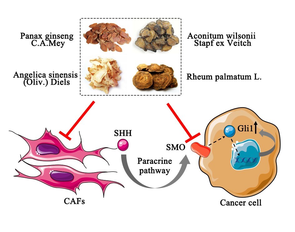
In traditional Chinese medicine (TCM), the formation of cancer is considered to be caused by the infiltration of a pathogen based on a deficiency of vital-qi [4, 5]. TCM can inhibit tumor growth and metastasis through overall regulation of the tumor microenvironment, thereby regulating and improving the clinical symptoms of patients, improving patients’ quality of life and extending their survival [6]. Therefore, the regulatory effect of TCM on the tumor microenvironment has become one of the main research directions of TCM against tumors. The tumor microenvironment (TME) consists of a complex network of heterogeneous cell types, including infiltrating immune cells, cancer associated fibroblasts (CAFs), granulocytes, extracellular matrix, endothelial cells, and other stromal components [7]. Among them, CAFs are the main components of stromal cells and can secrete a variety of cytokines to regulate tumor growth and metastasis [8]. Previous study has shown that CAF mediated activation of the Hh-Gli1 pathway is one of the most important molecular mechanisms for promoting the growth, invasion and metastasis of lung cancer [9].
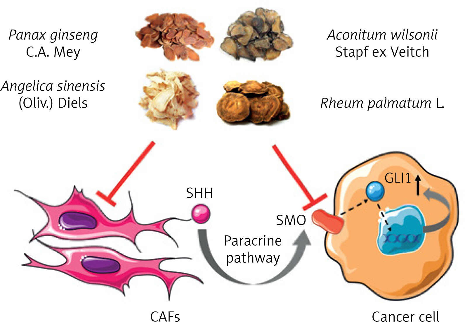
Wenxia Formula (WF) is a TCM formula consisting of four crude herbs: Rheum palmatum L., Aconitum wilsonii Stapf ex Veitch, Panax ginseng C.A. Mey and Angelica sinensis (Oliv.) Diels. The combination of the four herbs utilizes the methods of warming, tonifying, purgation and elimination, and achieves the effect of dispelling cold, restoring yangqi, purgation of stagnation and clearing blood stasis [10]. The results of modern pharmacological studies have shown that the four Chinese herbal medicines have significant antitumor effects [11–14]. Our previous studies have shown that WF can significantly inhibit the proliferation of lung adenocarcinoma cells, induce apoptosis and significantly reduce the expression of PCNA and cyclin D1 [15–18]. However, the specific antitumor components and mechanism of action of WF are not clear.
In order to facilitate the study of the material basis of WF, the WF was extracted with ethanol and oil ether, dichloromethane, ethyl acetate and n-butanol. Four extraction sites were obtained, and the effective antitumor extracts of WF were screened by a cell experiment in vitro. We selected human lung adenocarcinoma cells A549 and H1299, human lung squamous cell carcinoma H520, and human large cell lung cancer H460 to investigate their antitumor activities in vitro. By calculating the half-inhibitory concentration (IC50), it was found that the ethyl acetate extract from WF (WFEA extract) had the best antitumor activity. In this study, we aimed to illuminate the possible anti-cancer mechanisms of the WFEA extract.
Material and methods
Animals
BALB/c Nude mice (6-8 weeks, weighing 20 ±2 g) were purchased from Beijing Vital River Laboratory Animal Technology Co. Ltd (Beijing, China). They were housed in groups of 4-6 per cage in laminar flow hoods in a pathogen-free environment (55 ±10% humidity, 22 ±2°C and 12–12 h/light-dark cycle) with free access to a standard laboratory water and diet. The animals were acclimatized for 1 week before the experiment. The study was approved by the Institutional Animal Care and Use Committee (animal authorization reference number: SCXK2018-0034) at Shandong University of Traditional Chinese Medicine (Shandong, China). Animal welfare and experimental procedures were strictly in accordance with the Guide for the Care and Use of Laboratory Animals (the Ministry of Science and Technology of China, 2006). All efforts were made to minimize the suffering of animals and the number of animals used.
Preparation of WFEA extract
Rheum palmatum L., Aconitum wilsonii Stapf ex Veitch, Panax ginseng C.A. Mey and Angelica sinensis (Oliv.) Diels were purchased from Shandong Zhonglu hospital. According to the ratio of 9 : 12 : 12 : 6, 9750 g of Panax ginseng C.A. Mey, Rheum palmatum L., Aconitum wilsonii Stapf ex Veitch and Angelica sinensis (Oliv.) Diels dried herbs were weighed. The crushed herbs were extracted ultrasonically twice with 75% ethanol. After being filtered, the extracts were concentrated to have no alcohol taste, and finally the WFEA extract was obtained. Then, it was extracted three times with petroleum ether, dichloromethane, ethyl acetate and n-butanol, respectively. The extracts were combined and concentrated to obtain petroleum ether partial extract, chloroform partial extract, ethyl acetate partial extract and n-butyl alcohol partial extract, respectively.
Ultra-performance liquid chromatography quadrupole time-of-flight mass spectrometry (UPLC/Q-TOF-MS) analysis
A XDB-C18 column (4.6 × 100 mm, 1.8 µm) was utilized for separation with the column temperature at 35°C. The standards and samples were separated using a gradient mobile phase consisting of (A) acetonitrile and (B) 0.1% formic acid in water. The flow rate was at 0.5 ml/min. The injection volume of sample was 1 µl. Mass spectrometry was performed on a Bruker Q-TOF equipped with an electrospray ionization source in a positive ion mode. The temperatures of the electrospray source and desolvation were 200°C and 325°C, respectively. The flow rate of nitrogen was set to 8 l/min. The ESI capillary and cone voltages were set at 3,500 and 30 V, respectively. The MS/MS analysis was performed using a variable collision energy (15–45 eV), which was optimized for each individual constituent.
Cell culture
The A549 cell line was purchased from Shanghai Institute of Biochemistry and Cell Biology, Chinese Academy of Sciences. Luciferase (Luc) gene-labeled human lung adenocarcinoma A549 cells (A549-Luc) were purchased from Keygen Biotech (Jiangsu China). Human Lung Adenocarcinoma CAFs were purchased from iCell-CRO (Shanghai, China). A549 and A549-Luc were grown in RPMI-1640; the CAFs were cultured in DMEM. Medium was supplemented with 10% fetal bovine serum (FBS, Gibco, USA), 100 U/ml penicillin and 100 µg/ml streptomycin in an incubator at 37°C with a 5% CO2 atmosphere.
Xenograft model procedure
Approximately 4 × 10⁶ cells were suspended in FBS-free medium and mixed with Matrigel (200 µl). A549 and A549 plus CAFs (1 : 1) cells were respectively injected subcutaneously into the mouse right flank. When tumor nodules appeared at the injection site, groups of nude mice injected with A549 plus CAFs cells were divided into 5 groups (WFEA high, WFEA middle, WFEA low, cisplatin (CDDP) and control group). The CDDP group was given an intraperitoneal injection of 5 mg/kg cisplatin twice a week; the WFEA group was given an intragastric administration of WFEA extract at doses of 400 mg/kg, 200 mg/kg and 100 mg/kg, respectively; the control group and A549 group were given intragastric administration of equal volume normal saline. Each group was continuously intervened for 5 weeks.
Immunofluorescent staining
Tissue sections were dewaxed with xylene and dehydrated with graded alcohol. After washing with distilled water, tissue sections were heated at high pressure in ethylene diamine tetraacetic acid (EDTA) antigen repair buffer (pH 8.0) for antigen retrieval. After the slices were cooled at room temperature, sections were incubated with primary antibodies (1 : 500, 4°C, overnight) and secondary antibodies (37°C, 50 min), respectively. Then paraffin sections were incubated with 4ρ,6-diamidino-2-phenylindole (DAPI, Beyotime Biotechnology, China) for 10 min in the dark. Finally, paraffin sections were sealed in anti-fluorescence. Images were acquired on a laser scanning confocal microscope FV12-IXCOV (OLYMPUS, Japan). Red fluorescence represented the positive expression of genes. The mean optical density was calculated by Image-J to analyze the positive expression.
Immunohistochemistry
Tissue sections were deparaffinized with xylene and rehydrated with ethanol, followed by antigen retrieval. After the slices were cooled at room temperature, tissue sections were incubated with primary antibodies (37°C, 2 h) and secondary antibodies (37°C, 30 min), respectively. Following a final washing in PBS, α-SMA and FAP signals were visualized with a DAB kit (Sigma, St. Louis, MO, USA), counterstained with hematoxylin and mounted in Kaiser’s glycerol gelatine. For semi-quantitative measurements of α-SMA and FAP density, the slides were photographed and analyzed using a computer-aided image-analyzing system (Motic Images Advanced 3.2).
Hematoxylin and eosin (HE) staining
Sections were dewaxed in xylene and rehydrated in a series of alcohol solutions. After washing in distilled water for a short time, the sections were stained in Harris hematoxylin solution for 5 min, washed in tap water and counterstained in eosin-phloxine solution for 2 min.
Quantitative real-time polymerase chain reaction (qRT-PCR)
Total RNA was isolated using TRIzol reagent (Invitrogen, USA). The cDNA of Ptch1, Smo and Gli1 was prepared using the RevertAid First Strand cDNA Synthesis Kit. The qRT-PCR analysis was performed using the SYBR Green RT-PCR Kit (Takara Bio, Japan) on an ABI 7500 Fast Real-Time PCR System (Applied Biosystems, USA). β-Actin was used as the endogenous control to calculate the relative RNA levels. Data were calculated by the comparative cycle threshold (CT) (2−ΔΔCT) method. The PCR primer sequences were as follows:
(1) Ptch1 forward: 5’-GCAACACCTGGACTCAGCACTC-3’; and reverse: 5’-AACCTGTCTCCGTGATAAGTTCC-3’;
(2) Smo forward: 5’-GTGCTTATTGTGGGAGGCTACTTC-3’; and reverse: 5’-TTGGCATAGCACATAGTCCCG-3’;
(3) Gli1 forward: 5’-GACTTTCTGGTCTGCCCTTTTG-3’; and reverse: 5’-GAGAGAGCCCGCTTCTTTGTTA-3’;
(4) Shh forward: 5’-AAAGCAGAGAACTCCGTGGC-3’; and reverse: 5’-CACTAACTTGGTGCCACCCT-3’;
(5) β-Actin forward: 5’-GTGACGTTGACATCCGTAAAGA-3’; and reverse: 5’-GTAACAGTCCGCCTAGAAGCAC-3’.
Western blot
Tumor tissue lysates were prepared using a proper amount of RIPA lysate, and the BCA method was used for protein quantification. Equal amounts of proteins (20 µg) of each sample were separated by sodium dodecyl sulfate-polyacrylamide gel electrophoresis and then transferred onto the polyvinylidene fluoride (PVDF) membranes (Millipore, USA). PVDF membrane was blocked in 5% skim milk at 37°C for 1 h, and subsequently incubated at 4°C overnight with antibodies against SHH, PTCH1, SMO, GLI1 or ACTIN (all in 1 : 1000 dilutions, Santa Cruz, USA). Next, PVDF membrane was incubated with corresponding horseradish peroxidase-conjugated (HRP)-linked secondary IgG antibodies (anti-rabbit or anti-mouse, 1 : 2000 dilutions, Santa Cruz, USA) for 1 h at room temperature. The signal was detected with a Super Signal Protein Detection kit (Pierce Biotechnology, USA).
Statistical analysis
All statistical analyses were performed using SPSS version 22.0 (IBM Corp., IL, USA). Results from each experiment were expressed as the mean ± standard deviation of three separate experiments. Statistical significance was determined with Student’s t-test. One-way analysis of variance (ANOVA) was used to perform the between-group comparisons. Statistical significance was set at p < 0.05.
Results
Identification of constituents of WFEA extract
UPLC-Q-TOF/MS was applied to analyze and identify the constituents of WFEA extract. Through the standard comparison, literature review, and disassociation rules, 15 chemical components were identified, and the results are shown in Table I and Supplementary Figure S1.
Table I
Identification results of the constituents of WFEA extract
WFEA extract inhibits the tumorigenicity of lung cancer cells
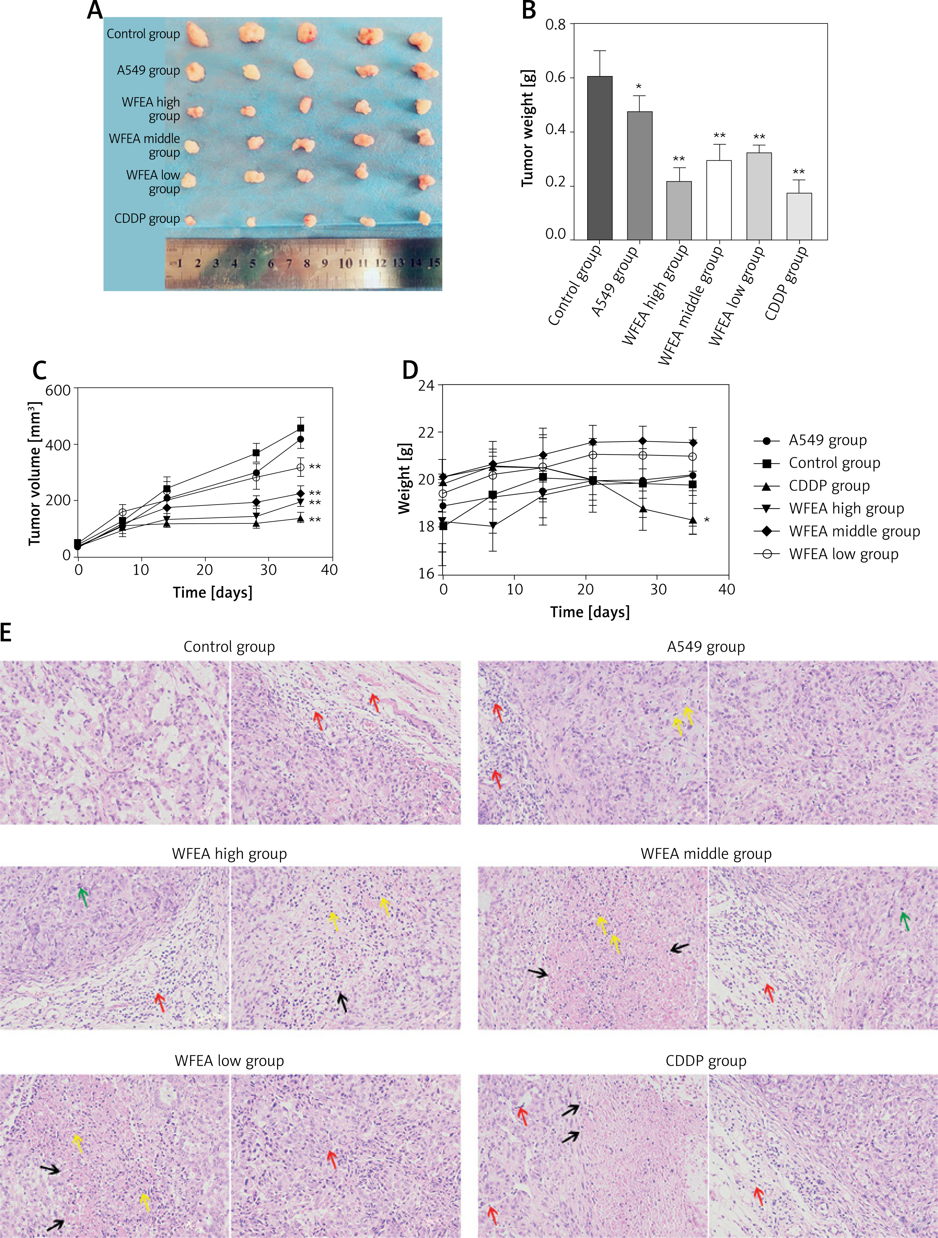
The antitumor activity in vivo of the WFEA extract was tested in lung cancer xenograft model. The results showed that the weight and volume of xenograft in the control group were greater than those in the A549 group, indicating that CAFs could promote lung cancer cells growth. The weight and volume of xenograft in the WFEA high/medium/low group were significantly lower than those in the control group, indicating that WFEA extract could inhibit lung cancer cells growth in vivo (Figures 1 A–C). In addition, we found that the body weight of mice in the A549 group, control group and WFEA group showed an increase trend, but there were no significant difference (all p > 0.05). Compared with the control group, the body weight of mice in CDDP group decreased significantly (Figure 1 D).Taken together, these results showed that WFEA extract could inhibit lung cancer cells growth in vivo and was safer than CDDP.
Figure 1
WFEA extract inhibits the tumorigenicity of lung cancer cells. A – optical image showing mouse tumors; B – weight of tumor in each group; C – quantification of tumor volume; D – body weight of mice in each group; E – HE staining (×200) of tumors in each group. The black arrow represents nuclear fragmentation or dissolution of necrotic tumors; yellow arrow represents neutrophil infiltration; red arrow represents inflammatory cell infiltration; green arrow represents karyokinesis. *p < 0.05, **p < 0.01 (compared with control group); CDDP – cisplatin
WFEA extract decreased expression levels of α-SMA and FAP
The immunohistochemistry results showed that the expression levels of a-smooth muscle actin (α-SMA) and fibroblast activation protein (FAP) in the xenograft of the control group were significantly higher than those in the A549 group (p < 0.05), indicating that the activation of CAFs could significantly increase the expression levels of α-SMA and FAP (Figure 2).
Figure 2
Digital images of xenograft tissue sections after FAP and α-SMA immunohistochemical staining. A – digital images of xenograft tissue sections after FAP immunohistochemical staining and quantification of the expression of FAP. B – digital images of xenograft tissue sections after α-SMA immunohistochemical staining and quantification of the expression of α-SMA. *p < 0.05, **p < 0.01 (compared with control group); #p < 0.05, ##p < 0.01 (compared with CDDP group); CDDP – cisplatin
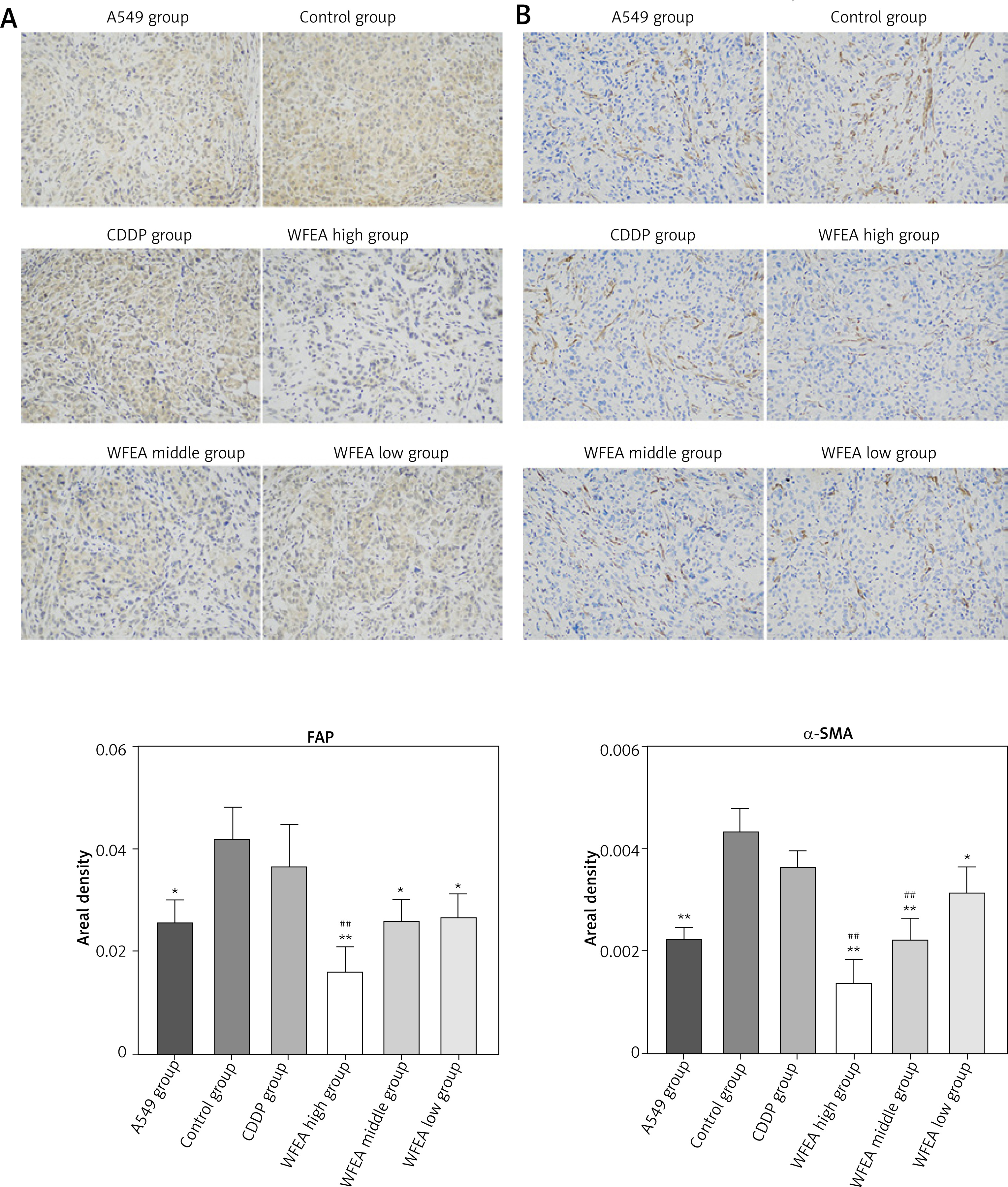
In addition, we found that the expression levels of α-SMA and FAP in the WFEA high/medium/low group were significantly lower than those in the control group, indicating that WFEA extract could significantly decrease the expression levels of α-SMA and FAP in the CAF mediated lung cancer xenograft model.
Effect of WFEA extract on E-cadherin and N-cadherin
In order to illuminate the possible anti-cancer mechanisms of the WFEA extract, we next examined the effects of WFEA extract on marker proteins of epithelial-mesenchymal transition (EMT). The immunofluorescence staining results showed that the expression of E-cadherin in the xenograft of the A549 group and WFEA group was significantly higher than that in the control group (Figure 3).
Figure 3
Digital images of xenograft tissue sections after E-cadherin and N-cadherin immunofluorescence staining. A – digital images of xenograft tissue sections after E-cadherin and N-cadherin immunofluorescence staining. Blue fluorescence represents cell nucleus and red fluorescence represents E-cadherin and N-cadherin. B – quantification of the expression of E-cadherin. C – quantification of the expression of N-cadherin. *p < 0.05, **p < 0.01 (compared with control group); #p < 0.05, ##p < 0.01 (compared with CDDP group); CDDP – cisplatin
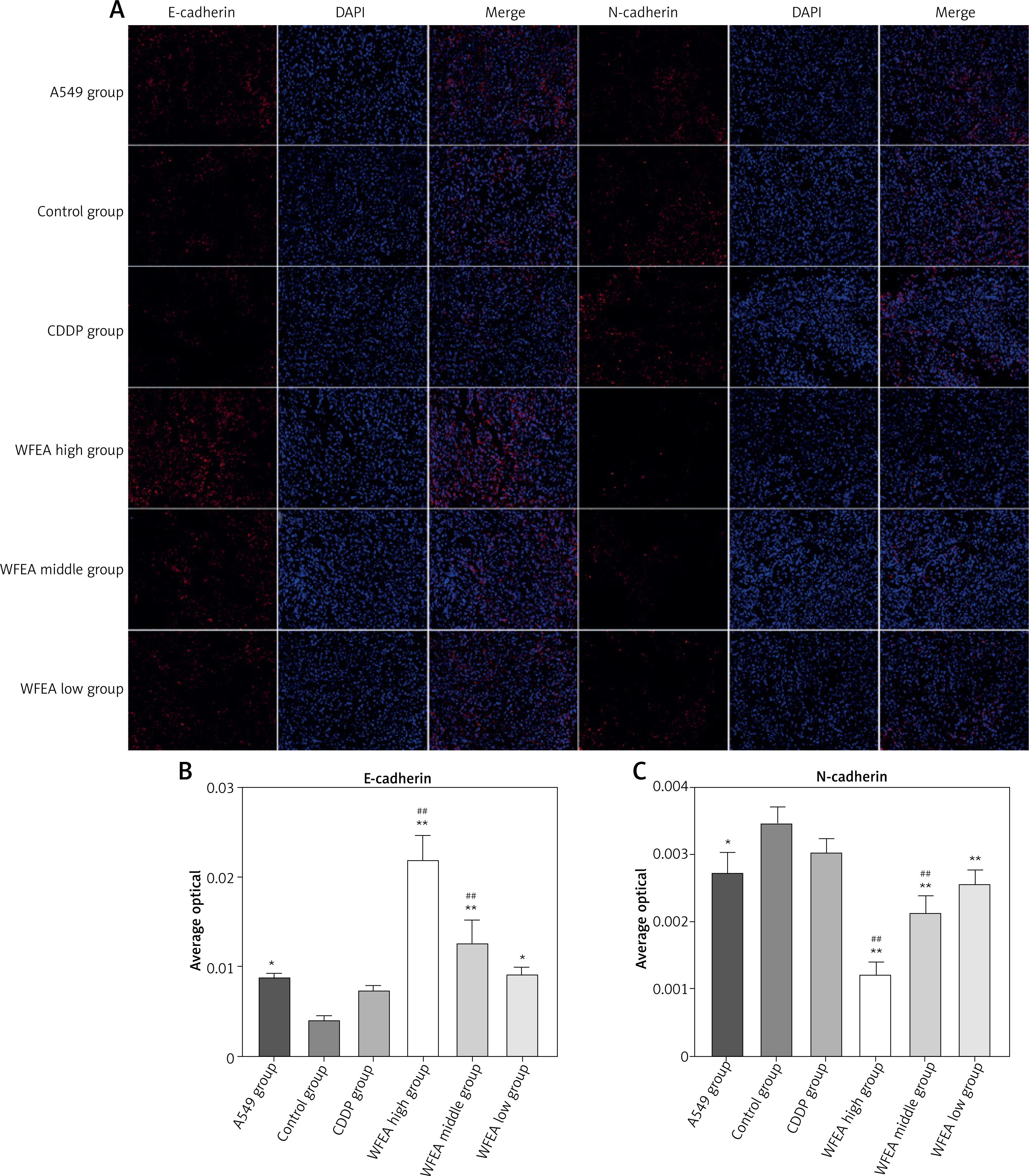
In addition, we found that the expression of N-cadherin in the xenograft of the A549 group and WFEA group was significantly lower than that in the control group. Taken together, these results showed that WFEA extract could inhibit EMT of lung cancer cells, thereby inhibiting tumor growth and metastasis.
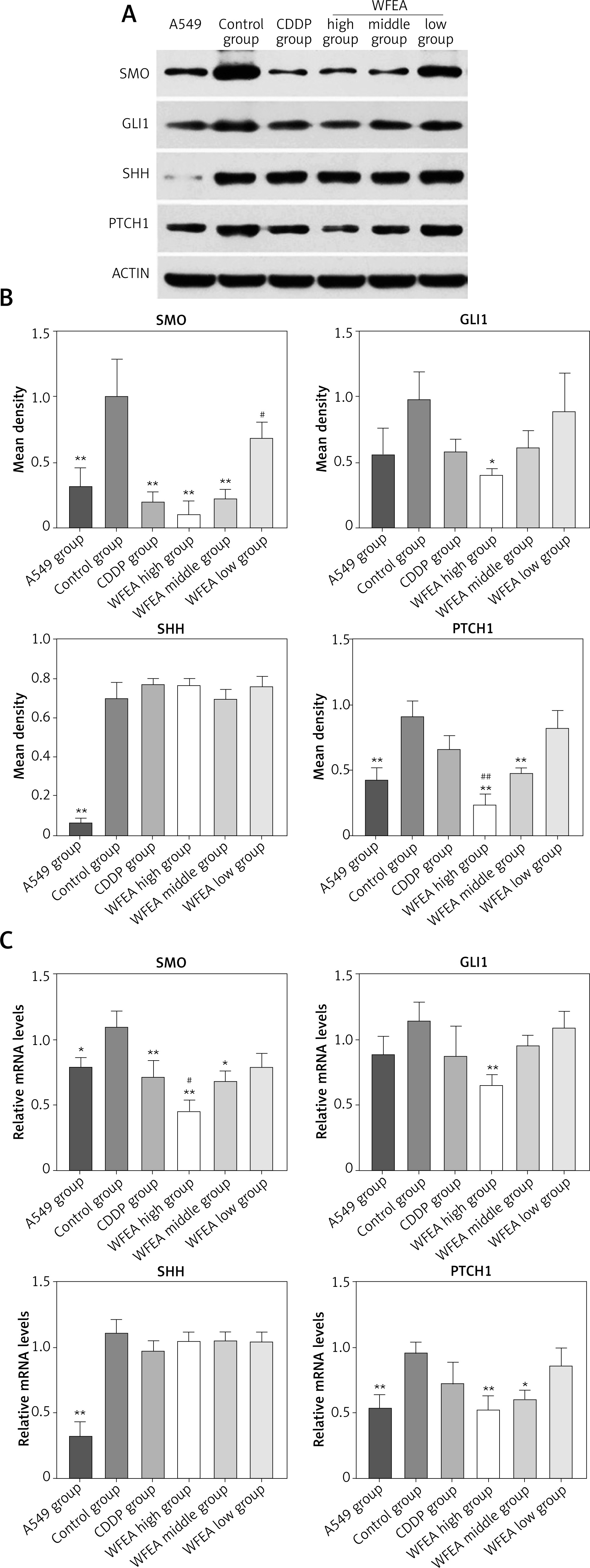
Effect of WFEA extract on Hh-Gli1 signal pathway
Western blot and qRT-PCR showed that the expression levels of related proteins and genes (SMO, GLI1, SHH and PTCH1) in the Hh-Gli1 signaling pathway of the A549 group and WFEA group were significantly lower than those in the control group (Figure 4). These results indicated that the WFEA extract could inhibit the Hh-Gli1 signaling pathway mediated by CAF activation in the lung cancer xenograft model (Figure 5).
Figure 4
Expression of related proteins and genes (SMO, GLI1, SHH and PTCH1) in Hh-Gli1 signaling pathway. A – Western blot analysis of SMO, GLI1, SHH and PTCH1 expression in xenograft. B – quantification of the expression of SMO, GLI1, SHH and PTCH1. C – real-time PCR analysis of Smo, Gli1, Shh and Ptch1 mRNA in xenograft. *p < 0.05, **p < 0.01 (compared with control group); #p < 0.05, ##p < 0.01 (compared with CDDP group); CDDP – cisplatin
Discussion
With the development of science and technology, the research on the extraction of effective anti-tumor components from Chinese herbal medicine and the compatibility of Chinese herbal medicine based on the theory of Chinese medicine has attracted increasing attention from the medical community [19]. Some traditional Chinese medicine (e.g. baicalein and Shen-Mai San) has been confirmed to be effective in treating tumors [20–22]. WF is a TCM formula consisting of four crude herbs: Rheum palmatum L., Aconitum wilsonii Stapf ex Veitch, Panax ginseng C.A. Mey and Angelica sinensis (Oliv.) Diels. Our previous studies have shown that WF can significantly inhibit the proliferation of lung adenocarcinoma cells, induce apoptosis and significantly reduce the expression of PCNA and cyclin D1 [15–18]. However, the specific antitumor components and mechanism of WF are not clear. In this study, we aimed to illuminate the possible anti-cancer mechanisms of the WFEA extract.
In this study, we found that the weight and volume of the xenograft in the WFEA high/medium/low group were significantly lower than those in the control group, indicating that the WFEA extract could inhibit lung cancer cell growth in vivo. These observations were consistent with our previous study [18], which showed that WFEA extract could significantly reduce the tumor volume from the 19th day and alleviate the tumor weight. In addition, we found that WFEA extract had no significant effect on the body weight of mice, while the CDDP group had significant weight loss, indicating that WFEA extract was safer than CDDP.
CAFs are the most important cells in the process of tumorigenesis, which could lead to the initiation and progression of malignant tumors. Among them, α-SMA and FAP are the most widely recognized markers of CAFs [23]. In this study, we found that the expression of α-SMA and FAP in the tumor tissues of mice co-injected with CAFs and A549 cells (control group) was significantly higher than that of mice injected with A549 alone (A549 group) and the expression levels of α-SMA and FAP in the WFEA high/medium/low group were significantly lower than those in the control group. These results indicating that WFEA extract could significantly inhibit the expression of α-SMA and FAP and affect the activation of CAFs in tumor tissues.
Epithelial-mesenchymal transition is a key process in tumor growth and metastasis, which includes the decrease of the epithelial cell marker E-cadherin and the up-regulation of N-cadherin and other mesenchymal cell-related markers, so as to reduce the adhesion of cells and increase the invasion and metastasis ability [24]. In this study, we found that the expression of E-cadherin in the xenograft of the A549 group and WFEA group was significantly higher than that in the control group (Figure 4).
In addition, we found that the expression of N-cadherin in the xenograft of the A549 group and WFEA group was significantly lower than that in the control group. Taken together, these results showed that WFEA extract could inhibit EMT of lung cancer cells, thereby inhibiting tumor growth and metastasis.
The Hh-Gli1 signaling pathway is important for inducing embryonic and stem cell development, and the abnormal activation of this pathway is closely related to the development of many malignant tumors [25]. CAF mediated activation of the Hh-Gli1 signaling pathway is one of the most important molecular mechanisms for promoting the growth and metastasis of lung cancer [9]. In this study, we found that the expression levels of related proteins and genes (SMO, GLI1, SHH and PTCH1) in the Hh-Gli1 signaling pathway of the A549 group and WFEA group were significantly lower than those in the control group. These results indicate that WFEA extract could inhibit the Hh-Gli1 signaling pathway mediated by CAF activation.
In conclusion, WFEA extract could inhibit the EMT of lung cancer, and affect the growth and metastasis of tumor tissue. Its mechanism may be related to the CAF mediated Hh-Gli1 signaling pathway. In this study, the antitumor effect and mechanism of WFEA extract were explored in vivo. The next step of our group is to further verify the antitumor effect of WFEA extract and its possible mechanism in vitro.