

Introduction
According to the World Health Organization, cardiovascular diseases (CVD) constitute the most common cause of death worldwide. In 2019, 17.9 million deaths due to CVD were recorded, accounting for 32% of the total number of deaths. Regardless of the region, the high mortality rate from cardiovascular diseases is largely determined by disorders underlying atherosclerosis [1]. There is a constant search for new predictors of CVD [2, 3].
An important role in the development of atherosclerosis is played by the immune process involving immune system cells and the mediators released by these cells. There is substantial evidence indicating the crucial, although not fully understood, role of neutrophils (polymorphonuclear neutrophils, PMNs) in the pathogenesis of atherosclerosis [4]. It has been proven that the presence of neutrophils within the atherosclerotic plaque is associated with both its formation and development as well as with destabilization. Neutrophils promote inflammatory responses and recruit immune-competent cells to atherosclerotic lesions, among other mechanisms, by releasing reactive oxygen species (ROS) and numerous enzymes [5].
The wave of experiments conducted on neutrophils was initiated by Brinkmann et al., who discovered an interesting aspect of neutrophil biology: the formation and release of neutrophil extracellular traps (NETs) into the extracellular space, through a form of cell death known as NETosis [6]. NETs are composed of numerous proteins and enzymes as well as strands of DNA and histones [7–11]. The results of many experiments have revealed that NET generation requires the activity of the NADPH oxidase complex that initiates the production of reactive oxygen species by reducing molecular oxygen to superoxide anion [12, 13].
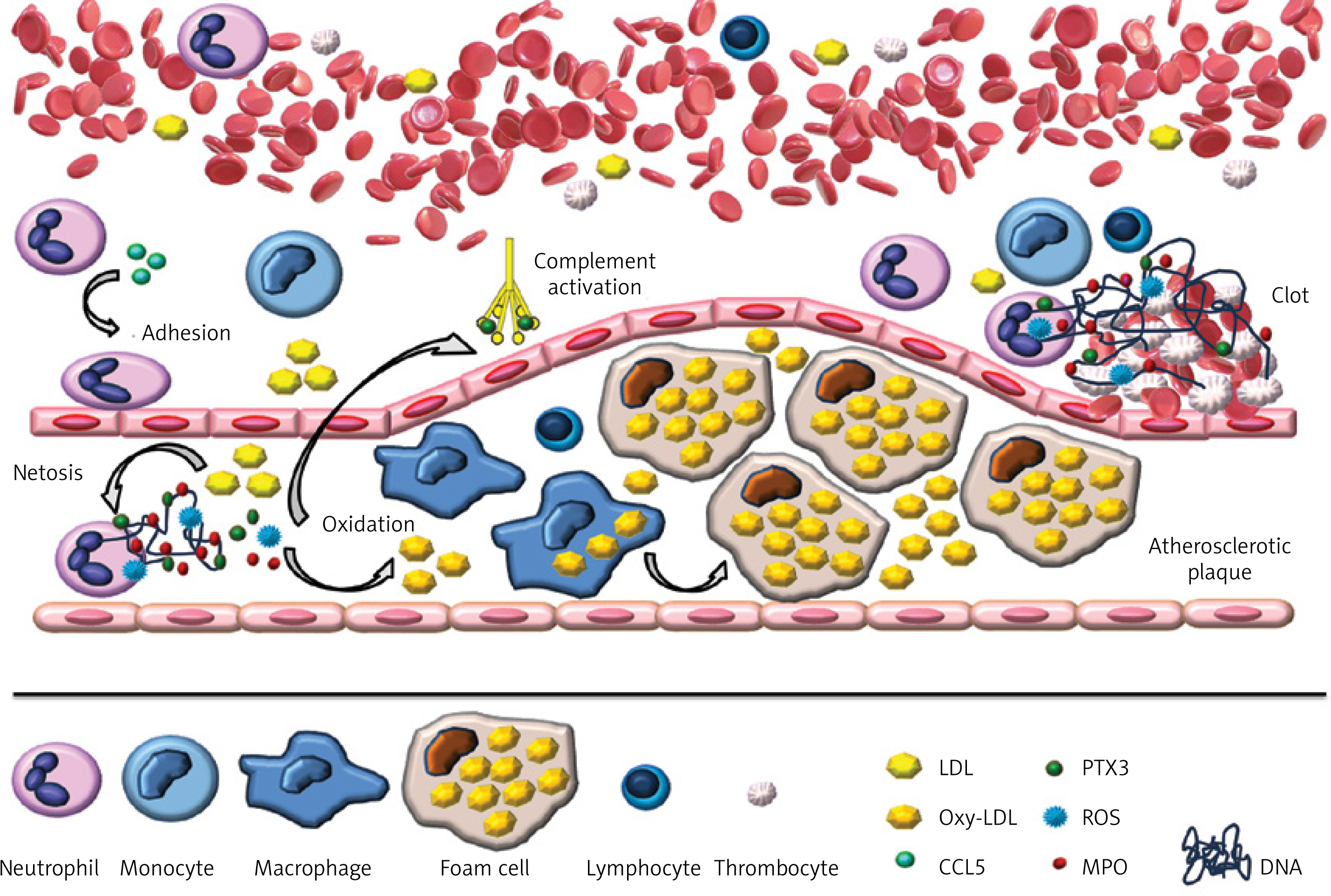
Numerous scientific studies have shown that NET generation plays a significant role in the body’s immune response to infection. NET structures are attributed with the ability to bind, neutralize, and eliminate a variety of pathogens, including bacteria, fungi, protozoa, and viruses [6, 11, 14–18]. By creating a physical barrier, NETs facilitate the degradation of ‘trapped’ pathogens and their virulence factors, thereby preventing the spread of microorganisms in the body [19]. Although the beneficial role of NETs in fighting pathogens has been proven, an excessive amount of traps may contribute to damage to surrounding tissues [20]. It has been observed that the increased NET generation, as well as a deficiency in DNase, which is responsible for the degradation of the traps, may be associated with the pathogenesis of autoimmune diseases as well as cardiovascular disorders, including atherosclerosis [21]. To date, studies analyzing the formation of NETs within the course of atherosclerosis are few and primarily involve experiments conducted on animal models [22]. Figure 1 presents a model of the involvement of neutrophils and NETs in the pathophysiology of atherosclerosis.
Figure 1
Model of the involvement of neutrophils and NETs in the pathophysiology of atherosclerosis. Cholesterol crystals induce the formation of NETs, which in turn affect LDL oxidation, activation of monocytes/macrophages, and the complement system. In addition, NETs participate in the formation of clots
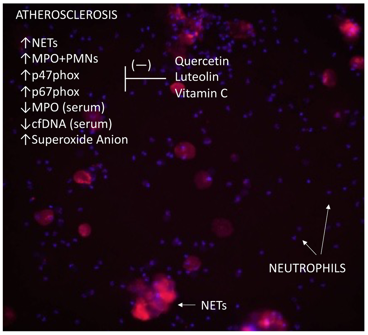
Given the consequences of excessive formation and release of NETs, it seems crucial to thoroughly understand the extent of this phenomenon and the potential for its control in patients with atherosclerosis, which is the objective of our study. The strategic role of NADPH oxidase and ROS in generating NETs has led us to investigate the potential use of substances with antioxidant properties, such as flavonoids and vitamin C, which could provide numerous beneficial effects in patients with atherosclerosis by limiting the release of NETs.
Material and methods
Materials
The study was conducted in a group of 22 patients with newly diagnosed atherosclerosis of the lower extremity arteries (11 women and 11 men), patients of the Academic Family Medicine Practice Clinic in Bialystok, with an age range of 49–79 years. The research procedures were carried out before the initiation of treatment. The patients exhibited symptoms of intermittent claudication, and the ankle-brachial index was < 0.9. The diagnosis was confirmed by Doppler ultrasound of the lower extremity arteries, which revealed arterial stenosis. Exclusion criteria included: the presence of chronic diseases other than hypertension; and the use of statins and dietary supplements containing vitamin C, other flavonoids, or antioxidants.
In the control group, 21 healthy individuals (10 women and 11 men) were enrolled, who were blood donors at the Regional Blood Donation and Blood Treatment Center in Bialystok, aged between 35 and 50 years, with a low likelihood of atherosclerotic disease. After obtaining written consents from the study participants, 9 ml of blood was drawn from the antecubital vein into a tube with the anticoagulant EDTA-K3, which was used for neutrophil isolation, and 6 ml was drawn into a tube with a clot activator to obtain serum.
Methodology
WBC count and blood smear analysis
The total leukocyte count (WBC) was assessed using the chamber method, direct counting with a light microscope, and a Bürker hemocytometer (Superior Marienfeld).
The peripheral blood smear was assessed in preparations made on glass slides, stained by means of the May-Grünwald-Giemsa method (Aqua-Med), under a light microscope.
Biochemical tests
In the group of patients with atherosclerosis of the lower extremity arteries (11 women and 11 men), a panel of biochemical tests was performed, which included the following parameters: total cholesterol, low-density lipoprotein (LDL), high-density lipoprotein (HDL), triglycerides, glucose, alkaline phosphatase, calcium, phosphorus, and homocysteine.
PMN isolation and incubation
Preliminary isolation of neutrophils was performed using the density gradient centrifugation method with the Polymorphprep reagent (Axis-Shield). The next step involved sorting the neutrophils using a MACS Separator with CD16 MicroBeads (Miltenyi Biotec). Neutrophil sorting was carried out according to the standard procedure outlined in the company’s research protocol attached to the device and reagents.
The isolated neutrophils were suspended in RPMI-1640 medium (Gibco, Life Technologies) supplemented with 4% fetal calf serum (PAN Biotech) and penicillin-streptomycin (Sigma Life Science). The cell suspension was then applied to 96-well plates (Falcon). The neutrophils were pre-incubated for 30 min with flavonoids: quercetin – 40 µM (ROTH) and luteolin – 10 µM (ROTH) as well as vitamin C (Sigma-Aldrich) at a concentration of 2 mM. Subsequently, the cells were stimulated with LPS at a concentration of 10 µg/ml (Sigma-Aldrich) for 1 h at 37°C with a 5% CO2 flow in an incubator (NUAIRE).
Protein isolation from PMNs
After incubation, the cell supernatants were collected from each well and stored at –20°C for further analysis.
After incubation, the protein was isolated from the neutrophils using sonication (SonicVibra Cell) in the presence of a protease inhibitor cocktail (Sigma-Aldrich). Protein concentration was determined by means of a fluorescence method with the Qubit Protein Assay Kit (Invitrogen), according to the manufacturer’s protocol, using a Qubit 2.0 Fluorometer (Invitrogen).
Visualization of NETs
The incubation of neutrophils at 37°C with a 5% CO2 flow in the incubation chamber was recorded by the In Cell Analyzer 2200 Microscope System (GE Healthcare Life Sciences). The main structures forming NETs were evaluated: DNA and myeloperoxidase. In order to stain the total DNA, Hoechst 33342 dye (Invitrogen) was used. Myeloperoxidase was stained by means of mouse monoclonal anti-MPO antibodies conjugated with fluorescein (Clone 8E6, MolecularProbes).
Assessment of neutrophil myeloperoxidase expression
After 1.5 h of incubation, neutrophils were permeabilized by means of FACS Permeabilizing Solution 2 (BD Biosciences) and labeled with monoclonal mouse anti-MPO antibodies (Clone 8E6, MolecularProbes). The data obtained on the FACSCalibur flow cytometer (BD Biosciences) were analyzed using FlowJo software (Tristar Inc.).
Quantification of circulating cell-free DNA (cfDNA)
The concentration of cfDNA in blood serum and in the supernatants obtained after neutrophil incubation was determined by means of the Circulating DNA Quantification Kit (Abcam) according to the provided instructions.
Determination of MPO concentration
The concentration of myeloperoxidase in the serum and in the supernatants obtained after neutrophil incubation was determined using the ELISA immunoassay (Human Myeloperoxidase Quantikine ELISA Kit, R&D Systems).
NADPH oxidase protein expression analysis
The expression of selected subunits of NADPH oxidase – p47phox, p67phox, Rac1 – was measured by Western blot in the obtained cell lysates.
The cell lysate suspended in Laemmli buffer was subjected to SDS-PAGE electrophoresis. In the next step, the fractions of separated proteins were transferred onto a nitrocellulose membrane (Bio-Rad Laboratories), which, after blocking non-specific sites, was incubated with appropriate primary antibodies: anti-p47-phox, anti-p67-phox, and anti-panRac (Santa Cruz Biotechnology). After washing with 0.1% TBS-T [(10x Tris-Buffered Saline) (Bio-Rad Laboratories) and Tween 20 – Electrophoresis Reagent (Sigma-Aldrich)], the nitrocellulose membrane was incubated with secondary antibodies conjugated with alkaline phosphatase (Jackson Immuno Research). The addition of the BCIP/NBT Liquid Substrate System (Sigma-Aldrich) allowed for the detection of immunoreactive protein bands, the intensity of which was assessed using the ImageJ program and expressed in arbitrary units.
Superoxide detection via cytochrome c reduction assay
Using the method developed by McCord and modified by Bhuyan [23], the oxidative burst was assessed based on the measurement of superoxide anion production by neutrophils.
A solution of cytochrome c in phosphate buffer (KH2PO4/K2HPO4) at pH 7.8 containing 0.1 mM EDTA was added to two parallel samples with isolated neutrophils. The concentration of cytochrome c was 15 mg/ml. Superoxide dismutase (SOD) with an activity of 5000 U/ml was added to the reference sample, and buffer to the test sample. Then, after the addition of quercetin – 40 µM (ROTH), luteolin – 10 µM (ROTH), vitamin C – 2 mM, and LPS – 10 µg/ml (Sigma-Aldrich) to both test samples, incubation was carried out at 37°C, and after 1.5 h, the absorbance was read at λ = 550 nm. The generation of superoxide anion was expressed as nmol of reduced cytochrome c, using the absorption coefficient of 21.1 mmol/l-1 cm-1 at 550 nm.
Statistical analysis
The obtained research results were processed by means of statistical tests with the Statistica 13.1 software. The results are presented as mean values, summarized in tables or graphs, with the standard deviation (± SD) indicated. The statistical significance of differences was tested using Student’s t-test and one-way analysis of variance (ANOVA). The normality of data distribution was assessed using the Shapiro-Wilk test. The Spearman method was applied to analyze the correlations between the parameters. The minimum level of significance was set at p < 0.05.
Results
WBC and leukogram
The mean WBC in the peripheral blood in the control group was 6.19 × 10³ cells/µl, while in patients with atherosclerosis it was 6.92 × 10³ cells/µl.
No significant differences were observed in the numbers of individual leukocyte populations in peripheral blood between patients with atherosclerosis and the control group. A summary of the mean values of leukocyte populations in the control group and in patients with atherosclerosis is presented in Table I.
Table I
Basic diagnostic parameters of patients and the control group
Biochemical test results in atherosclerosis patients
No significant differences were observed in the levels of the measured biochemical parameters between women and men. The mean levels of total cholesterol, LDL cholesterol, glucose, and homocysteine exceeded reference values in women, whereas in men only the glucose level exceeded the reference range. The case-by-case analysis revealed that 55% of women and 12% of men had elevated total cholesterol levels. LDL cholesterol levels were above the reference values in 45% of women and 34% of men. High triglyceride levels were observed in 23% of women, while in men, the levels were within the reference range. Furthermore, 55% of both women and men had elevated glucose levels. The level of alkaline phosphatase in women was within the normal range. Alkaline phosphatase levels were reduced in 12% of men. The levels of total calcium and phosphorus were normal in the study group of women and men. Elevated homocysteine levels above the reference values were found in 67% of women and 50% of men. The results are summarized in Table II.
Table II
Patients’ biochemical diagnostic tests
| Parameter | Patients n = 22 | References | |||
|---|---|---|---|---|---|
| Women n = 11 | Men n = 11 | ||||
| Mean | SD | Mean | SD | ||
| TC [mg/dl] | 213.67 | 42.84 | 173 | 35.87 | < 190 |
| LDL-C [mg/dl] | 121.33 | 42.22 | 101.11 | 25.37 | < 115 in the absence of risk factors; < 100 severe risk factors, diabetes without organ complications; < 70 with risk: disease |
| HDL-C [mg/dl] | 64.33 | 13.83 | 56.56 | 15.34 | > 40 |
| TG [mg/dl] | 116.33 | 65.78 | 88.67 | 21.01 | < 150 |
| Glc [mg/dl] | 105↑ | 14.35 | 105 | 9.03 | 70–100 |
| ALP [IU/l] | 58.89 | 14.95 | 53.63 | 17.38 | 37–123 |
| tCa [mmol/l] | 2.45 | 0.11 | 2.48 | 0.08 | 2.25–2.75 |
| PHOS [mmol/l] | 1.15 | 0.07 | 1.11 | 0.11 | 0.74–1.52 |
| HCY [µmol/l] | 16.49 | 6.42 | 13.24 | 4.37 | 4.44–13.56 |
NET formation analysis
The microscopic analysis has revealed that neutrophils in patients with atherosclerosis exhibit a significantly higher ability to spontaneously release extracellular traps as compared to neutrophils in healthy individuals (Table III).
Table III
Percentage of neutrophils forming NETs
| Control | Mean (%) SD | 5.03 1.95 | 9.07a 2.09 | 5.53b 1.18 | 3.51cf 0.57 | 5.55dg 1.32 | 4.93e 2.26 |
| Patients | Mean (%) SD | 8.66* 1.47 | 14.85*a 1.58 | 9.78b 5.14 | 9.69*c 4.82 | 10.24 5.9 | 11.57 8.69 |
| Incubation procedure | PMNs | + | + | + | + | + | + |
| LPS | − | + | + | + | + | + | |
| QCT | − | − | + | − | − | − | |
| LT | − | − | − | + | − | + | |
| Vit C | − | − | − | − | + | + |
In the presence of LPS, neutrophils from both atherosclerosis patients and the control group produced a significantly higher amount of NETs, with values higher in atherosclerosis patients than in controls.
In both groups, preincubation of neutrophils in the presence of quercetin and luteolin significantly reduced NET release following LPS stimulation. The amount of NETs released from neutrophils after preincubation with luteolin was higher in patients than in controls. Preincubation of neutrophils with vitamin C or with vitamin C combined with luteolin resulted in a reduction in NET release after LPS stimulation in the healthy group, whereas no significant changes were observed in the patient group.
Neutrophils from healthy individuals preincubated with luteolin generated a significantly lower amount of NETs as compared to cells preincubated with quercetin or vitamin C.
The analysis of the results with regard to sex in the control group revealed significantly higher amounts of NETs after LPS stimulation of neutrophils in women as compared to men.
Quantification of MPO-expressing neutrophils
Flow cytometry analysis (Figure 2) revealed a higher percentage of MPO-positive (MPO+) neutrophils among unstimulated PMNs in patients with atherosclerosis as compared to healthy individuals.
Figure 2
Percentage of MPO+ neutrophils in patient and control groups. Statistically significant differences (p < 0.5): *vs. control group, avs. unstimulated PMNs, bPMNs + LPS vs. PMNs + 30′QCT + LPS, cPMNs + LPS vs. PMNs + 30′LT + LPS, dPMNs + LPS vs. PMNs + 30′VitC + LPS, ePMNs + LPS vs. PMNs + 30′(VitC + LT) + LPS. PMNs – neutrophils, LPS – lipopolysaccharide, QCT – quercetin, LT – luteolin, Vit C – vitamin C.
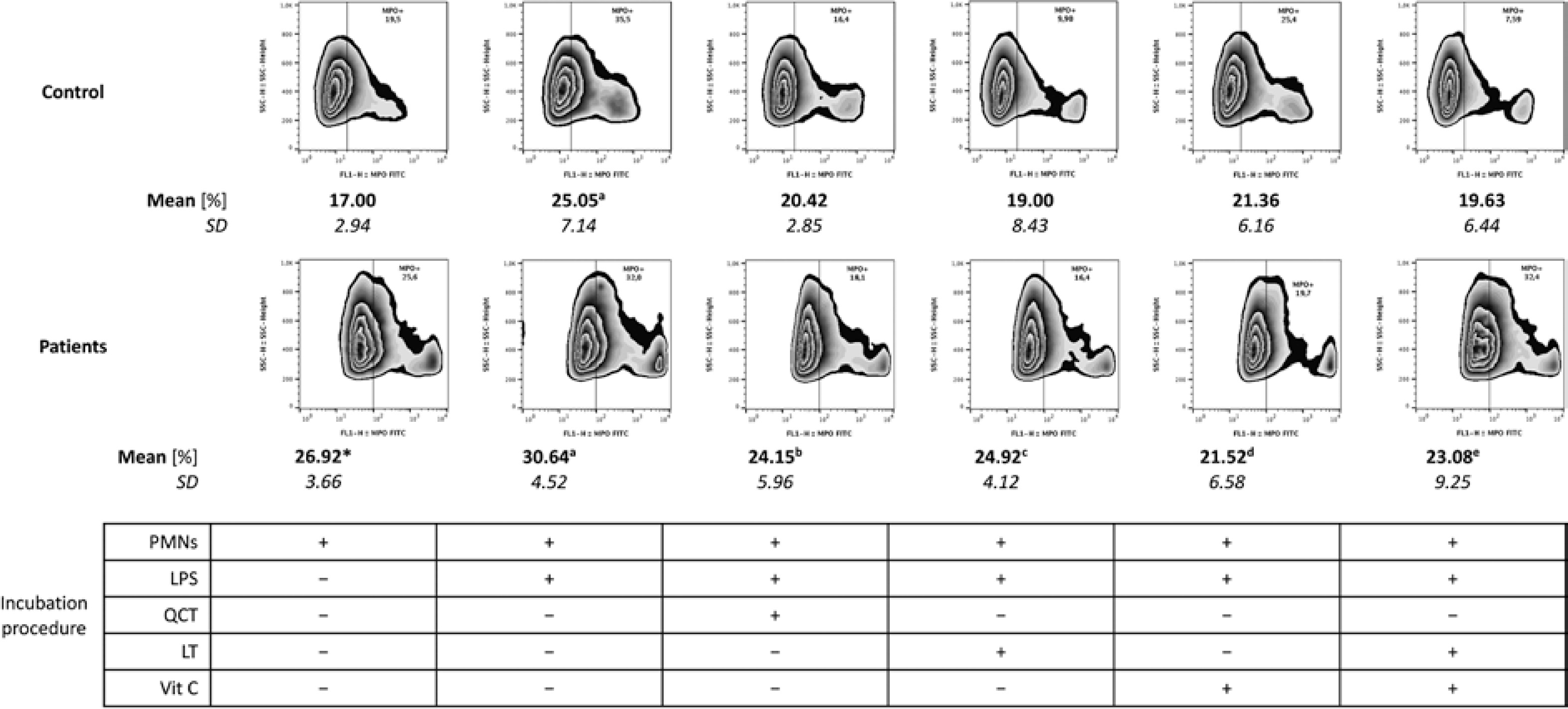
The study results revealed that in both the patient group and healthy individuals, LPS stimulation of cells significantly increased the percentage of MPO+ neutrophils as compared to unstimulated PMNs.
In patients, preincubation of neutrophils with flavonoids such as quercetin and luteolin, as well as vitamin C or vitamin C combined with luteolin, reduced the percentage of MPO+ neutrophils after LPS stimulation.
No differences were observed in the results between the female and male groups.
MPO concentration assessment
No significant differences were observed between the myeloperoxidase (MPO) concentration in the supernatants of unstimulated neutrophils from patients with atherosclerosis and the concentration in the cellular supernatants of healthy individuals (Table IV).
Table IV
Concentrations of MPO, cfDNA, and superoxide anion in patient and control samples
| Parameter | PMNs | PMNs + LPS | PMNs + 30′QCT + LPS | PMNs + 30′LT + LPS | PMNs + 30′Vit C + LPS | PMNs + 30′(Vit C + LT) + LPS | Serum |
|---|---|---|---|---|---|---|---|
| MPO [ng/ml] | |||||||
| Control | 28.08 4.25 | 38.57a 6.23 | 27.89b 3.68 | 28.19c 4.11 | 28.09d 4.20 | 27.73e 4.36 | 556.4 339.2 |
| Patients | 30.28 3.65 | 35.97a 8.01 | 27.69b 4.62 | 29.11c 4.17 | 28.76d 4.07 | 27.71e 4.04 | 273.3* 108.2 |
| cfDNA [ng/ml] | |||||||
| Control | 24.36 5.93 | 39.20a 17.01 | 28.52 8.50 | 27.90c 11.91 | 28.87d 7.40 | 28.10 16.51 | 27.91 6.29 |
| Patients | 30.42* 7.82 | 36.39a 10.19 | 27.33b 10.19 | 29.57 12.72 | 27.27d 7.06 | 31.25 7.70 | 24.30* 2.94 |
| Superoxide anion radical [mmol/l-1cm-1] | |||||||
| Control | 2.62 1.14 | 4.52a 1.42 | 2.87 1.86 | 3.68 0.19 | 1.27dg 0.48 | 3.13 2.35 | – |
| Patients | 2.89 0.60 | 7.63*a 2.38 | 4.34b 1.73 | 4.48c 1.54 | 4.06*d 1.51 | 1.82eijk 0.61 | – |
LPS stimulation of neutrophils resulted in a significant increase in MPO levels in the cellular supernatants, both in patients with atherosclerosis and in healthy individuals, as compared to the supernatants of unstimulated neutrophils.
In both studied groups, preincubation of PMNs with quercetin, luteolin, vitamin C, and vitamin C combined with luteolin led to a significant reduction in MPO concentrations in the cellular supernatants as compared to the supernatants of PMNs after LPS stimulation.
The analysis of myeloperoxidase levels in blood serum revealed significantly lower concentrations of that protein in the serum of patients with atherosclerosis as compared to the values obtained in the serum of healthy individuals.
No differences in MPO concentrations were observed between women and men.
cfDNA concentration assessment
The assessment of cfDNA levels revealed significantly higher concentrations in the supernatants of unstimulated neutrophils from patients with atherosclerosis compared to the values obtained in the control group (Table IV).
Both in patients with atherosclerosis and in healthy individuals, the cfDNA level in the supernatants of PMNs after LPS stimulation was significantly higher than that in the supernatants of unstimulated neutrophils.
In both studied groups, preincubation of neutrophils with vitamin C contributed to a decrease in cfDNA concentration in the supernatants of those cells after LPS stimulation. In patients with atherosclerosis, lower cfDNA concentrations were also observed in the supernatants of neutrophils preincubated with quercetin. However, in the healthy group, preincubation of PMNs with luteolin resulted in a reduction of cfDNA levels in the cellular supernatants.
The analysis of cfDNA in the whole blood serum revealed lower concentrations of the NET marker in the serum of patients with atherosclerosis as compared to the values obtained in the serum of healthy individuals.
No significant differences were observed in cfDNA concentrations between the values in women and men.
Assessment of superoxide anion (O2•−) generation
No significant difference was observed in the amount of superoxide anion in the supernatants of unstimulated neutrophils from patients as compared to the control group (Table IV).
Both in patients with atherosclerosis and in healthy individuals, PMNs released higher amounts of O2•− after LPS stimulation as compared to unstimulated cells, with significantly higher values in patients.
Preincubation of PMNs from patients with antioxidant compounds such as quercetin, luteolin, vitamin C, or vitamin C combined with luteolin led to a reduction in the release of O2•− by those cells as compared to PMNs after LPS stimulation.
In the control group, the amount of O2•− in the supernatants of neutrophils preincubated with vitamin C was lower than that in the supernatants of PMNs preincubated with luteolin.
In the patient group, neutrophils preincubated concurrently with vitamin C and luteolin generated a smaller amount of superoxide anion as compared to cells preincubated with quercetin, luteolin alone, or vitamin C alone.
No difference was observed in the amount of generated O2•− between the female and male groups.
NADPH oxidase subunit expression
Unstimulated neutrophils from patients with atherosclerosis showed higher expression of the p47phox and p67phox subunits of NADPH oxidase as compared to unstimulated PMNs from healthy individuals (Figure 3).
Figure 3
Expression of NADPH oxidase subunits in neutrophils of patients and healthy individuals. Statistically significant differences (p < 0.5): *vs. control group, avs. unstimulated PMNs, bPMNs + LPS vs. PMNs + 30′QCT + LPS, cPMNs + LPS vs. PMNs + 30′LT + LPS, dPMNs + LPS vs. PMNs + 30′VitC + LPS, ePMNs + LPS vs. PMNs + 30′(VitC + LT) + LPS, fPMNs + 30′LT + LPS vs. PMNs + 30′QCT + LPS, gPMNs + 30′LT + LPS vs. PMNs + 30′VitC + LPS, hPMNs + 30′QCT + LPS vs. PMNs + 30′VitC + LPS. PMNs – neutrophils, LPS – lipopolysaccharide, QCT – quercetin, LT – luteolin, Vit C – vitamin C
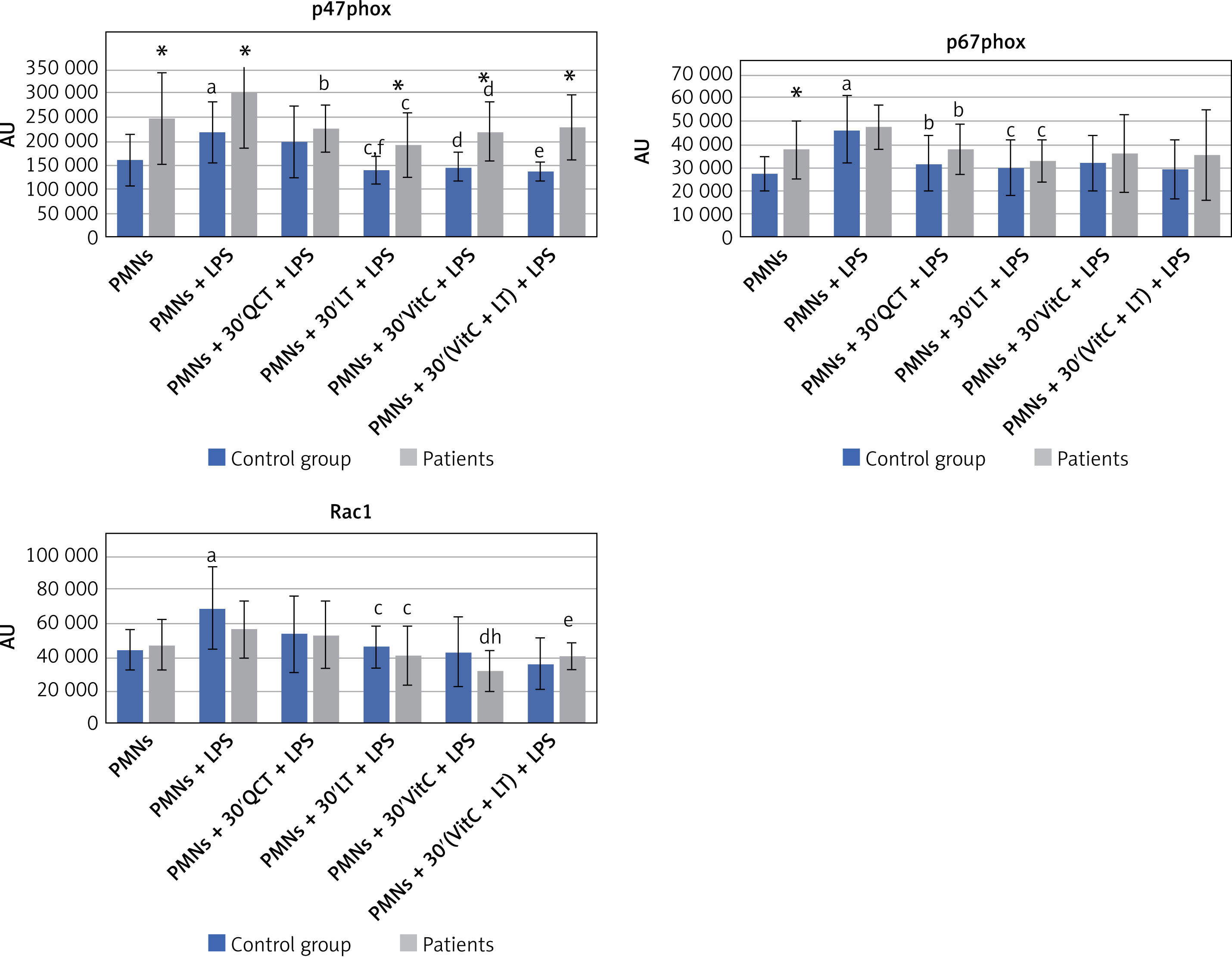
No significant difference in Rac1 protein expression was observed in unstimulated neutrophils from patients with atherosclerosis compared to unstimulated cells from healthy individuals.
LPS stimulation of neutrophils from healthy individuals resulted in a significant increase in the p47phox and p67phox expression as compared to unstimulated cells. In neutrophils from patients with atherosclerosis, expression levels of these proteins were also higher; however, the difference was not statistically significant. After LPS stimulation, the p47phox and p67phox expression was significantly higher in patients as compared to the expression in cells from healthy individuals. In the case of Rac1, LPS stimulation led to a significant increase in the expression of that protein only in healthy individuals.
In both study groups, preincubation of neutrophils with luteolin or vitamin C contributed to a reduction in the expression of p47phox and Rac1 in PMNs incubated with LPS as compared to cells incubated only with LPS. Additionally, in patients with atherosclerosis, significantly higher expression of p47phox was observed in neutrophils preincubated with luteolin and vitamin C as compared to cells from healthy individuals. Neutrophils from patients preincubated with quercetin showed lower p47phox expression as compared to cells stimulated with LPS. In the control group, preincubation of neutrophils with vitamin C and luteolin reduced the expression of the studied protein as compared to LPS stimulation. The p47phox expression was significantly higher in neutrophils preincubated with vitamin C and luteolin in patients with atherosclerosis as compared to the expression in healthy individuals.
In the control group, significantly lower expression of the p47phox protein was also observed in neutrophils preincubated with luteolin as compared to cells preincubated with quercetin.
In both groups, preincubation of neutrophils with quercetin or luteolin resulted in a reduction of the p67phox expression as compared to neutrophils stimulated with LPS.
In patients with atherosclerosis, a reduction in Rac1 expression was also observed in neutrophils preincubated with vitamin C and vitamin C combined with luteolin as compared to cells stimulated with LPS.
In the patient group, lower expression of the Rac1 protein was observed in neutrophils preincubated with vitamin C as compared to cells preincubated with quercetin.
No differences were found in the expression of the p47phox, p67phox, and Rac1 proteins between the female and male groups.
Assessment of parameter correlations
A positive correlation was observed between the concentrations of cfDNA and MPO in the supernatants of unstimulated neutrophils in the control group (r = 0.63; p = 0.002) (Figure 4 A). No such correlation was observed in patients with atherosclerosis.
Figure 4
Correlations. Correlations between concentrations of: A – cfDNA and MPO in supernatants of unstimulated neutrophils in the control group; B – cfDNA and MPO in serum in the control group; C – cfDNA in neutrophil supernatants and cfDNA in serum in the control group; D – MPO in neutrophil supernatants and MPO in serum in the control group; E – MPO in neutrophil supernatants and MPO in serum in patients with atherosclerosis
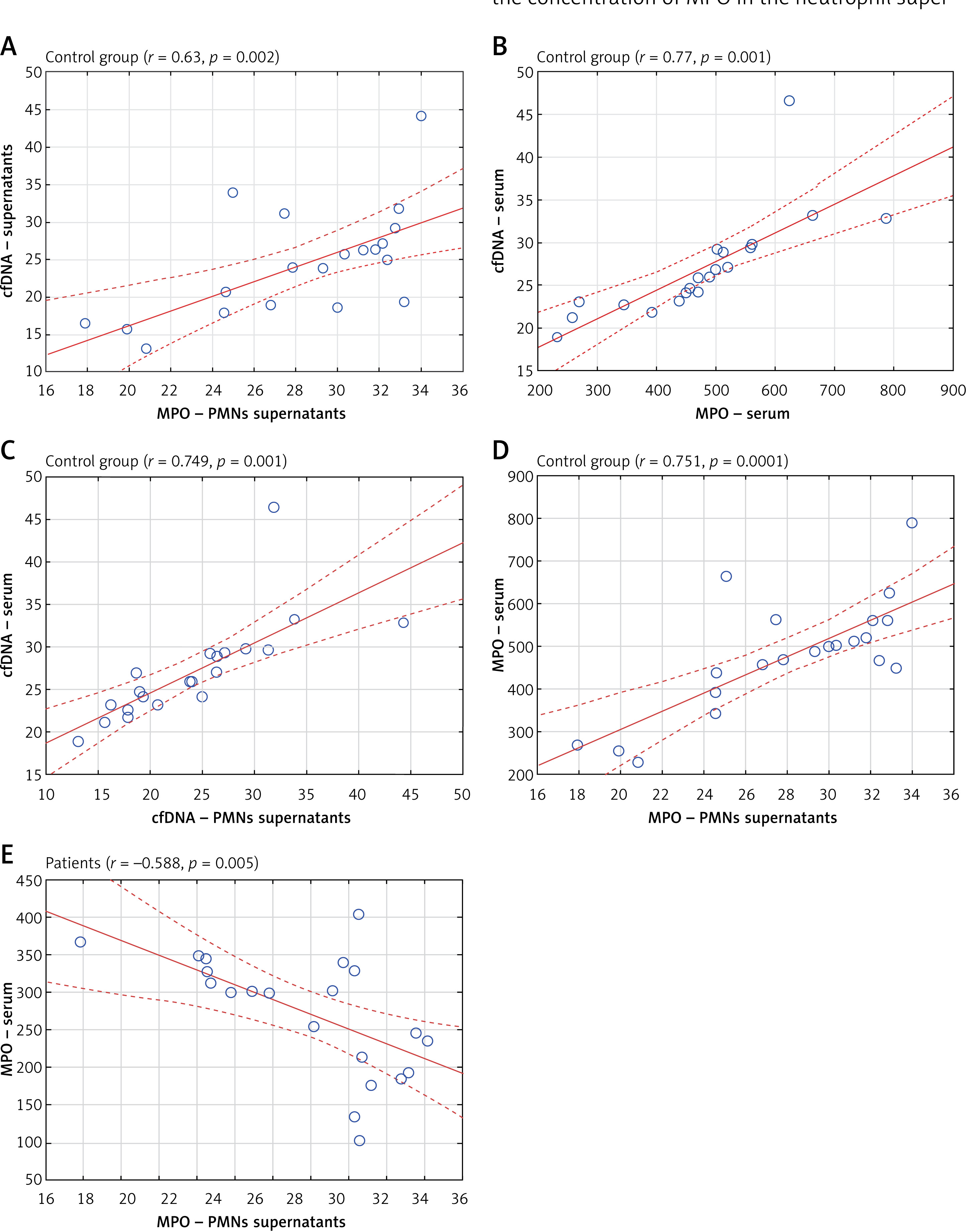
A positive correlation was found between the amount of cfDNA and MPO in the serum of healthy individuals (r = 0.77; p = 0.001) (Figure 4 B). No such correlation was found in the group of patients with atherosclerosis.
A positive correlation was found in the healthy group between the concentration of free circulating DNA in the supernatants of PMNs and its concentration in serum (r = 0.749; p = 0.001) (Figure 4 C). No such correlation was observed in the patient group.
In healthy individuals, a positive correlation was found between the concentrations of myeloperoxidase in the supernatants of PMNs and in the serum (r = 0.751; p = 0.0001) (Figure 4 D). In contrast, in the group of patients with atherosclerosis, an inverse correlation was found between the concentration of MPO in the neutrophil supernatants and the concentration of that protein in the serum (r = –0.588; p = 0.005) (Figure 4 E).
No correlation between age and the analyzed parameters was found in any of the study groups.
Discussion
Studies have suggested that NET generation constitutes a potential factor in the development of atherosclerosis [24]. Knight et al. reported that neutrophils isolated from mice with atherosclerosis were more prone to NET formation than healthy animals [22]. Research on NET formation in humans was conducted by Megens et al., who detected NETs in patients undergoing endarterectomy [25]. Our findings also suggest a potential role of NETs in the course of atherosclerosis, demonstrated by increased release of NETs observed under the microscope and a rise in the levels of the MPO and cfDNA. The observed changes were not related to the age of the patients, which is confirmed by previous studies [26].
A high number of neutrophils containing MPO and the enhanced ability of those cells to release MPO may directly influence the development of atherosclerosis. It is known that hypochlorous acid, formed in a reaction catalyzed by MPO, is responsible for the oxidative modification of LDL. Those modified LDL particles may be taken up by macrophages, leading to their transformation into foam cells that are part of atherosclerotic plaques [27]. It has also been shown that elevated levels of MPO are associated with an increased risk of coronary artery disease [28], and high MPO levels in plasma may even serve as a biomarker for acute myocardial infarction [29]. Excessive MPO levels may also lead to increased activation of the NETosis process [30–32]. Similar unfavorable consequences may be associated with a significant increase in cfDNA concentrations. Jylhävä et al. found that increased cfDNA levels correlated with a group of cardiometabolic risk factors, including high blood pressure, unfavorable lipid metabolism profile, and systemic inflammation [33]. Free circulating DNA may also contribute to the intensification of coagulation processes or impaired fibrinolysis, which is crucial in the pathogenesis of cardiovascular diseases [34].
The role of MPO and cfDNA in the development and progression of atherosclerosis is also supported by the studies of Borissoff et al., who observed that levels of double-stranded DNA (dsDNA), nucleosomes, and MPO-DNA complexes were significantly elevated in patients with severe coronary atherosclerosis [35]. Lower levels of cfDNA and MPO observed in the blood serum of the studied patient group, along with higher levels in the supernatants of PMNs, may be a result of their binding in the atherosclerotic plaque.
The significance of NET formation in the course of atherosclerosis is also highlighted by the studies indicating vascular damage in atherosclerosis, preceded by intense recruitment and activation of neutrophils [36]. It has been observed that direct co-culture of activated endothelial cells with neutrophils induces NET formation, and leads to vascular damage [36, 37].
Warnatsch et al. reported that NETs may stimulate macrophages to release IL-1β, which activates Th17 lymphocytes [38]. Th17 lymphocytes, by enhancing the recruitment of other immune cells to atherosclerotic plaques, may contribute to the development of inflammation in blood vessels. Furthermore, NETs may affect the activation of plasmacytoid dendritic cells present in atherosclerotic plaques that release large amounts of IFN-α, also promoting the development of atherosclerotic lesions [39].
It has been demonstrated that the activity of NADPH oxidase, which is also essential for the formation and release of NETs, is a factor influencing the development of atherosclerosis. This is further supported by the results of studies on neutrophils from patients with chronic granulomatous disease, characterized by a genetic defect in NADPH oxidase [12, 40–42]. There is evidence that ROS cause a series of molecular changes that lead to the infiltration of the endothelium by inflammatory cells, ultimately promoting the formation of an atherosclerotic plaque. ROS also affect arterial dysfunction by inactivating nitric oxide, a potent vasodilator [43]. Our findings support the relationship between increased NET formation and elevated NADPH oxidase activity in neutrophils from approximately 50% of patients with atherosclerosis.
The causes of increased NET formation in patients with atherosclerosis may have various origins. One of them appears to be the elevated levels of total cholesterol and LDL, observed in a significant percentage of the studied patients. Warnatsch et al., in a mouse model of atherosclerosis, demonstrated that cholesterol crystals influence the induction of NETosis [38].
Brinkmann et al. indicated that the generation of NETs may also be regulated by PMA or LPS [6]. Our own studies confirm that, in patients with atherosclerosis, LPS stimulates NET production, as confirmed by an increase in the number of MPO-positive neutrophils and a rise in cfDNA concentration in the supernatants of cells.
Given the adverse consequences of increased NET formation within the course of atherosclerosis, it seems important to explore ways to regulate their generation [44–49]. Mohammed et al. observed increased NET formation in animals with ascorbic acid deficiency. Furthermore, human neutrophils stimulated with PMA, supplemented with vitamin C, had a reduced ability to generate NETs [48].
There are data regarding the regulation of different neutrophil functions through flavonoids, such as quercetin and luteolin [50–53]. However, there are limited data on the role of these compounds in the regulation of NET formation. One of the few examples is the study by Yang et al., who observed that luteolin significantly impaired NET formation by human neutrophils [54].
Our findings showed that in patients with atherosclerosis, the presence of both quercetin and luteolin, as well as luteolin combined with vitamin C, led to reduced formation of NETs by neutrophils stimulated with LPS.
Similar results were obtained by Pečivová et al., who observed that quercetin leads to a reduction in MPO release by neutrophils after PMA stimulation [55]. Yang et al. reported that the anti-inflammatory effect of luteolin is associated with the removal of ROS and inhibition of the Raf1-MEK-1-ERK signaling pathway, which regulates the formation of NETs [54]. It prompted us to investigate the combined effect of luteolin and vitamin C, which, in the case of superoxide anion generation, was more effective than the presence of either luteolin or vitamin C alone. It should be noted here that the effect of flavonoids on human health is not clear; some studies confirm the protective effect of flavonoids on cardiovascular diseases or cancer, while other data indicate no effect, and some reports demonstrate potential harm [56].
This study has several limitations. First, the number of patients included was relatively small, which may limit the statistical power and generalizability of the findings. The recruitment was constrained by the duration of the project and strict inclusion/exclusion criteria. Second, the experimental procedures were conducted exclusively in vitro, which, while useful for mechanistic insight, may not fully reflect the complex in vivo environment. Therefore, the observed effects of flavonoids and vitamin C on NET formation and oxidative responses in neutrophils should be interpreted with caution. Further research involving larger patient cohorts and in vivo models is necessary to validate these findings and assess their clinical relevance.
In conclusion, the results suggest that high NET formation may contribute to the development and progression of atherosclerosis. Our findings also indicate that quercetin, luteolin, and vitamin C can inhibit the release of NETs under the study conditions, highlighting their potential relevance in limiting the development of this disease. These observations support further research on strategies aimed at preventing excessive NET formation as a possible approach in atherosclerosis.

