

Pituitary adenomas (PAs) are common benign endocrine tumours, ranking third in incidence among intracranial tumours and accounting for approximately 10% of all intracranial tumours. However, about 40% are locally invasive and may not be surgically cured, and about 1% of them demonstrate an aggressive clinical behaviour [1]. Additionally, some tumours demonstrate functional activity. Clinical symptoms of functioning tumours typically result from excessive hormone secretion [2]. For example, when a tumour leads to excessive GH secretion, it may cause acromegaly in adults, characterised by progressive enlargement of the hands, feet, head, chest, and limbs, prominent forehead, and a significantly protruding jaw; in children before puberty, it may cause gigantism, which is manifested by height and weight far exceeding that of their peers. Furthermore, when the tumour invades or compresses adjacent structures, it can cause related symptoms: compression of the optic nerve may result in visual field defects, compression of the oculomotor nerve may lead to oculomotor nerve palsy, and when the tumour progresses to a certain extent, it may even cause hydrocephalus or pituitary apoplexy [3]. Imaging plays a crucial role in assessing the invasiveness of pituitary tumours. On X-ray plain films, invasive pituitary adenomas (IPAs) can exhibit destruction of surrounding bone structures, such as the sella floor and dorsum sellae. Computed tomography (CT) scans further reveal the extent of infiltration toward the parasellar region. Magnetic resonance imaging (MRI) preoperatively provides a clearer visualisation of the invasion of IPAs toward neighbouring structures from three different planes [4, 5]. Tumour tissue pathology evaluation is the cornerstone for predicting the progression of pituitary tumours. Ki-67 index, mitotic count, and P53 immunohistochemical staining are recognised as important assessment indicators and are also the gold standard for diagnosing IPAs [6]. The final diagnosis of IPAs is made by combining clinical manifestations, imaging characteristics, and pathological evaluation. For the treatment of IPAs, specific methods include surgical treatment, radiotherapy, and drug therapy. However, single-modality treatment is not ideal for the prognosis of patients with IPAs because it is difficult to achieve a radical cure without causing severe complications. Therefore, multiple treatment modalities are now commonly combined, with the most frequent approach being surgical resection combined with drug therapy.
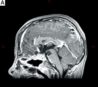
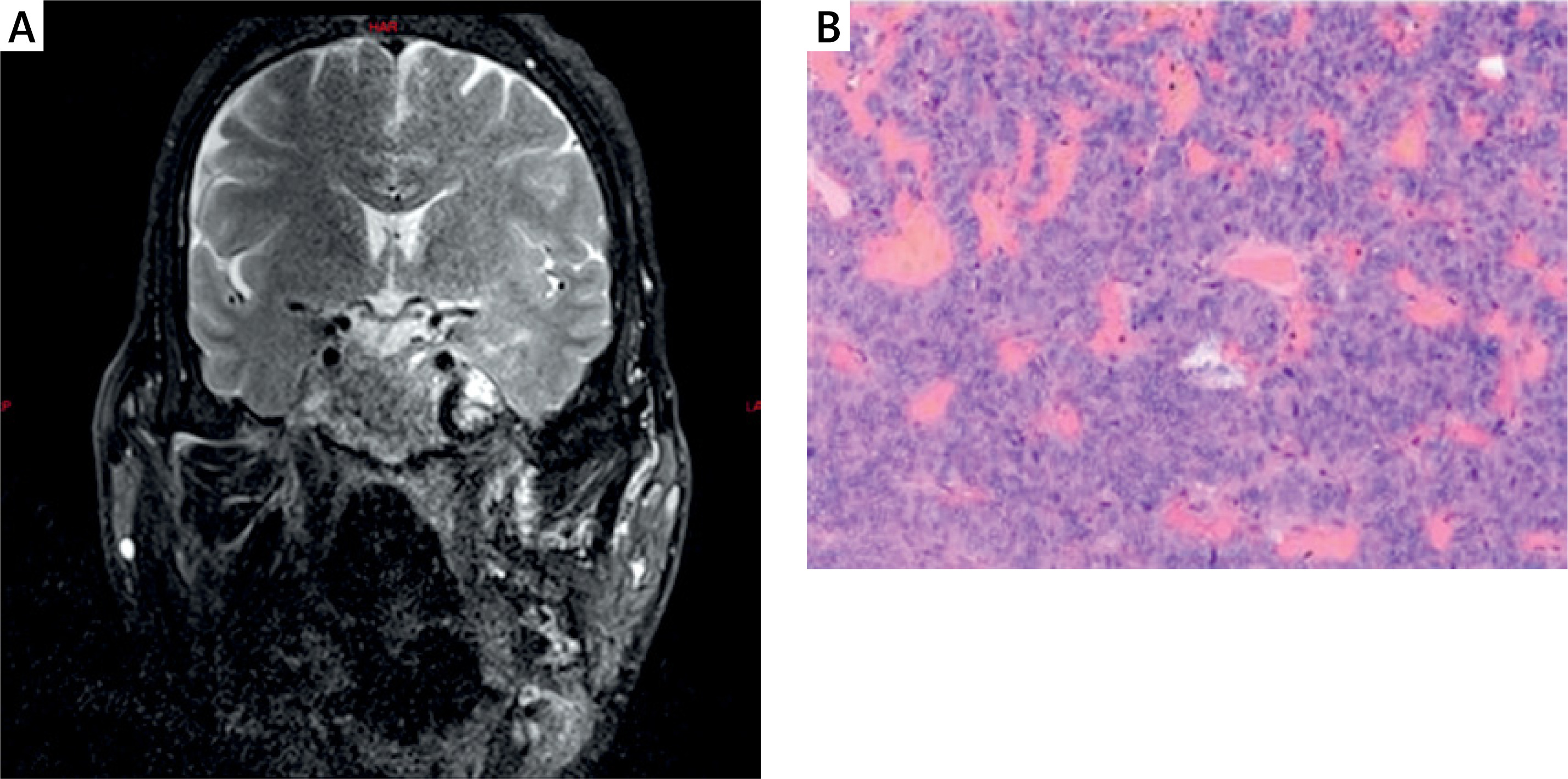
Case 1. A 48-year-old male patient was admitted for ptosis of the left upper eyelid lasting over 2 months. Physical examination revealed prominent brow ridges, protruding cheekbones, thickened nasolabial folds, altered facial features, mandibular prognathism, ptosis of the left upper eyelid, thickened and enlarged fingers, widened toes, thickened feet, and a barrel chest. Head CT revealed a mass lesion in the sella turcica region with adjacent skull base bone destruction and possible involvement of bilateral internal carotid arteries at segments C3-5. Calcified plaques were noted in the siphon segments of both internal carotid arteries, with localised mild luminal stenosis. Preoperative MRI revealed a mass in the sellar region with iso-signal intensity on both T1- and T2-weighted imaging, showing possible lesion involvement of the pituitary stalk and sella floor depression (Figure 1 A). The patient underwent transsphenoidal pituitary lesion resection via neuroendoscopy under general anaesthesia. Pathological findings suggest a pituitary neuroendocrine tumour (PIT-1 positive) with a growth hormone-secreting prolactinoma component (Figure 1 B). Postoperative follow-up at 1 month showed normalised GH levels and alleviated neural compression symptoms. However, due to household financial constraints and the patient’s age, the follow-up ended prematurely.
Figure 1
A – Preoperative MRI reveals a mass-like T1/T2 isointense lesion in the sella turcica measuring approximately 39 × 66 × 41 mm. Contrast-enhanced imaging shows slightly heterogeneous, marked enhancement with irregular margins. The lesion encases the internal carotid artery, with poorly defined pituitary stalk structures and sella turcica depression. B – Pathological findings: Pituitary neuroendocrine tumour consistent with the PIT-1 lineage, growth hormone-secreting prolactinoma. Immunohistochemistry: PIT-1(+), T-PIT(-) ER(-) GATA3 (scattered +) Syn(+) CK8/18 (perinuclear +) Ki67 (about 2%+) GH (about 100%+) PRL (approximately 20%+), TSH (-), ACTH (-), LH (-), FSH (-), SSTR2 (3+), P53 (approximately 10% with variable intensity+)
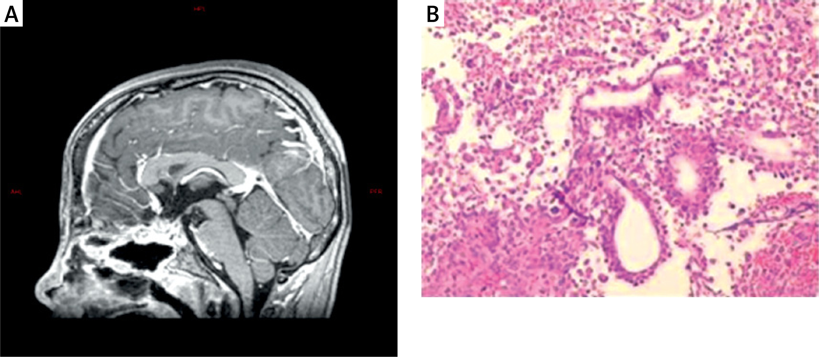
Case 2. A 13-year-old male patient was admitted for blurred vision lasting over 4 months. Physical examination revealed height and weight significantly exceeding his peers, decreased visual acuity in both eyes, bilateral temporal visual field defects, and incomplete development of secondary sexual characteristics. Head CT revealed an enlarged sella turcica with a collapsed sella floor and adjacent bone thinning. A slightly hyperdense mass was visible within the sella turcica, extending upward into the suprasellar cistern and growing laterally to involve the cavernous sinus. Head computed tomography angiography (CTA) demonstrated mild compression and rightward displacement of the right internal carotid artery’s cavernous sinus segment. Internal carotid artery siphon segments, bilateral anterior cerebral artery A1 segments, and the left posterior communicating artery were closely related to the sella turcica mass. Head MRI revealed a solid mass lesion within and above the sella turcica. The pituitary stalk was compressed and poorly visualised, the optic chiasm was displaced upward due to compression, and the right internal carotid artery was encircled. The patient underwent transnasal-transsphenoidal pituitary lesion resection under general anaesthesia using a neuroendoscope. Postoperative MRI showed absence of the pituitary fossa and surrounding structures in the sella region (Figure 2 A). Pathological examination suggested a high likelihood of an immature PIT-1 lineage tumour (Figure 2 B). The patient is currently undergoing regular weekly treatment with lanreotide and has attended multiple postoperative follow-ups in the outpatient clinic. Laboratory results indicate that GH and IGF-1 levels are within the normal range. However, due to the surgical removal of the pituitary gland, the patient is experiencing gonadotropin deficiency and is receiving treatment with human chorionic gonadotropin.
Figure 2
A – Postoperative MRI demonstrated absence of the pituitary fossa and adjacent structures within the sella region, along with partial defects in the ethmoid and sphenoid sinuses, showing ring-like enhancement after contrast administration. B – Pathological examination suggested a high likelihood of an immature PIT-1 lineage tumour.Immunohistochemistry: Tumour cells PIT-1 (+), T-PIT (-), SF-1 (-), ER (scattered +), GATA3 (+), Syn (+), CK8/18 (scattered +), Ki67 (approximately 8%+), GH (approximately 60% with variable intensity +), PRL (approximately 10%+), TSH (–), ACTH (–), LH (–), FSH (–), SSTR2 (3+), and P53 (approximately 10% with variable intensity+)
Pituitary tumours are common among intracranial tumours, with most being benign functional tumours. However, a small subset exhibits clinical invasive behaviour, characterised by infiltration and destructive actions on surrounding tissues, such as destroying surrounding bone, invading the cavernous sinus, and compressing surrounding nerves when the tumour is large, leading to intracranial hypertension, pituitary apoplexy, and a high postoperative recurrence rate. Clinical manifestations include headaches, visual field defects, endocrine symptoms, and other symptoms such as visual field loss and psychiatric symptoms. CT and MRI are advantageous for assessing this condition, showing tumour size, invasion extent, and basilar bone destruction. The density (CT) and signal (MRI) of IPAs can be uniform or heterogeneous. The latter is seen in intratumoral cystic changes, haemorrhage, necrosis, or enhancement after calcification, presenting as uniform or heterogeneous density or signal increase. MRI can most intuitively assess tumour invasion extent, and Ki-67 index and p53 expression are considered related to the degree of invasiveness. According to the latest WHO classification (2022), pituitary tumours are classified into Pit-1 lineage PitNETs, TPIT-lineage PitNETs, SF1-lineage PitNETs, and PitNETs of undefined cell lineage. Based on pathological testing, Case 1 was classified as a Pit-1 lineage pituitary neuroendocrine tumour, specifically a mixed-growth hormone and prolactin-producing adenoma. Case 2 was identified as an immature Pit-1 lineage tumour.
Due to the pivotal role of the pituitary’s endocrine function in maintaining homeostasis in the human body, patients often experience various endocrine functional disorders for a long time before and after surgery. Therefore, the development of treatment plans for patients requires meticulous attention. As a result, patients with IPAs need multidisciplinary, strict follow-up and the development of multimodal treatment plans.
Aggressive IPAs grow rapidly, currently have no specific drugs for their treatment, and treatment at this stage primarily involves surgical resection. Somatostatin analogues may be used as adjuvant therapy or as primary treatment in selected cases, including presurgical therapy in patients with large invasive macroadenomas, immediate relief of symptoms and reduction of growth hormone hypersecretion in patients awaiting surgery or those with recurrent disease, morbidity in elderly patients, and patients who refuse surgical treatment [7]. Treatment with somatostatin analogues or bromocriptine should also be initiated when previous surgical therapy has not achieved biochemical remission [8]. Radiosurgery and other adjuvant medical therapies may be employed in selected patients if standard first- and second-line therapies fail [9]. Therefore, the treatment approaches for IPAs secreting growth hormone can be broadly categorised into drug treatment, preoperative use of somatostatin followed by surgical resection, and surgical resection combined with postoperative somatostatin adjuvant treatment, as the main first-line treatment strategies. Among the first three strategies, a special drug – somatostatin – has been introduced. The first-generation somatostatin analogues octreotide and lanreotide are currently the medical treatment of choice in acromegaly as both adjuvant and first-line therapy, and they have demonstrated efficacy in controlling GH and IGF-1 levels and in reducing pituitary tumour volume [10]. The anti-proliferative effects of somatostatin analogues in pituitary adenomas may be mediated by somatostatin receptors, activation of which can induce apoptosis, cell cycle inhibition, and inhibition of growth factor effects [11, 12]. In the PRIMARYS study of lanreotide 120 mg in patients with treatment-naive macroadenomas, ≥ 20% tumour volume reduction was achieved in 54% at 12 weeks and in 63% at 48 weeks or the last post-baseline visit available [13]. Therefore, somatostatin analogues can be used alone, and for patients with macroadenomas or widely IPAs, preoperative use of somatostatin analogues can effectively reduce tumour volume and increase the rate of surgical resection. Surgical tumour debulking prior to medical therapy can be considered in appropriate candidates if the patient cannot be surgically cured (MQ), if a substantial amount of the mass can be successfully removed, and/or there are symptoms of mass effect [14]. Radiotherapy (RT) induces tissue sclerosis and fibrosis, which would theoretically increase their consistency, blurring surgical dissection planes with the surrounding tissues. Specifically, in PAs, previous RT has been associated with an increase in tumour consistency. Nishioka et al. described a diffuse increase in fibrosis in two previously irradiated adenohypophysis cases. This higher consistency would lead to a more challenging surgery, with lower cure rates, complete excisions, and increased rates of complications [15]. In chemotherapy, the only chemotherapy with established activity in the treatment of pituitary tumours is the alkylating agent temozolomide. At most, 50% of patients exhibit an objective response to temozolomide and the median time to progression is short; thus, there remains a significant unmet need for effective treatments within this patient population. Several targeted agents have reported activity in this tumour type – including small molecule inhibitors, checkpoint inhibitors, and other biologics – but remain investigational at this time [16].
In summary, individualised treatment plans for aggressive IPAs must first address unique clinical manifestations and select appropriate methods to alleviate symptoms directly related to the tumour. For patients with large tumours causing neuropathy or other symptoms, surgical treatment should be considered as a first-line approach based on patient evaluations and physical status. For patients with serious comorbidities or older age unable to tolerate surgery, radiation therapy or hormone therapy should be considered to ease symptoms using highly sensitive approaches where possible. Additionally, long-term follow-up and timely adjustments to treatment plans based on follow-up outcomes are essential to ensuring maximum patient quality of life and longevity. For example, supplementing hormone therapy in patients with reduced endocrine function due to pituitary resection is necessary to meet the body’s functional needs.
Among patients opting for surgical treatment, early assessments and choosing appropriate surgical approaches reduce physical trauma, improve post-surgical quality of life, and contribute to long-term survival outcomes. While traditional surgical methods have certain limitations, newer techniques have been developed based on research findings. For instance, research from Professor Hong Tao identifies the distribution patterns of type A KG4PA tumours primarily in the posteroinferior and lateral regions, type B KG4PA in the anteroinferior region, and type AB KG4PA involving multiple areas with features of both type A and B. The study describes multiple fibres connecting the horizontal segment of the internal carotid artery (ICA) to the abducens nerve. The fibres, the sympathetic nerve, and the inferior lateral trunk form a partition-like structure in the lateral compartment of the cavernous sinus (LCCS) named the abducens nerve–ICA complex (AIC), and the LCCS can be divided into the superolateral and inferolateral compartments by the AIC. The superolateral compartment (SLC) is located at the posterosuperior part of the LCCS and is a very narrow space limited laterally by the lateral wall of the cavernous sinus (CS), medially by the C-shaped ICA, superiorly by the roof of the CS, and inferiorly by the AIC. The inferolateral compartment (ILC) is located at the anteroinferior part of the LCCS, bounded superiorly by the AIC, inferiorly by CN V2 (maxillary nerve), and laterally by the lower half of the lateral wall, and it communicates medially with the inferior compartment (IC). Accordingly, the lateral approach was subclassified into the lateral superior (LS) approach and the anterior inferior (AI) approach. The LS approach was mainly used to resect type A KG4PAs, whereas the AI approach was used to resect type B KG4PAs, and a combination of the two was used to resect type AB KG4PAs. For type A KG4PAs, the medial approach was used to remove the tumour in the sellar area posterosuperior compartment (PSC) of the CS. The LS approach was used in combination to remove the tumour in the SLC. During the LS approach, the medial part of the supraorbital fissure and the lower part of the optic strut needed to be resected to fully expose the SLC. Medial displacement of the cavernous segment of the internal carotid artery (csICA) was necessary to achieve satisfactory manoeuvrability in the SLC. In addition to dissecting the lateral portion of the proximal ring and the caroticoclinoid ligament (CCL) located posterior to the proximal ring, sharp dissection of these fibres between the distal csICA and abducens nerve (CN VI) was necessary to mobilise the csICA medially, and sometimes even electrocoagulation and sacrifice of inferior lateral trunk (ILT) was also required. The LS approach was sometimes required in combination if the tumour extended significantly into the oculomotor triangle, involved the temporal region through the Parkinson’s triangle, and could not be resected via the medial approach alone. For type B KG4PAs, immunological analysis methods are employed for IC and ILC tumours. For type AB KG4PAs, a combination of three approaches is utilised to achieve maximal tumour resection [17].
In Case 1, an in-depth examination of the patient’s imaging reports revealed that the tumour had a wide range of invasion. After extensive discussion, it was decided that surgical removal of the tumour would be performed using a transnasal approach. Given the tumour’s extensive infiltration, expanded access was required. The septum was incised, and the anterior wall of the sphenoid sinus was opened to expose the sinus cavity, where a tumour protruding into the cavity was observed. The tumour had invaded the anterior ethmoidal sinus, posterior clivus, and lateral pterygoid fossa, accompanied by skull base bone destruction and tumour infiltration. Using a drill, the sella floor, tuberculum sellae, and part of the sphenoid bone platform were resected to expose the corresponding dura mater. The tumour was highly vascularised. After opening the dura of the sellar floor and tuberculum sellae, it was found that the tumour was reddish-grey, soft in texture, poorly defined, and highly vascularised. The tumour measured approximately 39 × 66 × 41 mm, displaced the cavernous sinus laterally, and invaded the cavernous sinus, encasing the internal carotid artery. Under Doppler monitoring of the internal carotid artery, the sellar portion of the tumour was resected via the medial approach. For cavernous sinus tumours, the LS and AI approaches were employed in combination. The anterior wall of the cavernous sinus was cut to gain access. Special care was taken to locate and gently displace the internal carotid artery medially, separating its fibres connected to the abducens nerve where possible to expand the operative space. After tumour removal, the site was repeatedly rinsed with warm physiological saline, and haemostasis was achieved using fibrin glue and gelatin sponge. The sellar diaphragm was reconstructed with artificial dura mater secured using bio-glue, and the skull base was repaired further using artificial dura mater, bio-glue, and nasal septum mucosal flaps to seal off cerebrospinal fluid leakage. In Case 2, imaging reports suggested encasement of the right internal carotid artery. After consultation among multiple physicians at different levels, a decision was made to proceed with surgery. Using a transnasal approach, both nasal cavities were accessed, and the anterior wall of the sphenoid sinus was opened to enter the cavity. The floor of the sella, tuberculum sellae, and part of the sphenoid bone platform were removed using a drill, exposing the dura mater of the area. Haemorrhaging from the cavernous sinus was observed and controlled through the application of fibrin glue. Upon opening the dura of the sellar floor and tuberculum sellae, the sellar lesion was identified, which appeared grey-red, soft in texture, poorly demarcated, and highly vascularised. The tumour measured approximately 30 × 40 × 32 mm and extended superiorly through the sellar diaphragm to compress the optic chiasm, displacing it upward, while laterally displacing the left cavernous sinus and encasing the right internal carotid artery. The tumour portions located in the sella and above the sellar diaphragm were excised first to relieve the compression of the optic nerve. Leakage of cerebrospinal fluid through the sellar diaphragm opening was noted and subsequently addressed with gelatin sponge packing. The tumour invading the right cavernous sinus was then resected by incising the medial wall of the right cavernous sinus via the medial approach under Doppler monitoring of the internal carotid artery. After tumour resection, the surgical site was irrigated with warm saline, and haemostasis was achieved using fibrin glue and gelatin sponge. A 5 cm longitudinal incision was made on the proximal lateral side of the right thigh to harvest subcutaneous fat and fascia lata from the muscle surface. The sellar diaphragm was reconstructed using artificial dura mater secured with bio-glue, and the sellar cavity was packed with a small amount of autologous fat. The skull base was further reinforced using the artificial dura, fascia lata, and bio-glue, covered by nasal septal mucosal flaps to repair the cerebrospinal fluid leakage.
In general, complete resection of IPAs secreting growth hormone should be pursued whenever possible to achieve biological cure. However, achieving total resection is often challenging for large tumours. In the case of patient, the tumour had relatively low consistency but was extremely large, invading the cavernous sinus and encircling the internal carotid artery, making total resection extremely difficult. Nevertheless, to achieve the best possible outcome, every effort should be made to resect as much of the tumour as feasible. Patient was young, necessitating exceptionally high surgical standards. During the procedure, protecting the internal carotid artery – which was encircled by the tumour and invaded the cavernous sinus – was paramount. Conventional techniques including neuro navigation, intraoperative Doppler monitoring, and EEG monitoring were employed to maximise safety. Under Doppler monitoring of the internal carotid artery, we resected the tumour within the cavernous sinus. Postoperative neurological compression symptoms in both cases have been alleviated. Case 2 is currently receiving regular treatment and undergoing periodic follow-ups, with serum GH and IGF-1 levels remaining within the normal range, indicating effective endocrine control. Based on individualised treatment plans, for Case 1, due to advanced age, a history of smoking, thoracic deformities, and respiratory function concerns, close monitoring should be emphasised. For Case 2, given the patient’s young age, issues related to gonadal development post-surgery should be addressed through collaborative treatment with the endocrinology department.
Herein, we discuss 2 cases of IPAs. The clinical manifestations and imaging features of the patients were significant, but the formulation of treatment plans for the patients requires careful discussion. When facing IPAs, early differentiation and the development of treatment plans specifically for IPAs are essential. In addition to monitoring the patient’s hormone levels, it is also crucial to consider imaging findings, the patient’s symptoms, family circumstances, surgical approach, and other factors, as well as to focus on the patient’s postoperative recovery. Through multidisciplinary discussions, the most appropriate treatment plan should be developed to improve the patient’s prognosis and quality of life.

