

Introduction
Chronic obstructive pulmonary disease (COPD) is a highly prevalent respiratory disease, affecting 200 million people worldwide and causes three million deaths each year [1]. Chronic obstructive pulmonary disease includes chronic bronchitis and emphysema, and is characterized by reduced lung function, progressive airflow obstruction as well as persistent symptoms of dyspnea, cough and sputum production [2]. This severity is also evident during an exacerbation, which is a transient period of increased symptoms, usually associated with additional medical treatment (e.g., prescription of oral steroids and/or antibiotics) and often hospitalization [3, 4].
Current management of COPD includes pharmacological interventions, such as bronchodilators, anti-inflammatory agents [5] and antibiotics, in particular macrolides [6]. Non-pharmacological intervention includes cessation of smoking, which is one of the major causes of COPD, pulmonary rehabilitation, vaccination and diet [5]. All these strategies are effective in stabilizing the disease, yet there have been limited advances in cure and reverse of its pathogenesis and the deterioration of lung function [7].
The mechanisms by which stable COPD and exacerbation develop are numerous, but the interest of the scientific community in the microbiota interactions is rapidly growing. It is well known that bacterial diversity and richness is one of the hallmarks of healthy gut microbiota [8]. In the intestinal tract, almost 95% of the total phyla is composed of Firmicutes and Bacteroidetes, whereas the remaining 5% is attributed to Proteobacteria and Actinobacteria [9]. These bacteria produce numerous metabolites, the most widely recognized being short-chain fatty acids (SCFAs), which can regulate the immune system and inflammation, leading to beneficial effects on the host.
Nowadays, apart from the gut, it is also well accepted that even the healthy respiratory tract is a low-biomass environment in which the commensal microbial density decreases from the upper to the lower respiratory tract in a gradient [10], whose composition can vary among individuals and across regions [11–13]. New evidence shows that any disease in the lungs also affects the gut and this is a bi-directional condition [14–20]. The gut-lung axis provides for continuous communication through the bloodstream between the two sites that, thereby, would be able to modulate the local immune response to each other and the composition of the respective microbiota. However, despite it being conclusively proved that metabolites from the intestinal microbiota are key determinants of host-microbe mutualism and they consequently provide the health or disease of the intestinal tract, the influence on inflammation by this host-microbe crosstalk in peripheral tissues, such as the lung, is still poorly understood [21].
Microbial alteration is known as dysbiosis and has been strongly associated with pathogenesis of the lung. There is evidence that many factors cause dysbiosis of the gut microbiota. Thus, it is likely that there is dysbiosis of the gut microbiota in COPD patients [7]. It is also predictable that, since specific nutritional components within the diet can positively influence the gut microbiota, giving a positive impact on COPD outcomes [22, 23], gut dysbiosis may well be another mediator in COPD pathogenesis, which can be treated with nutritional support and dietary supplementation. A diet poor in fermentable fiber, as in the Western diet, results in malnourishment of the microbiota, which can lead to gut-lung dysbiosis and promotion of local and systemic chronic inflammation [24]. Consequently, dietary fiber intake may potentially have a great impact on repairing dysbiosis. This review aims to highlight the importance of nutrition and dietary supplements affecting the gut-lung microbiota axis to improve stable COPD and prevent exacerbations.
Gut-lung axis microbiota
The gut microbiota is divided into phyla. In general, the analysis by phylum in the adult shows the presence of two dominant groups that together make up 95% of microbial populations [8]. The most abundant phyla in the intestinal tract are Firmicutes (40–45%) and Bacteroidetes (50–55%), whereas the remaining 5% is attributed to Proteobacteria (2–4%) and Actinobacteria (0.5–1%). Other, less abundant phyla are Tenericutes, Verrucomicrobia, Fusobacteria and Cyanobacteria (< 1%) [9]. Among these phyla, specific gut colonization is ascribable to Lactobacillus, Bifidobacterium, Streptococcus, Enterococcus and Lachnospiraceae [25]. In contrast, the intestines of a newborn and an infant, if born in natural childbirth and breastfed, have a microbiota whose most abundant phylum is Actinobacteria, where the genus Bifidobacteria represents 40–50% of the total neonatal microbiota. Actinobacteria are Gram-positive bacteria whose presence opposes the dominance of Proteobacteria, which in contrast are proinflammatory Gram-negative species [26]. The physiological switch from the gut microbiota of the child to that of the adult is believed to occur after 30–36 months.
The gut microbiota is certainly the most widely known because it can be easily analyzed with simple sampling. In contrast, that of the lungs has only been recently identified, due to the difficulty to demonstrate it by culture examination. The theory of sterility of the lower airways in physiological conditions represents, indeed, one of the longest lasting dogmas in the history of medicine. However, since 2010, with the spread of new generation sequencing (NGS) techniques and their application in microbiology, this theory has been disproved by the detection of bacterial communities in the lower airways of healthy subjects, not detectable by classical cultivation techniques [27]. The new sequencing techniques applied to bacteriology include the amplification and sequencing of the gene encoding for 16S ribosomal RNA, i.e. a small trait of bacterial genome encoding for the minor subunit of ribosomes, highly preserved in prokaryote cells: this gene is used as a barcode, whose analysis through specific databases allows the identification of bacterial communities by describing their taxonomic composition by phyla and by genera [27]. These methods were used for the first time by Hilty et al. on bronchial samples, studying 24 adults (5 with COPD, 11 asthmatics and 8 controls) and 20 children (13 with severe asthma and 7 controls), all clinically stable [28, 29]. Nasal and pharyngeal swabs were performed in all subjects, while in 23 subjects, bronchoscopy with bronchial brushing and/or bronchoalveolar lavage (BAL) was also performed: the results showed that in the airways of healthy subjects there were germs belonging to the phylum Bacteroidetes and, in particular, the genus Prevotella (anaerobic Gram-negative bacteria), which are part of the normal oral and vaginal flora. In subjects with COPD and asthma, instead, the prevalent phylum was that of Proteobacteria, of which the genera Haemophilus, Moraxella and Neisseria were the main ones. For the first time, it was therefore demonstrated that the bronchial tree of healthy subjects possesses its own bacterial flora. Subsequent studies have confirmed this hypothesis, clarifying that the phylum Firmicutes (especially Veillonella and Streptococcus) was also highly represented in healthy subjects [30] and demonstrating that the pulmonary microbiota is quite similar to the upper airway microbiota, which would, therefore, be the main source of lower airway colonization [30].
The gut-lung axis is the crosstalk between the gut and the lung [15]. Their direct communication is perhaps not surprising as these two mucosal sites have the same embryonic origin and are anatomically and functionally similar [31]. The composition of the lung microbiota is determined by the balance of migration of the bacteria from inhalation, mucosal dispersion or micro-aspiration and the microbial elimination by innate and adaptive host defenses, cough and mucociliary clearance [32, 33]. Unlike the gut, the lung is oxygen-rich and it contains numerous lipid-rich surfactants that have bacteriostatic effects on selected bacterial species [34].
In healthy individuals, the gut is the most densely colonized surface, in which Bacteroidetes represent the most abundant phylum, followed by Firmicutes [35, 36]. Bacteroidetes, Faecalibacterium and Bifidobacterium are the most abundant genera [35, 36]. In contrast, the lower respiratory tract is one of the least-populated surfaces of the body. Similar to the intestine, the predominant detected phyla are Firmicutes and Bacteroidetes, whereas Proteobacteria, Actinobacteria and Fusobacteria are minor constituents of the airway microbiota [28, 37, 38].
The majority of commensal bacteria inhabiting the gastrointestinal tract are known to regulate the human immune system utilizing control of the local mucosal defenses against pathogens. This occurs through the production of antimicrobial peptides [39] and depletion of nutrients by the microbial pattern of the microbiota to prevent the growth of potential pathogens [40, 41]. Numerous studies have shown that in the absence of signals derived from commensal microbiota, the host is more susceptible to pulmonary viral infection [42–44] and that any diseases in the lung also affect the gut and vice versa [14–20]. However, the regulation of immunity to systemic infection by microbiota at sites outside the intestine is still limited. A very recent study [45] showed that bacterial signals from the gut have a great impact on establishing the levels of antibacterial defenses in distal tissues. In particular, using a variety of in vivo and ex vivo models, the author showed that early defenses against respiratory infection by Klebsiella pneumonia in the lung are enhanced by bacterial peptidoglycan, which is recognized by the Nod-like receptors within the intestine that lead to the production of reactive oxygen species (ROS) in alveolar macrophages [45]. Thus, the role of the gut-lung microbiota in pathogenesis, exacerbations and, more generally, in the natural history of respiratory diseases is not yet clear, but there is evidence of its key role in maintaining the homeostasis of the immune response. Hence it is logical to expect that a dysbiosis can facilitate the onset of the disease, and it is likely that the immune response can influence the composition of the lung microbiota at the same time [46].
Microbiota dysbiosis and chronic obstructive pulmonary disease
The microbiota present in the respiratory tract acts as a doorkeeper to respiratory health by providing resistance to colonization by the potential pathogens of the respiratory system [47]. The respiratory microbiota is also associated with maturation and maintenance of lung physiology and immunity [48]. In healthy subjects, local factors cause the pulmonary environment to be inhospitable, resulting in a reduced bacterial load and reduced replication rates, while in the case of respiratory pathologies, local conditions become crucial, evolving to create permissive niches that can promote bacterial replication, leading to chronic colonization in the more advanced stages, often difficult to eradicate [49]. In patients with chronic disease, there is a dramatic perturbation of this commensal flora, in favor of the outgrowth of harmful bacteria, such as Clostridium and Escherichia [37].
Many studies have reported a shift in the microbial species in stable COPD towards potentially pathogenic microorganisms, particularly those belonging to the Gammaproteobacteria class (e.g., Pseudomonas, Haemophilus influenzae, Streptococcus pneumoniae, Moraxella catarrhalis). There is also evidence that a higher bacterial load of these pathogens correlates with more severe airflow obstruction in stable disease [50]. Other preliminary evidence is found in the sputum of those patients with more severe airflow limitation, where there is a lack of commensal microbial communities in the upper respiratory tract, including Dolosigranulum spp. and Corynebacterium spp., which appear to be related to respiratory problems [51–54].
The lung microbiota also changes during COPD exacerbation conditions compared to stable disease samples [55–57], where the microbial composition shifts toward an abundance of Proteobacteria and decrease in Firmicutes [58]. In particular, Hemophilus influenzae increased [3] whereas the relative abundance of Streptococcus pneumonia species decreased. Other reports have determined colonization by Pseudomonas aeruginosa during exacerbations [33, 59, 60]. Also, a significant increase of Moraxella catarrhalis was seen between exacerbation versus non-exacerbation samples [58]. A very recent study [61] provided further evidence that some microbial species, potentially those dominated by Moraxella, may be associated with an increased risk of future lung disease as well as an abundance of the Proteobacteria class in general, which seems to be correlated also with the presence of disorders such as ulcerative colitis and Chron’s disease [62]. Notably, there was a significant positive correlation between Moraxella catarrhalis and the percentage of sputum neutrophils [58], suggesting the possible involvement of the host immune response [33].
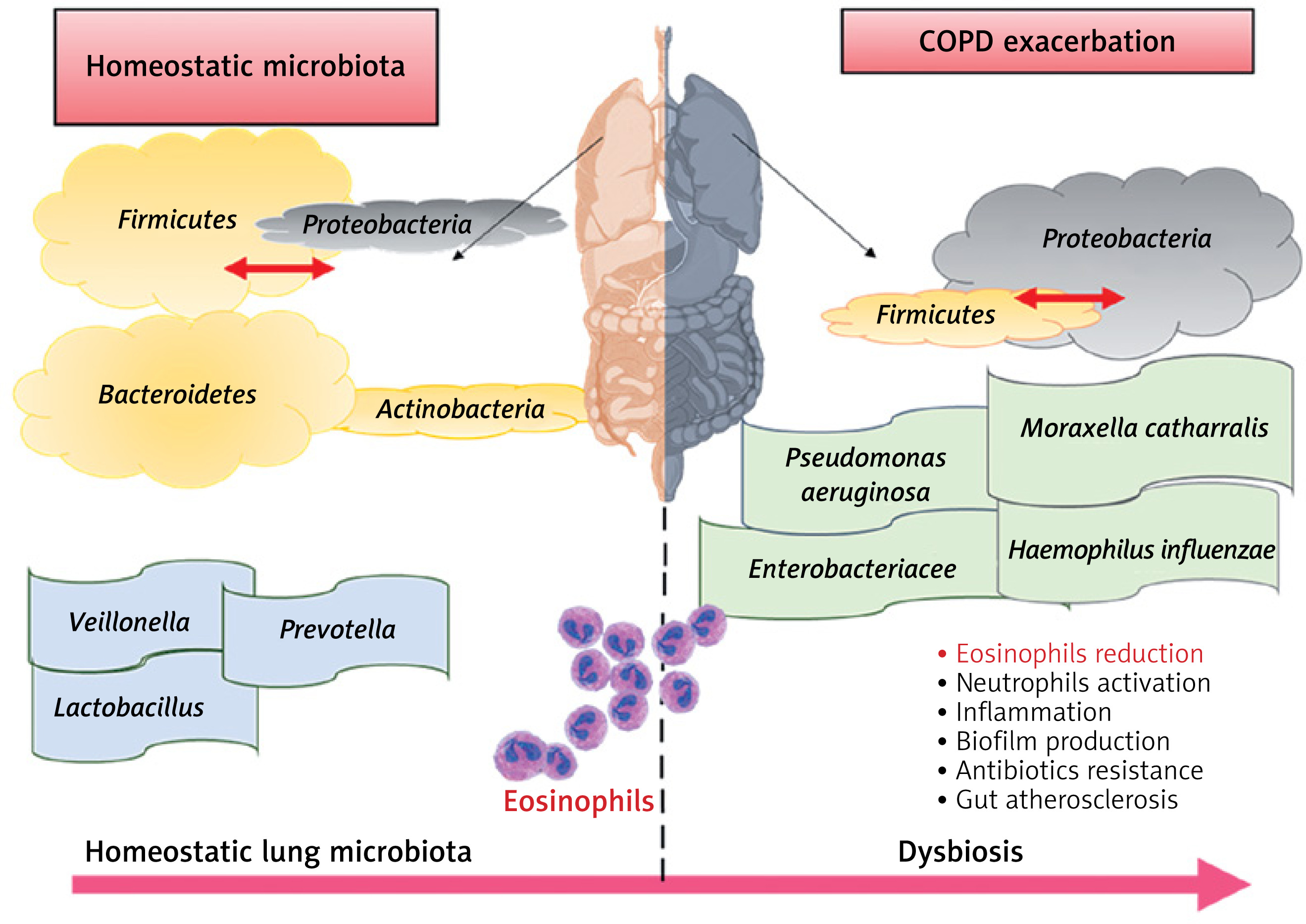
Personalized treatments based on risk stratification by the dominant organism in the microbiota may be beneficial [63]. As we have already described [33], differences in the microbiota composition between healthy and exacerbation are illustrated in Figure 1.
Figure 1
Differences in the lung microbiota composition between healthy (left side) and chronic obstructive pulmonary disease (COPD) exacerbations (right side). In physiological condition, the most abundant phyla are Firmicutes (e.g., Veillonella, Streptococcus spp.) and Bacteroidetes (e.g., Prevotella); with a Firmicutes : Proteobacteria ratio in favor of Firmicutes. In COPD exacerbation, the most abundant phylum is Proteobacteria (e.g., Haemophilus influenzae, Pseudomonas aeruginosa, Moraxella catarrhalis and Escherichia spp.), and the Firmicutes : Proteobacteria ratio is in favor of Proteobacteria. In addition, eosinophil levels seems to be reduced in exacerbations compared to homeostatic condition, with an increase of inflammation, biofilm production, immune system activation, gut atherosclerosis and possible antibiotic resistance [45]
Main factors associated with dysbiosis
There could be different variables that affect the lung microbiota, such as bacterial composition, host immune response, lifestyle, diet, cigarette smoking and the use of antibiotics and corticosteroids that are the standard therapy for COPD exacerbation [33]. Cigarette smoking, in particular, contributes to impaired lung innate immunity through the alterations in ciliary function, mucus, cell phagocytosis, and the increase of bacterial virulence (e.g., enhanced biofilm formation) [64, 65]. Smoking damages airway epithelia and epithelial tight junctions, also causing bronchitis. Also, regular exposure to tobacco smoking provokes changes of the microbiota in healthy smokers, leading to dysbiosis [34].
As we have previously reported [33], a reduction of microbiota diversity adds an increment of the Proteobacteria/Firmicutes ratio in subjects treated with corticosteroids alone, and the reversed trend seen in patients who received antibiotics suggests that the current standard of care therapy for COPD exacerbation can alter the lung microbiota. In agreement with previous reports on the limited efficacy and the greater side effects of steroids [66–68], it is also probable that corticosteroid treatment alone could potentially affect the microbial species. Also, treatment with steroids may negatively influence the eosinophilic inflammation in COPD exacerbation [33], which could be considered as a potential biomarker for identifying subgroups of patient who may respond to therapy [69, 70]. Recent investigations in persistent asthma patients showed that after 1 year of azithromycin therapy, bacterial diversity was decreased compared to asthmatics receiving the placebo control [71], and COPD patients treated with the same antibiotics showed lowered alpha-diversity without altering the total microbial burden [72]. At the same time, patients who require a higher dose of corticosteroids and those exhibiting more severe airway obstruction showed higher pathogenic species in the microbiota compared to asthmatic patients with a better controlled disease [73]. It seems possible that corticosteroid treatments alter the lung microbiota by promoting the growth of potential pathogens and thereby contributing unresponsiveness to corticosteroids [28, 74–76]. All these considerations suggest that the recurrent use of antibiotics and corticosteroids during exacerbations is controversial, because it may have adverse consequences on the lung microbial species by driving the loss of diversity that may lead to a higher risk of other exacerbations or disease progression [33].
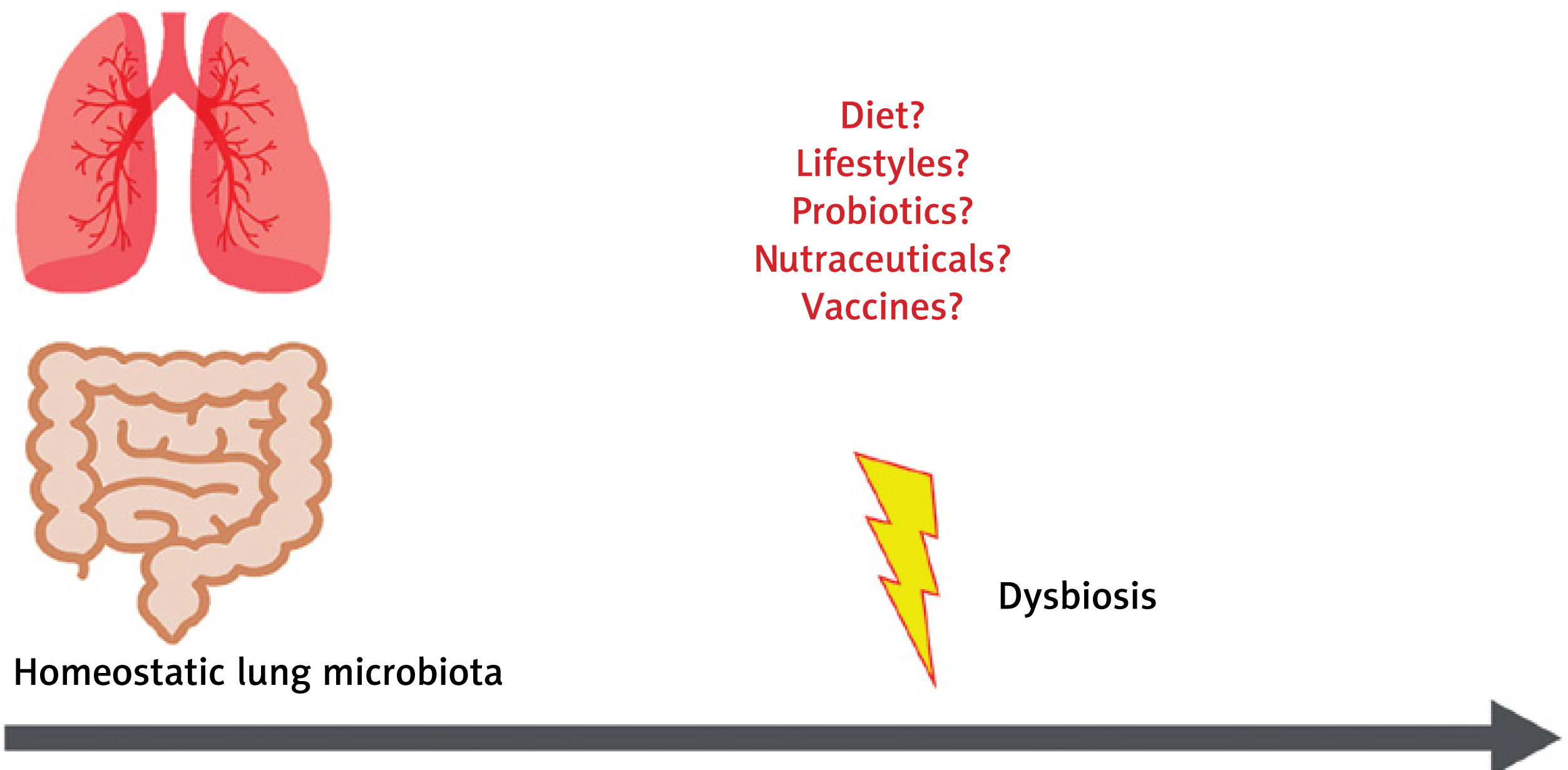
Clinicians should be aware of these potential risks, prescribing such treatments only when necessary, especially in the case of infants and young children [29]. Furthermore, it has not yet been clarified whether it is possible to treat the lung microbiota by diet or by probiotics and nutraceuticals [33, 77]. Figure 2 shows events that may revert microbiota alterations.
Disease and nutrition
An association between malnutrition and lung status of COPD patients has also been shown [78]. Low fat-free mass (FFM) is a common problem found in these patients, especially with emphysema manifestation, which is associated with a reduction in respiratory muscle and skeletal muscle masses, resulting in declines in strength and endurance [79]. Low weight patients have higher gas trapping, lower exercise capacity and decreased health-related quality of life compared to normal weight patients with the same airflow limitation [80]. Also, this loss of body cell mass, which reflects the amount of skeletal muscle mass, is associated with shorter survival due to impaired health status and less skeletal muscle strength [81]. Notably, confirmations come from biopsies of the muscles, which showed alterations in the number and function of mitochondria [82, 83]. Also, it is reported that in chronic diseases, such as COPD, there could be a deficit of elements such as coenzyme Q10 or ubiquinone that are essential for the production of ATP and energy by mitochondrion promotion [84–86].
As malnutrition is an important factor in COPD, nutritional support might be a strong component of disease management and prevention. Poor nutrition is strongly linked with chronic diseases, many of which have an inflammatory nature [87–89]. The Western diet is often nutrient deficient, as it is characterized by frequent intake of red meat, saturated fat, refined grains and processed sugar [90, 91]. In contrast, a diet that includes vegetables, fruit, olive oil, cereals, legume, whole grains, rice/pasta, fish, low-fat dairy, poultry and water has an inverse relationship with the risk of COPD in men and women [23, 92].
Epidemiological studies have reported that a diet with higher fiber content was associated with decreased risk of COPD and better lung function [93, 94]. Supplementation of dietary fiber, in particular cereal and fruit fiber, was associated with a 30% lower risk of COPD in a cohort of 35,339 Swedish women [95]. Another study showed that consumption of fruits and vegetables was associated with lung function improvement [96].
Thus, based on new evidence, a healthy balanced diet, mainly characterized by high consumption of fruits, vegetables, whole grains, plant oils and fish, low alcohol assumption (preferably wine), and avoidance of high-saturated fat foods, refined sugar, red meats and sugar-containing beverages, would be recommended for managing respiratory health and COPD, along with physical exercise [97].
Nutrition and dietary supplements could affect microbiota dysbiosis
An unbalanced diet causes an insufficient intake of important nutrients that might not only inhibit physical development in the infant and adolescent phase [98] but also increase the risk of diet-dependent diseases. The most favored dietary regimen is the Mediterranean diet, which is characterized by increased consumption of vegetables, fruits, legumes, olive oil and fish and low consumption of red meat, dairy products and saturated fats [99]. Adherence to this diet is consistently associated with reduced mortality and incidences of several diseases as well as increased antioxidant and anti-inflammatory activities [100]. The effect of nutrition and dietary supplements on microbiota changes is still under investigation.
Recent studies conducted in mice have pointed out that the intake of dietary fiber and some of its fermentation products have a strong impact on gut microbiota health [101, 102]. Dietary fiber comprises complex carbohydrates consisting of both soluble and insoluble components. The soluble forms can be fermented by certain species of gut bacteria, leading to the production of SCFAs, physiologically active bio-products that are an energy source for certain bacterial species [103–105]. Short-chain fatty acids are organic molecules mainly composed of acetate, propionate and butyrate, which regulate host metabolism, the immune system and cell proliferation [106]. They are subjected to bloodstream absorption and transported to the peripheral circulation through the portal vein to act on the liver [107] and peripheral tissues, where they regulate inflammation by acting as signaling molecules [106], releasing anti-inflammatory cytokines, inducing apoptosis, and reducing chemotaxis and adherence in immune cells [37, 106]. Notably, these molecules are not restricted to the intestinal tract but can disseminate in the blood and be absorbed systematically [108]. Epidemiological studies documented that fiber intake was associated with lower C-reactive protein serum levels and cytokines, including IL-6 and TNF-α, and higher levels of adiponectin, an adipocytokine with anti-inflammatory properties [109]. Other studies found that higher dietary intake of fiber was able to reduce by about 40% the risk of COPD, and it was also negatively associated with lung function decline, incidence and prevalence of the disease [110–112]. Trompette et al. [21] also reported that dietary fiber content influences gut microbiota and thus the circulating levels of SCFAs, which enhance the hematopoiesis of dendritic cells (DC) precursors from bone marrow, and exhibit an impaired ability to activate TH2 effector cells in the lung, which act as a humoral response against extracellular bacteria, parasites, and toxins. In another recent study, Ghosh et al. [113] applied a novel leave-one-out-cross-validation machine-learning methodology and observed that the Mediterranean diet was able to increase specific taxa of the gut microbiota in older people that were positively associated with several markers of lower frailty and cognitive function, and negatively associated with inflammatory markers including C-reactive protein and IL-17. All these findings supporting the concept of intervention strategies through diet are a valuable approach for not only intestinal but also for respiratory inflammatory diseases.
Notably, wholegrain intake was reported to have a beneficial effect on lung function and against mortality from chronic respiratory disease [114–116]. The protective role could be associated with the fact that whole grains are rich in phenolic acids, flavonoids, phytic acid, vitamin E, selenium and other essential fatty acids [97].
The consumption of fruit and vegetables is considered essential for the maintenance of a correct state of health and physical efficiency due to the presence of essential substances such as vitamins and minerals, soluble and insoluble fiber and molecules with antioxidant action. Their contribution to the lung function may be partially associated with their high content in vitamins and antioxidants. The diet is the primary source of vitamins, since our bodies cannot synthesize all of them. However, certain vitamins are synthesized by the intestinal microbiota [117].
Patients with COPD showed lower fruit and vegetable intake compared to healthy subjects [118], as well as lower intake of several micronutrients, such as minerals and vitamins, e.g. iron, calcium, potassium, zinc, folate, vitamin B6, retinol and niacin, mostly in association with obesity [119, 120]. These findings suggest an increased risk of malnutrition in these patients.
Lower serum vitamin E levels were attributed to COPD exacerbation compared to stable disease [121]. Vitamin E, or tocopherol, is a lipid-soluble antioxidant acting with vitamin C in synergy to break the lipid peroxidation chain reaction. The role of vitamin E in influencing the microbiota has been reported in several recent studies [122–124] and may derive from its natural antioxidant effect against excessive free radicals and by promoting cellular and humoral immune responses [122]. It was reported that a high-level intake of vitamin E is associated with an increased ratio of Bacteroidetes to Firmicutes compared to low-level consumption and controls [124]. An increase of the Bacteroidetes/Firmicutes ratio at the phylum level as well as an overall increase in microbial diversity due to a higher intake of vitamin E was also reported in a mouse model of ileal pouchitis. In this study, vitamin E along with selenium and retinoic acid showed enrichment of the gut microbes in favor of anti-inflammatory patterns and reduction of mucosal inflammation [123]. In another cross-sectional Spanish study, dietary intake of vitamin E, along with vegetables and olive oil rich in vitamin E and polyphenols, was found to be inversely correlated with oxidative stress markers in the serum, especially in current smokers [125]. Although vitamin E was believed to protect the lung against oxidative damage [126, 127], randomized trials of its supplementation have reported mixed results, both protective [128] and without effects on the risk of disease development [129].
Another important studied vitamin is vitamin D. Over time, more and more evidence suggests that vitamin D may be involved in the alteration of the gut microbial composition. Vitamin D can develop a healthy intestinal microbiota, maintain the integrity of the gut barriers and allow beneficial bacteria to outcompete opportunistic pathogens [130]. The immunomodulatory properties of vitamin D may explain its potential effects on microbial colonization of the gut microbiome. In particular, vitamin D3 supplementation shows a positive influence on gastrointestinal diseases such as inflammatory bowel disease, infection from bacteria [131–133], gastrointestinal inflammatory disease, Crohn’s disease [134, 135] as well as malignant tumors [136–138]. The administration of vitamin D3 was also reported to increase the abundance of the genus Lactococcus and decrease that of the Veillonella genus and the Erysipelotrichaceae family, some members of which were found to be potential pathogens, in the gut microbiota of cystic fibrosis patients [139].
Another randomized control trial showed that weekly administration of vitamin D over a 1-year period increased the enrichment of SCFA-producing genera and the subsequent SCFA fecal levels [140]. The relationship between vitamin D deficits and respiratory infections was hypothesized many years ago, when it was observed that people with rickets had a high risk of lower respiratory tract infections. Vitamin D, which is mainly absorbed after sun exposure in addition to diet, has numerous immunoregulation properties, capable of affecting both innate and adaptive immunity. For example, 1,25(OH)2D has been shown to stimulate the secretion of antimicrobial peptides, such as cathelicidin, by epithelial cells and macrophages in respiratory infections. A recent meta-analysis [141] of data extracted from 25 randomized controlled trials, with a total of 11,321 participants aged 0 to 95 years, showed that vitamin D supplementation was effective in reducing the risk of developing acute respiratory tract infections. Jolliffe et al. [142] promoted randomized controlled trials of vitamin D to prevent COPD exacerbations with conflicting results: vitamin D supplementation was able to safely reduce the rate of moderate/severe COPD exacerbations in patients with baseline 25-hydroxyvitamin D levels equal to or less than 25 nmol/l but not in those with higher levels. Vitamin D deficiency was also correlated with genetic variants in the vitamin D-binding gene, highly prevalent in COPD [143]. Although still controversial, vitamin D supplementation may be beneficial for the highly prevalent osteoporosis condition found in COPD patients.
Other dietary factors with a potential protective role are polyphenols, the most abundant antioxidants and anti-inflammatory molecules present in vegetables. Polyphenols, which include phenolic acids, flavonoids, stilbenes, lignans and secoiridoids, seem to positively influence the human gut microbiota through inhibition of potential pathogens such as Helicobacter pylori and Staphylococcus species and favoring the enrichment of other potential beneficial species, such as Lactobacillus and Bifidobacteria [144–149]. Polyphenols have also been associated with chronic disease, cancer and neurodegenerative disease prevention [150–153].
A nutraceutical can be defined as a dietary fortified supplement that may provide health benefits in addition to its basic nutritional value [84], delivered for therapeutic purposes in a higher concentration than what is available from a normal diet. Several studies have been addressed to modulate muscle protein synthesis and energy state in COPD patients. Coenzyme Q10 and creatine were previously reported to be clinically effective in chronic heart failure (CHF) and COPD patients [84, 85], because they were associated with their capacity to regulate numerous metabolic pathways, including mitochondrial energy depletion in the skeletal muscles, oxidative stress and calcium overload, leading to cytoskeleton alteration, cellular dysfunction, apoptosis and necrosis. Mitochondrial dysfunction is the key determinant of these mechanisms, with defective energy production and increased oxidative stress that contributes to altering the structure of DNA, proteins and lipids [79]. Coenzyme Q10 is known for its role in oxidative phosphorylation, acting as a key element for the mitochondrial respiratory chain. Although it is known for its antioxidant properties, gene expression modulation, and oxidative phosphorylation, its role was hypothesized in clinical practice as a food supplement [86]. However, coenzyme Q10 has limited solubility in water, poor bioavailability and chemical instability [154].
Creatine was also shown to increase exercise performance and fat-free mass in normal individuals [155] when used as a dietary supplement. Creatine kinase plasma levels are also a marker of extreme exercise. A study on the impact of exercise on gut microbial diversity highlighted that athletes, besides higher plasma creatine levels, showed a higher gut microbial diversity (22 distinct phyla) compared to controls, with a beneficial impact in terms of inflammatory and metabolic markers [156]. Creatine seems also to protect against adenosine triphosphate (ATP) depletion, stimulate protein synthesis and biological membranes and reduce protein degradation. However, this alone might be insufficient to restore all energy metabolism [79, 82, 83].
The impact of dietary fats, such as poly-unsaturated fatty acids (PUFAs), on the microbiota is less well defined. PUFAs are considered nutritionally essential due to the low synthesis in our bodies. They are mainly found in seafood. The n-3 (omega-3) PUFAs, which include α-linolenic acid, have anti-inflammatory properties with beneficial effects in numerous chronic disease [157], whereas n-6 PUFAs (linoleic and arachidonic acid), mostly found in vegetable oils, dairy, egg and grain-fed animals, display opposite effects. In particular, they produce eicosanoids, such as thromboxane A2, prostaglandin E2, and leukotriene, which are pro-inflammation, favoring thrombosis and vasoconstriction [158]. In the Western diet, we are assisting the dietary shift from n-3 PUFA consumption to an n-6 PUFAs intake, a trend that is thought to raise chronic inflammatory disease [157]. Consumption of 3-PUFAs should be encouraged as they show a beneficial impact in regulating intestinal microbial composition and inflammation [159]. 3-PUFA administration was found to revert the microbiota composition in inflammatory bowel disease and increase the production of anti-inflammatory compounds, such as SCFAs [160]. Also, omega-3 fatty acids help to maintain the intestinal wall integrity in animal model studies and interact with host immune cells [161]. For example, the constitutive production of 3-PUFAs in transgenic mice has been shown to contribute to the prevention of metabolic syndrome by promoting changes in the gut microbiota [162]. PUFA supplementation also led to the improvement in mass muscle strength in COPD patients with wasted muscle, by increasing the muscle oxidative capacity and energy state [163]. As underlined in a recent systematic review, the ability of PUFAs to modulate COPD has also been assessed, with controversial results [164]. Interestingly, fish consumption seems to reduce COPD risk not alone, but within the whole diet, in particular when the intake of n-3 PUFAs from plants is high [165].
Another dietary component is curcumin or diferuloylmethane, a phenol capable of modulating the inflammatory response, down-regulating the activity of cyclo-oxygenase 2 (COX-2), lipoxygenase, and iNOS and inhibiting the production of inflammatory cytokines such as TNF-α, IL-1, IL-6 and IL-12. These characteristics gave it the potential to modulate inflammatory phenomena by acting on the inflammatory cascade [166]. There are several enteric bacteria capable of metabolizing curcumin, e.g. some Escherichia coli, Bifidobacteria and Lactobacillus strains [167, 168]. Increasing evidence shows that oral administration of curcumin can improve microbial richness, diversity, and composition of the gut microbiota, and can provide benefit by restoring dysbiosis of the gut microbiome [169]. For example, curcumin supplements considerably increase Bifidobacteria and Lactobacilli and reduce potential pathogenic bacteria such as Enterobacteria, Enterococci, Coriobacteriales and Prevotellaceae [170–173]. In an Asian cross-sectional study, curcumin was significantly associated with improved lung function, and this improvement was greater in smokers with the highest curcumin intake than smokers not consuming it [174].
Overall, all this evidence suggests that a new strategic area of investigation is modulation of the microbiota through dietary intervention because nutrition and dietary supplements are important factors in the pathogenesis and prevention of COPD and exacerbations, probably by influencing our gut-lung microbial population. These considerations provide support for specific dietary modifications to promote lung health. More studies are needed to confirm the effectiveness of diet in preventing and improving disease outcomes.
Probiotics for microbiota dysbiosis improvement
Probiotics are defined as “live microorganisms which, when administered in adequate amounts, confer a health benefit to the host” [175]. The association between probiotics and health was described at the beginning of the twentieth century by a Nobel Prize Winner in Physiology [176]. The U.S. probiotics market was estimated to be worth over 40 billion dollars in 2017, whereas in Europe, it is trailing behind [177], probably due to the stricter regulation for the nutrition and health claims on food supplements (Regulation (EC) No. 1924/2006) [178].
Further evidence from animal models and human studies has brought to light the positive action of probiotics in the alteration of the healthy gut microbiota composition [179–182]. In pediatric subjects, the microbiota is particularly rich in Bifidobacteria, a very heterogeneous species ascribable to the phylum Actinobacteria, considered protective and necessary in infants and young children. In nature, Bifidobacteria are transmitted to the baby during childbirth and subsequent breastfeeding directly from the mother, who derives them from her intestine, because they are essential to the newborn and infant to metabolize HMO (human milk oligosaccharides) carbohydrates present in breast milk, and mucin glycans [183]. Bifidobacteria of this type are the species B. longum infantis, B. bifidum and B. breve, which promote an anti-inflammatory capacity [184]. Children with a higher risk of Bifidobacteria deficiency are those born by caesarean section and/or not breastfed. The same subjects are also those considered at higher risk of allergies, inflammation and obesity. A recent clinical study has shown that allergic children with Bifidobacterium and Enterococcus deficient states show reduction of disease symptoms by about 50% if they are corrected in their dysbiosis with probiotics [160]. Treating intestinal dysbiosis in advance would favor milder atopic symptomatology, with less need for drugs, corticosteroids and antihistamines. Therefore, identifying children with dangerous bacterial presence or microbiota deficiency could allow intervention with targeted dietary choices or using probiotics, also for prophylactic purposes [185].
Several functional effects of multiple probiotic strains on the intestinal microbiota composition have also been well documented in the adult disease state. The hypothesis is that the use of probiotics in clinical settings may reduce the inflammatory response and form a protective barrier to prevent and even treat carcinogenesis in mice [186, 187], in human gastrointestinal tumors [188, 189], as well as in irritable bowel syndrome [190] and metabolic dysfunction such as non-alcoholic fatty liver disease and lipid metabolism [191, 192]. For example, in a recent Chinese study [193], probiotics treatment and dietary intervention raised the gut microbial diversity of high-fat diet subjects. The most increased species belonged to two butyrate-producing families Ruminococcaceae and Lachnospiraceae, whereas the majority of the species with reduced populations belonged to the Bacteroidaceae family [193]. Potential new treatments based on probiotics might not only reduce pathogenic species colonization but also increase commensal bacterial growth in the respiratory tract. Iwase et al. reported that commensal bacterial species can directly suppress the outgrowth of potential pathogens belonging to the same genus or family, suggesting the importance of microbiota interactions in maintaining homeostasis [194, 195]. Concerning this funding, Actinobacteria are Gram-positive bacteria, whose presence may also oppose the dominance of the phylum Proteobacteria, consisting of Gram-negative bacterial species [26]. The substances produced by some commensal bacteria, capable of inhibiting the growth of pathogens, can be antimicrobial peptides, such as bacteriocins, but also metabolic products, such as organic acids, hydrogen peroxide or biosurfactants. A recent study has shown that Streptococcus salivarius K12, a commensal of the oral cavity of some subjects, is capable of producing two antimicrobial peptides, salivaricin A2 and salivaricin B. These substances have been shown to strongly inhibit the growth of oral pathogens such as Streptococcus pyogenes, Haemophilus influenzae, Streptococcus pneumoniae, Moraxella catarrhalis and Candida albicans, and also promote the production of immune-stimulating cytokines. The preventive use of K12 as an oral probiotic has already been shown to reduce recurrences of pharyngotonsillitis of bacterial and viral origin, but also to reduce the percentage of acute middle ear infections, both in children and adults, with a considerable saving of antibiotics [196–199]. Further studies have shown that Streptococcus salivarius K12 can inhibit the production of biofilm of S. epidermidis and S. aureus [200]. Biofilm is a matrix produced by some bacteria, often consisting of extracellular polymeric substances of polysaccharide proteins, also enriched with lipids and nucleic acids, through which the bacterial colony fixes on the mucosal surfaces of the host. Within the biofilm, bacteria are protected from attack by the host immune defenses and the bactericidal action of antibiotics. Haemophilus influenzae, Moraxella catarrhalis and Pseudomonas aeruginosa are known to produce biofilms protecting them from the host immune system and antibiotics. The formation of biofilm is a normal behavior of the colonization process of some bacteria; therefore understanding what mechanisms regulate the formation of biofilm would allow us to know better the bacterial physiology, providing a more effective basis for developing drugs or alternative strategies to prevent bacterial infections.
We previously hypothesized the possible role of probiotics in augmenting the treatment response of COPD patients who receive frequent antibiotic therapies [33]. It is already well known that modulation of the intestinal microbiota through probiotics is an accepted method for restoring intestinal flora and to improve host defense [201]. Fermented foods that are produced through a fermentation technology process can harbor several probiotic bacterial species and strains. These are considered to be a natural source of live microorganisms that are consumed in large amounts worldwide. Despite the extensive literature focusing on characterizing these bacteria in fermented foods, it is still not fully understood how they interact with the gut microbiota [202]. Indeed, ingested bacteria need to first survive the barriers of the gut before exerting their beneficial effects, provided that they can survive and become part of the gut complex environment. A very recent large-scale genome-wide analysis [203] reported that lactic acid bacterial species identified in food only partially match those found in the gut and their concentration is generally low and variable, and this variability mostly depends on age and lifestyle. The study confirmed that food is likely the major source of lactic acid bacteria, with the most prevalent species being Streptococcus thermophilus and Lactococcus lactis [203]. Their abundance might be explained by the widespread use of the former for yoghurt making and its use as starter cultures for many kinds of cheese obtained by thermophilic fermentation, while the second is widespread in cheeses produced by mesophilic fermentation. The beneficial effect of Streptococcus thermophiles and its resistance to the gastrointestinal barriers are still questioned and debated [204]. In contrast, Lactococcus lactis has been shown to survive in the gut and it seems to boost the immune system by promoting antimicrobial activity through bacteriocin production [205, 206]. Interestingly, it has been reported that human macrophages can phagocytose Lactobacillus species, reducing cigarette smoking-related inflammation and thus smoking-related lung diseases, including COPD [207]. Lactobacillus species have already been associated with anti-inflammatory properties in COPD patients [207] and protection against viral infections [208]. For example, Lactobacillus rhamnosus species seem to modulate the anti-viral response within the lungs of animal models infected with a respiratory syncytial virus (RSV) prior to infection [209]. All this knowledge may also support the possibility to use vaccines or antibacterial drugs targeted against pathogens to prevent lung microbiota alterations.
Overall, new evidence suggests that the lung microbiota is critical to disease, and that manipulation of these microbial patterns could be an attractive new scenario for future treatments for stable COPD and in preventing exacerbation. Describing the causes and consequences of the lung dysbiosis and finding a possible solution to correct and revert microbiota alterations may influence the choice of adequate therapy of COPD, especially for exacerbations [210].
Conclusions
There is accumulating evidence that a bidirectional cross-talk exists between the gut and the lung, the so-called gut-lung axis, important for the maintenance of immune homeostasis. Exogenous factors, such as cigarette smoking, lifestyle, diet, the use of antibiotics and corticosteroids, as well as endogenous factors, including bacterial composition and host immune response, can surely affect the gut-lung microbiota compositions and thus facilitate the onset of diseases. Hence it can be expected that dietary intervention, probiotics or bacterial lysates could interfere via the gut-lung axis to revert microbial dysbiosis. Understanding the role of diet and dietary supplementation affecting the gut-lung microbiota axis, with respect to stable COPD and exacerbation, is of great interest for the development of prevention strategies and recommendation of therapies for combatting this respiratory disease.
Among all dietary supplements, fermentable fiber can change the composition of the gut microbiota, especially by altering the Firmicutes/Bacteroidetes ratio. The gut microbiota metabolizes the fiber, consequently increasing the circulating levels of SCFAs, which can boost the immune system in the lung, lastly protecting against inflammation. Besides fermentable dietary fiber, fruit, vegetables and whole grain showed a beneficial effect on lung function and against mortality due to chronic respiratory diseases. These positive effects might be ascribed to the content of micronutrients, including vitamins, antioxidants and polyphenols.
Nutraceuticals were also hypothesized to provide health benefits if administered in a higher concentration than what they are available from the diet. Curcumin, along with coenzyme Q10, creatine, and PUFAs, showed the ability to regulate numerous metabolic pathways, including the improvement in mass muscle strength in COPD patients with wasted muscle, mitochondrial energy depletion in the skeletal muscles, oxidative stress and calcium overload.
The association between probiotics and health has also been extensively assessed. Bacteria used for probiotics mainly belong to lactic acid bacteria, and it is known that these bacterial strains and products exert several beneficial effect. The use of probiotics in a clinical setting might not only reduce pathogenic species colonization but also increase commensal bacterial growth in the respiratory tract.
In conclusion, a balanced diet rich in substances able to counter the pathogenic processes of COPD such as oxidative stress, systemic inflammation, dysmetabolism, also potentially acting at the level of the gut-lung microbiota, may significantly affect the risk of developing COPD, its clinical progression and its comorbidities. Preventive and therapeutic strategies to counteract microbiota dysbiosis and restore a healthy microbial pattern by diet, dietary supplements, probiotics and bacterial lysates have not arrived in a clinical setting so far. Thus, many further studies are needed to explore the impact of microbiota composition and function on COPD pathogenesis and in general for lung disease, to subsequently refine new treatments that can prevent and treat this respiratory disease, as well as the effect of diet on different clinical phenotypes. Definitely, the current scientific evidence is sufficient to develop dietary recommendations that represent an opportunity, along with other lifestyle interventions including smoking cessation, to improve preventive and therapeutic approaches in COPD.

