

Introduction
Bile acids are produced by hepatocytes and facilitate lipid absorption in the intestinal lumen. They are associated with vascular diseases through various mechanisms, including direct involvement in lipid metabolism, modulation of inflammatory responses, and regulation of immune cell expression [1–4]. Research has indicated that both cerebrovascular disease and coronary artery plaque are associated with reduced fecal bile acid excretion (BAE) [1, 5–9]. The potential connection between gut microbiota and cardiovascular health has also drawn attention to the role of bile acids [2, 6, 10, 11]. Since serum total bile acid (TBA) levels are strongly correlated with various metabolic diseases and cardiovascular risk [5–9], TBA serves as a practical and efficient biomarker for screening and monitoring cardiovascular disease (CVD) and metabolic health across diverse populations.
CVD is escalating into a major global health issue, accounting for nearly one-third of annual deaths worldwide [12–17]. Coronary artery calcification (CAC), an early lesion of coronary atherosclerosis that progresses with the disease, is recognized as a robust marker of adverse cardiovascular events and exhibits the highest predictive value among assessed risk factors [18, 19]. CVD risk is typically quantified using the CAC score [20, 21]. Multi-detector computed tomography (CT) is commonly employed for coronary artery disease diagnosis [22–25]. Although BAE and gut microbiota-related bile acids are established as independent CVD risk factors [5, 26, 27], their integration into traditional risk assessment frameworks remains limited. Several studies have investigated the association between serum bile acid level and coronary artery disease (CAD) in subjects with suspected CAD [28] or the association between serum bile acid and high-risk coronary artery plaques in asymptomatic population [29]. However, the relationship between serum TBA levels and CAC risk has not received much research attention. A growing body of evidence indicates that the bile acid network is intricately linked to the pathogenesis of vascular calcification [30].
We hypothesized that the serum TBA level might also be associated with CVD risk. The correlation between CAC and TBA among the general Chinese population was examined in this study.
Material and methods
Participants
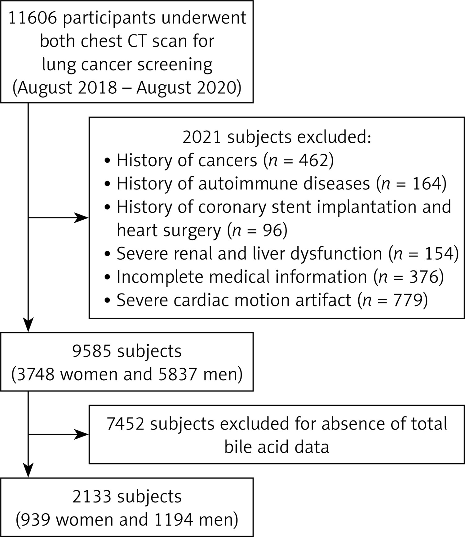
Participants who underwent CT lung cancer screening between 2016 and 2020 were initially considered. Our medical record system identified 11,606 individuals. Inclusion criteria were: age ≥ 18 years and high-quality cardiac CT scans suitable for CAC assessment. Exclusion criteria comprised: history of cancer, autoimmune disease, prior cardiac surgery or coronary stenting, severe renal or hepatic insufficiency, incomplete medical records, or severe cardiac motion artifacts. Ultimately, 2,133 participants who had undergone serum bile acid testing were enrolled (Figure 1). The Institutional Academic Committee approved this retrospective cross-sectional study (approval No. 2019-NL-158). Informed consent was waived by the Institutional Ethics Committee owing to the retrospective design. The study was conducted in full accordance with the Declaration of Helsinki.
Data collection
Data on characteristics – including age, sex, and body mass index (BMI) – were collected, along with laboratory test results such as TBA, blood glucose, high-density lipoprotein cholesterol (HDL-C), low-density lipoprotein cholesterol (LDL-C), total cholesterol (TC), triglycerides (TG), creatinine, aspartate transaminase (AST), serum albumin, and uric acid. Medical history information, including diagnoses of diabetes mellitus, cancer, chronic renal disease (defined as an estimated glomerular filtration rate [eGFR] < 60 ml/min/1.73 m2), and blood pressure, was also obtained. Blood samples were collected from participants in the morning after a minimum 8-hour overnight fast. TBA (µmol/l) was measured using an automated biochemical analyzer with the enzymatic cycling method. This information has been included in the revised manuscript. All laboratory tests were performed on the same day as the chest CT scan.
Definition of coronary artery calcification
Numerous studies have demonstrated the feasibility of determining CAC using non-electrocardiogram (ECG)-gated chest CT [22, 23]. In this study, CAC was evaluated using nonenhanced, non-ECG-gated CT, as described in our earlier publication [31]. Briefly, CT examinations were performed using three scanners: OptimaCT660, GE, USA; Brilliance 64, Philips, Netherlands; SOMATOM go.Top, Siemens, Germany. The scanning parameters included a slice thickness of 0.625–1.25 mm, a tube voltage of 120 kV, automatic tube current modulation, and no ECG gating. Two radiologists (XC and MC), each with 5–10 years of experience in cardiac radiology, independently assessed the presence of CAC. Both radiologists were blinded to the clinical data. In cases of disagreement, a senior cardiac radiologist (JW) was consulted to reach a consensus. An example of CAC detected on non-gated chest CT is provided in Supplementary Figure S1. CAC was defined as the presence of calcification with a density greater than 130 Hounsfield units in any coronary artery. Agatston scoring was not performed due to the presence of calcification and motion artifacts in non-ECG-gated chest CT images.
Statistical analysis
For analysis of statistics, IBM SPSS (version 29.0) and R (version 4.4.1) were implemented. Participants with missing data accounted for less than 5% of the total sample. Missing values were handled using mean imputation for continuous variables. Quantitative data that were not normally distributed are presented as the median (interquartile range), while normally distributed data are presented as the mean ± standard deviation. For categorical variables, data are expressed as numbers (percentages). The Mann-Whitney U test was performed to determine whether the nonnormally distributed data differed significantly between groups. To compare categorical variables, the χ2 test was applied. The inter-rater reliability for CAC evaluation was analyzed using the kappa test. To ascertain the relationship between TBA and the risk of CAC, multivariable logistic regression analysis was employed. The quartiles of TBA were Q1 < 1.90, Q2 1.90–2.80, Q3 2.80–4.30, and Q4 > 4.30. Demographic information, factors related to CAC and TBA were considered as confounders. Age, sex, and body mass index were taken into account in Model 1; liver and renal function, diabetes mellitus, uric acid, albumin, blood pressure, and hepatic CT attenuation were further taken into account in Model 2; and LDL-c, TC, and HDL-c were further taken into account in Model 3. Male and female individuals over 50 with or without diabetes or hypertension were subjected to subgroup analysis. The dose-response relationship regarding the TBA index and the risk of CAC was evaluated by limited triple splines after variables were taken into consideration. Differences with a p value less than 0.05 were considered statistically significant.
Results
Participant characteristics
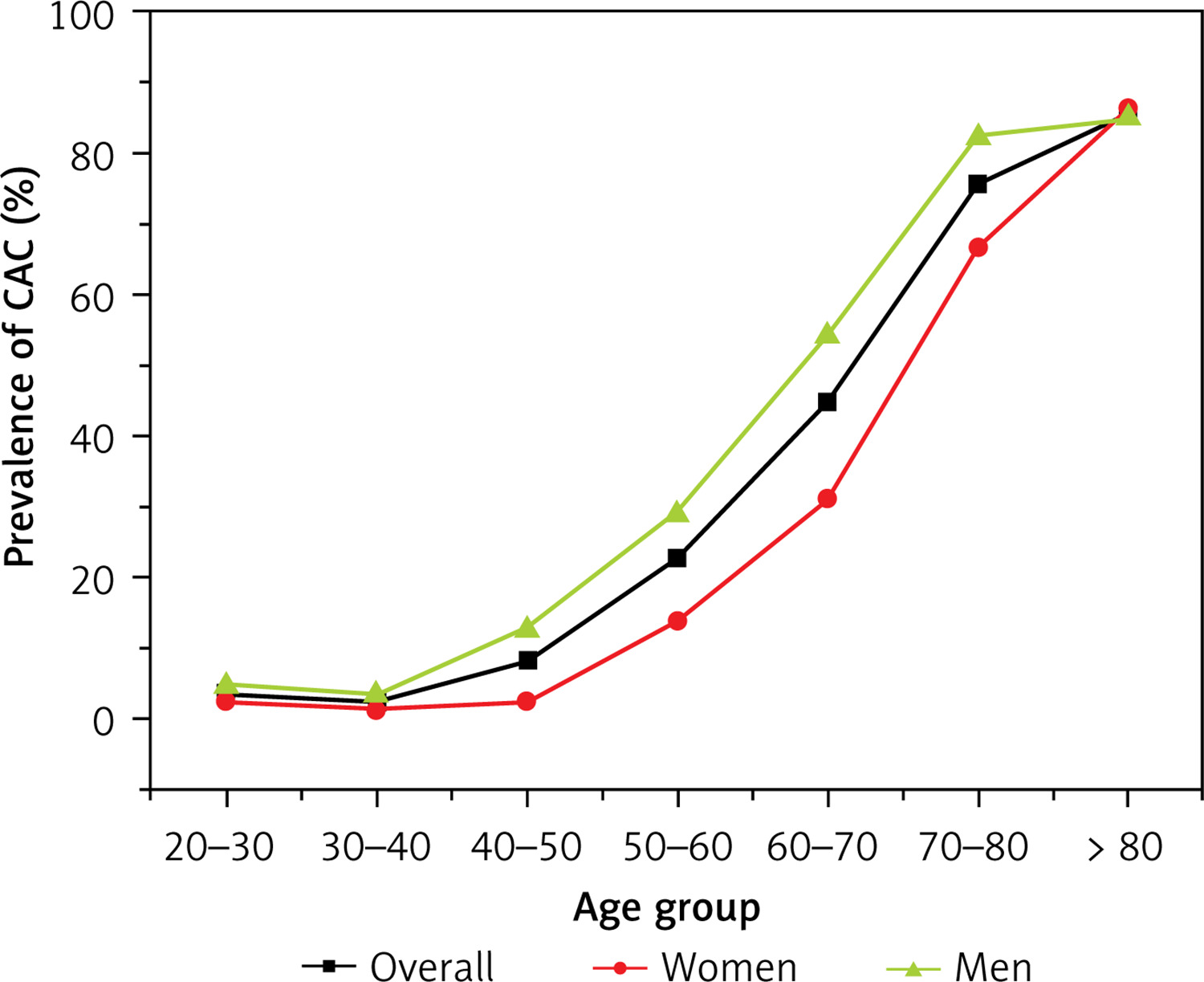
The mean age of the participants was 48.7 ±14.2 years, and 56.1% were male. Among the 2133 participants, 426 had CAC. Inter-rater reliability for CAC assessment was high (k = 0.99). Participants were categorized into quartiles based on their TBA levels. Baseline characteristics are summarized in Table I. Higher TBA levels were associated with lower albumin and HDL-C levels (p < 0.05), and with higher AST, creatinine, and uric acid levels (p < 0.01). Additionally, compared with the first TBA quartile, the fourth quartile showed a higher prevalence of CAC and diabetes mellitus (p < 0.01). Figure 2 illustrates the prevalence of CAC in the overall population and sex-specific subgroups. In the overall population, CAC prevalence increased with age, rising notably between the ages of 40 and 50, and was particularly high in the 60–80-year age group. Between ages 40 and 80, males had a higher prevalence of CAC than females.
Table I
Characteristics of subjects across total bile acid quartiles
[i] AST – aspartate aminotransferase, BMI – body mass index, CKD – chronic kidney disease, CT – computed tomography, DBP – diastolic blood pressure, HDL-C – high-density lipoprotein cholesterol, HU – Hounsfield unit, LDL-C – low-density lipoprotein cholesterol, SBP – systolic blood pressure, TC – total cholesterol, TG – triglyceride.
Association between TBA and CAC
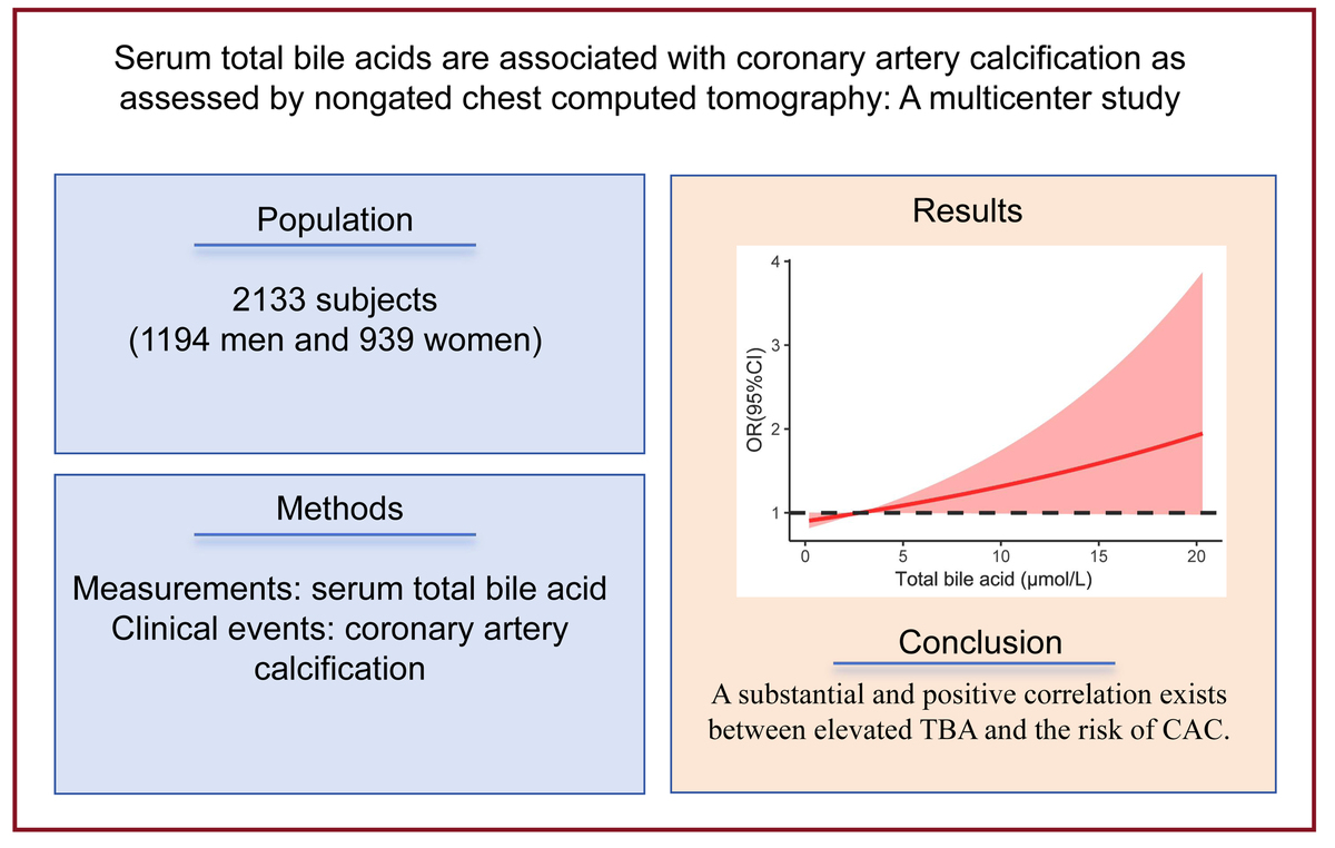
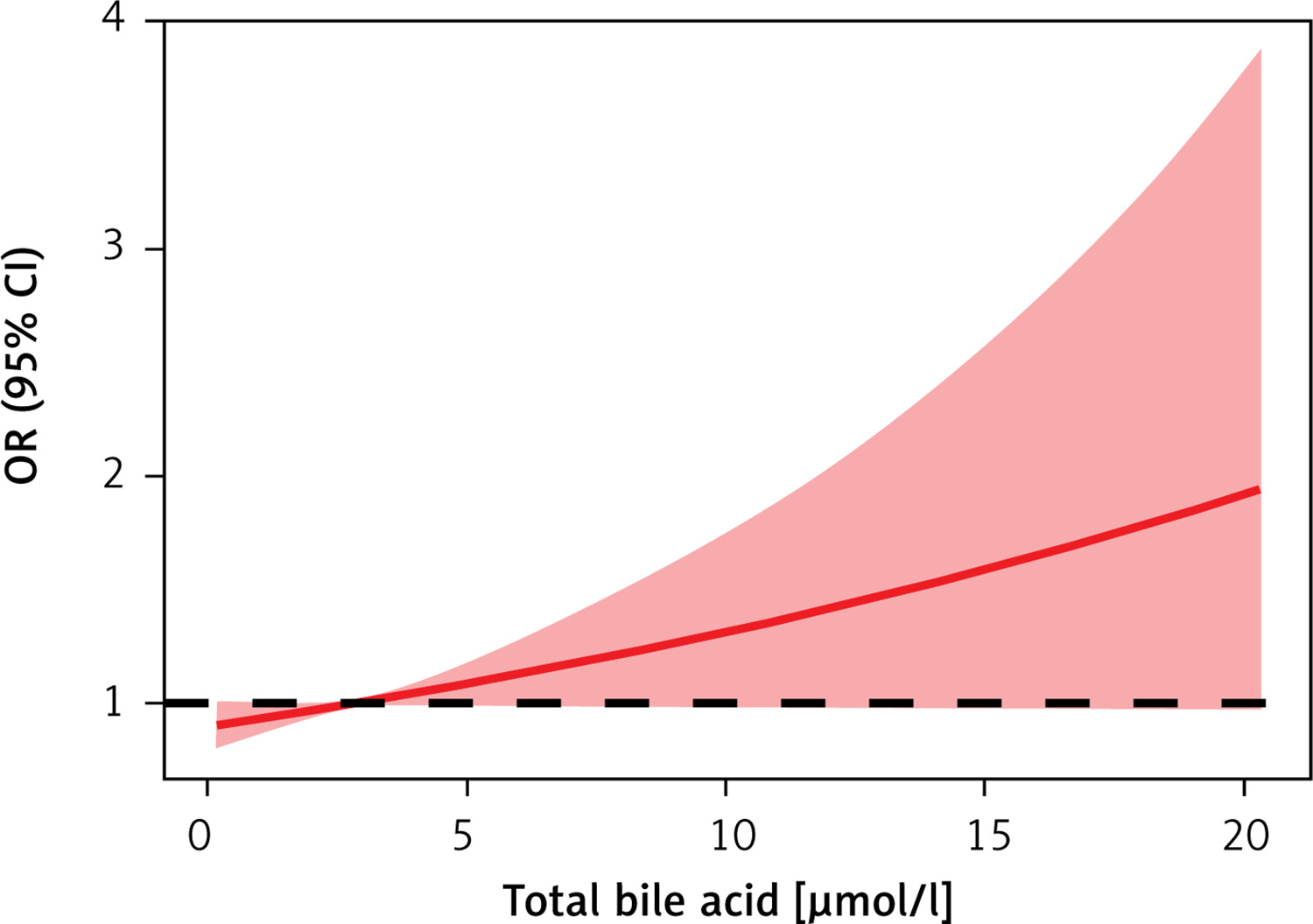
To examine the association between TBA and coronary artery calcium (CAC), we performed multivariable logistic regression analyses (Table II). After adjusting for age, sex, BMI, liver function, renal function, diabetes mellitus, albumin, uric acid, blood pressure, liver CT attenuation, and serum lipid levels, TBA remained independently associated with CAC when analyzed as a continuous variable (odds ratio [OR] = 1.05, 95% confidence interval [CI]: 1.00–1.10). A positive dose-response relationship between TBA and CAC risk was observed using restricted cubic spline curves (Figure 3).
Table II
Association between total bile acid and risk of coronary artery calcification
[i] Model 1 was adjusted for age, sex, and body mass index; Model 2 was further adjusted for liver function, renal function, diabetes, albumin, uric acid, blood pressure, and liver CT attenuation. Model 3 was further adjusted for low-density lipoprotein cholesterol, total cholesterol, and high-density lipoprotein cholesterol. CI – confidence interval, HDL – high-density lipoprotein cholesterol, OR – odds ratio.
Figure 3
Restricted cubic splines show the multivariable- adjusted odds ratio for risk of coronary artery calcification according to total bile acid (TBA). Age, sex, and body mass index, liver function, renal function, diabetes, uric acid, blood pressure, liver CT attenuation, low-density lipoprotein cholesterol, total cholesterol, albumin, and high-density lipoprotein cholesterol were adjusted. A dose-response relationship was observed between serum TBA and CAC risk
CI – confidence interval, OR – odds ratio.
We further evaluated the relationship between categorical TBA levels and CAC. After full adjustment for the same covariates, the highest TBA quartile was significantly associated with a higher prevalence of CAC (OR = 1.76, 95% CI: 1.02–3.06) compared with the lowest quartile.
Subgroup analyses were then conducted on participants who were above 50 years of age (Table II), male or female (Supplementary Table SI), and had or did not have hypertension or diabetes. The fully adjusted model (controlling for age, sex, BMI, diabetes mellitus, hepatic and renal function, blood pressure, serum uric acid, albumin levels, and lipid levels) showed that the fourth TBA quartile was significantly predictive of CAC in subjects aged ≥ 50 years (OR = 1.66, 95% CI: 1.03–2.68), without hypertension (OR = 1.74, 95% CI: 1.16–2.63), without diabetes (OR = 1.62, 95% CI: 1.07–2.45), and those who were male (OR = 1.92, 1.18–3.14).
Discussion
Many studies have shown that bile acids are associated with cardiovascular disease [6, 32]. Several have also demonstrated an association between TBA and coronary artery diseases [28, 29]. However, to our knowledge, the relationship between CAC and total serum bile acid levels in the general population is rarely reported. It seems that only Zhang et al. have demonstrated an association between serum bile acid and high-risk coronary artery plaques in an asymptomatic population [29]. According to our data, TBA is associated with CAC. Compared to participants with low TBA levels, those with high TBA levels were at higher risk of developing CAC.
An association between reduced BAE and vascular diseases has been reported [5]. Charach et al. found that patients with coronary artery disease had significantly lower BAE levels than controls, and those with high BAE levels (> 415 mg/day) exhibited lower cardiovascular mortality than those with low BAE levels [33]. A recent study also suggested that high BAE levels may have a therapeutic effect against peripheral vascular disease [5]. The relationship between bile acids and coronary artery calcium (CAC) has been examined in several studies [34, 35]. Jovanovich et al. reported that circulating deoxycholic acid (DCA), a secondary bile acid, was associated with CAC volume score [34], but not with the prevalence, incidence, or progression of CAC in patients with chronic kidney disease (CKD) [35]. Our data also indicated that TBA was not associated with CAC in subjects with hypertension or diabetes. While many studies have found no significant association between serum bile acid levels and coronary artery disease [28, 36], limited research has specifically investigated the relationship between serum bile acid levels and CAC. However, little research has been conducted on the connection between serum bile acid and CAC. Zhang et al. determined that the degree of coronary artery stenosis and high-risk coronary arterial plaques was correlated with a high serum TBA level in an asymptomatic population [29]. These results were confirmed by our investigation, which also showed an association between TBA and CAC.
Bile acids may influence cardiovascular health through multiple mechanisms, primarily involving cardiovascular signaling pathways and modulation of gut microbiota [10, 37, 38]. Bile acid receptors include G protein-coupled receptors (GPCRs) and nuclear receptors [39], which are implicated in cardiomyocyte apoptosis, cardiac injury, and vascular metabolism [40–44]. The role of gut microbiota (GM) in cardiovascular disease has gained increasing attention [11, 45–47]. As metabolites of GM [48, 49], bile acids can act on various ion channels and receptors in the myocardium and vasculature to modulate cardiac function. For instance, ursodeoxycholic acid, the most hydrophilic bile acid, has been shown to prevent refractory arrhythmias [50]. Additionally, bile acids help maintain intestinal barrier function and support cardiovascular health through several pathways: they disrupt bacterial integrity, directly modulate GM composition, inhibit bacterial overgrowth, and prevent bacterial translocation and endotoxemia via FXR-mediated activation of gut-protective genes.
Our research has several advantages. We may be the first to investigate the association between serum TBA and CAC in the general population. Secondly, we have a sizable sample size. There are also some limitations to our study. First, we only reported the existence or absence of CAC without evaluating its severity because calcification artifacts during non-ECG-gated chest CT may have affected the accurate evaluation of Agatston scoring. Future studies are needed to evaluate the association between TBA and Agatston score. Second, the analysis did not account for certain confounding factors, such as dietary habits, drinking and smoking habits, medications, and physical activity. Diabetes and hypertension are the two main chronic diseases which may be related to drinking and smoking habits and medications use. In our subgroup analyses, we found that TBA was also associated with CAC in subjects without diabetes and hypertension, possibly suggesting that TBA may be associated with CAC independently of those factors. Third, the sample size in subjects with hypertension and diabetes may be insufficient, increasing the likelihood of a type I error. The association between TBA and CAC in subjects without hypertension and diabetes should be investigated in further studies with a large sample size. Fourth, this was a cross-sectional observational study. Longitudinal research is required to establish the causal link between TBA and CAC risk. Fifth, our population was Chinese. Other populations should be used to confirm that the study’s findings are generalizable. In addition, although electrocardiogram-gated (ECG) CT is the standard for CAC evaluation, studies have shown that non-ECG chest CT performs comparably in assessing CAC severity [23, 51]. Lastly, this study did not investigate the potential correlation between aberrant bile acid levels and other cardiovascular conditions such as heart failure or unstable angina.
In conclusion, our study demonstrated that serum TBA was independently associated with CAC, even after comprehensive adjustment for lipid profiles. Elevated TBA levels were significantly correlated with a higher prevalence of CAC. This association exhibited sex-specific patterns and was particularly evident in participants under 50 years of age without diabetes or hypertension. Our findings identify TBA as a factor potentially associated with CAC. Monitoring and modulating TBA levels may therefore have value in assessing CAC or cardiovascular disease risk, possibly complementing conventional risk factors. Further longitudinal studies are warranted to establish the causal relationship between TBA and CAC risk. In addition, experimental research using animal models is warranted to elucidate the precise biological mechanisms through which specific bile acids influence vascular calcification.

