
Extramedullary disease (EMD) is an aggressive form of multiple myeloma (MM). The reported incidence ranges from 0.5% to 4.8% among patients with newly diagnosed MM, and from 3.4% to 14% among those with relapsed/refractory MM (RRMM) [1]. Central nervous system (CNS) involvement is an even rarer form of EMD, reported in approximately 1% of MM cases, and isolated CNS relapse without concurrent systemic disease is exceptionally uncommon [2]. These patients face a dismal prognosis, with median survival often less than 6 months [3]. Treatment is challenging because the blood-brain barrier (BBB) limits drug penetration, and no standardized approach currently exists.
Teclistamab, a B-cell maturation antigen (BCMA)-directed bispecific T-cell engager (BiTE), has demonstrated efficacy in RRMM, but its role in CNS involvement remains underexplored [4]. Herein, we reported the case of ultrahigh-risk MM with isolated CNS relapse following autologous stem cell transplantation (ASCT) that was successfully treated with teclistamab. This case study was approved by the Institutional Review Board of Army Medical University, Chongqing, China.
A 70-year-old male presented in December 2024 with fatigue and bone pain. Bone marrow aspirate revealed 17.4% monoclonal plasma cells. Immunofixation identified an IgA κ monoclonal protein (M-protein, 10.7 g/l). Karyotyping demonstrated a complex karyotype. Fluorescence in situ hybridization probes revealed multiple abnormalities, including 17p deletion, 1q21 amplification, and IgH/MAF rearrangement. Whole-spine MRI showed a lobulated soft tissue mass at the left neural foramen of T9/T10, measuring approximately 3.4 × 3.5 cm, which exhibited marked enhancement on post-contrast imaging. Accordingly, the patient was diagnosed with ultrahigh-risk MM.
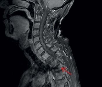
After developing a rash to lenalidomide, the patient received four cycles of daratumumab, bortezomib, and dexamethasone (DVd), achieving a very good partial response (VGPR). Subsequently, autologous hematopoietic stem cell collection was performed, with a CD34+ cell yield of 2.48 × 10⁹/l. In May 2025, M-protein and free light chain levels remained negative, but bone marrow abnormal plasma cells abruptly increased to 94.5%. Following an 8-week bridging therapy with teclistamab (escalating from 0.06 mg/kg to 0.3 mg/kg and then to 1.5 mg/kg, administered once weekly), bone marrow minimal residual disease (MRD) became negative again, and the vertebral mass decreased by 90%. In September 2025, the patient underwent ASCT. His post-transplant recovery was complicated by pulmonary aspergillosis, which delayed the initiation of maintenance therapy. In November 2025, maintenance therapy with KPd (carfilzomib, pomalidomide, and dexamethasone) regimen was initiated. One month later, the patient gradually developed acute neurological manifestations, including left upper limb weakness, finger numbness, left ptosis, diplopia, and right eye deviation, with concurrent mild blood pressure elevation. Given the absence of a prior history of hypertension, the insidious progression of symptoms (without sudden headache, epilepsy, or altered consciousness), and the findings on/of cranial imaging, hypertensive encephalopathy was reasonably excluded. Comprehensive evaluation revealed no evidence of systemic relapse of myeloma (M-protein negative, MRD negative by flow cytometry). However, contrast-enhanced spine MRI revealed extensive leptomeningeal enhancement along the entire spinal cord (Figure 1 A) and an occupying lesion within the left neural foramen at the T5/6 level (Figure 1 C), suggestive of tumor invasion. The cerebrospinal fluid (CSF) showed a total cell count of 0.13 × 10⁹/l, total protein of 1.27 g/l, and glucose of 1.92 mmol/l. CSF flow cytometry demonstrated that abnormal plasma cells accounted for 80% of nucleated cells (Figure 2), confirming isolated CNS relapse. Peripheral blood lymphocyte subset analysis revealed a CD4+ T-cell count of 0.22 × 10⁹/l and a CD8+ T-cell count of 0.38 × 10⁹/l.
Figure 1
Contrast-enhanced MRI of the whole spine showed that leptomeningeal enhancement throughout the spinal cord was markedly reduced after teclistamab treatment (A and B), and the space-occupying lesion in the left neural foramen at T5/6 also significantly shrank (C and D)
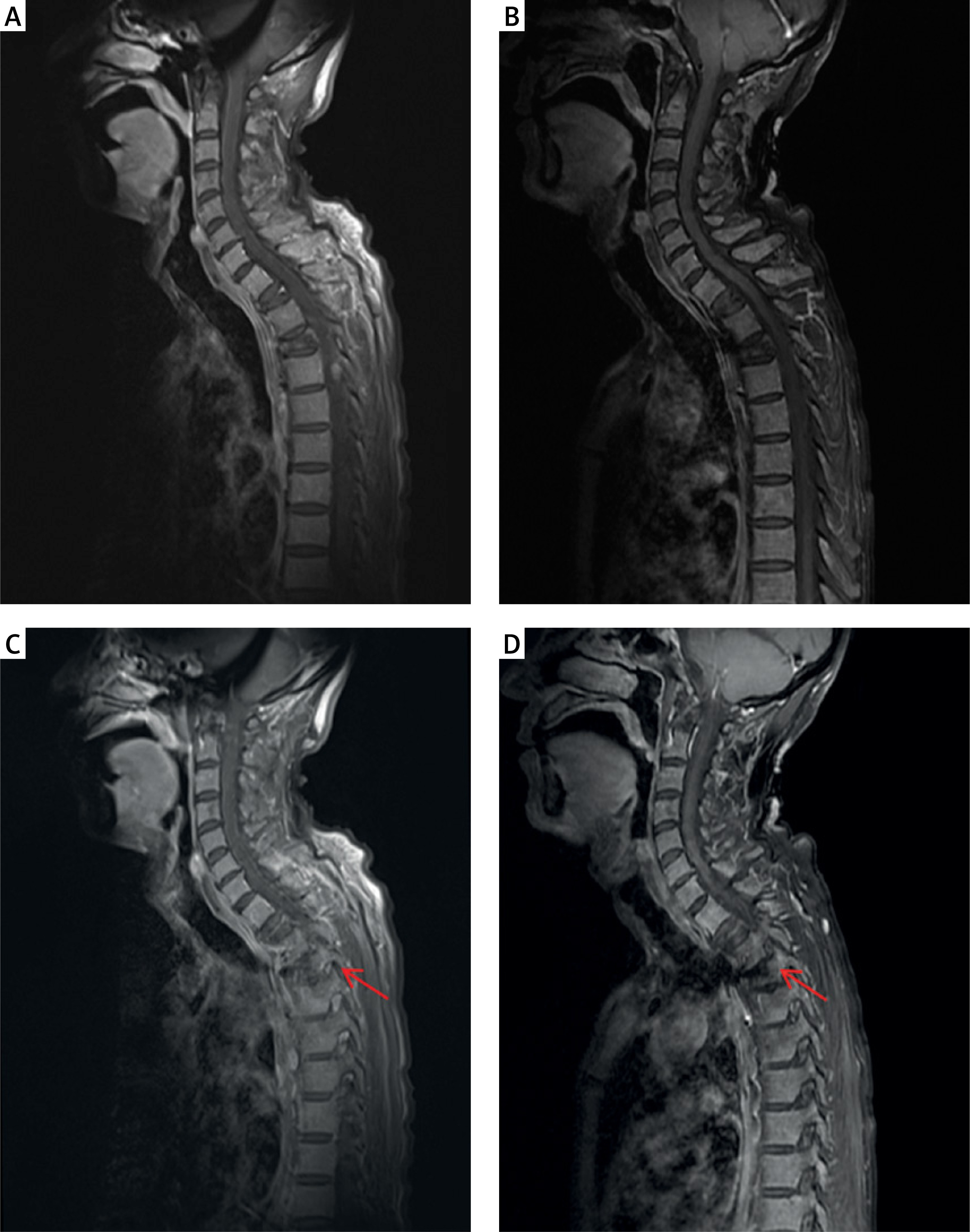
Figure 2
A – Flow cytometric analyses of CNS fluid at the time of diagnosis showed CD38 and CD138 positive kappa clonal plasma cells. B – Flow cytometry analysis of CNS fluid showed that kappa clonal plasma cells positive for CD38 and CD138 became negative after teclistamab treatment
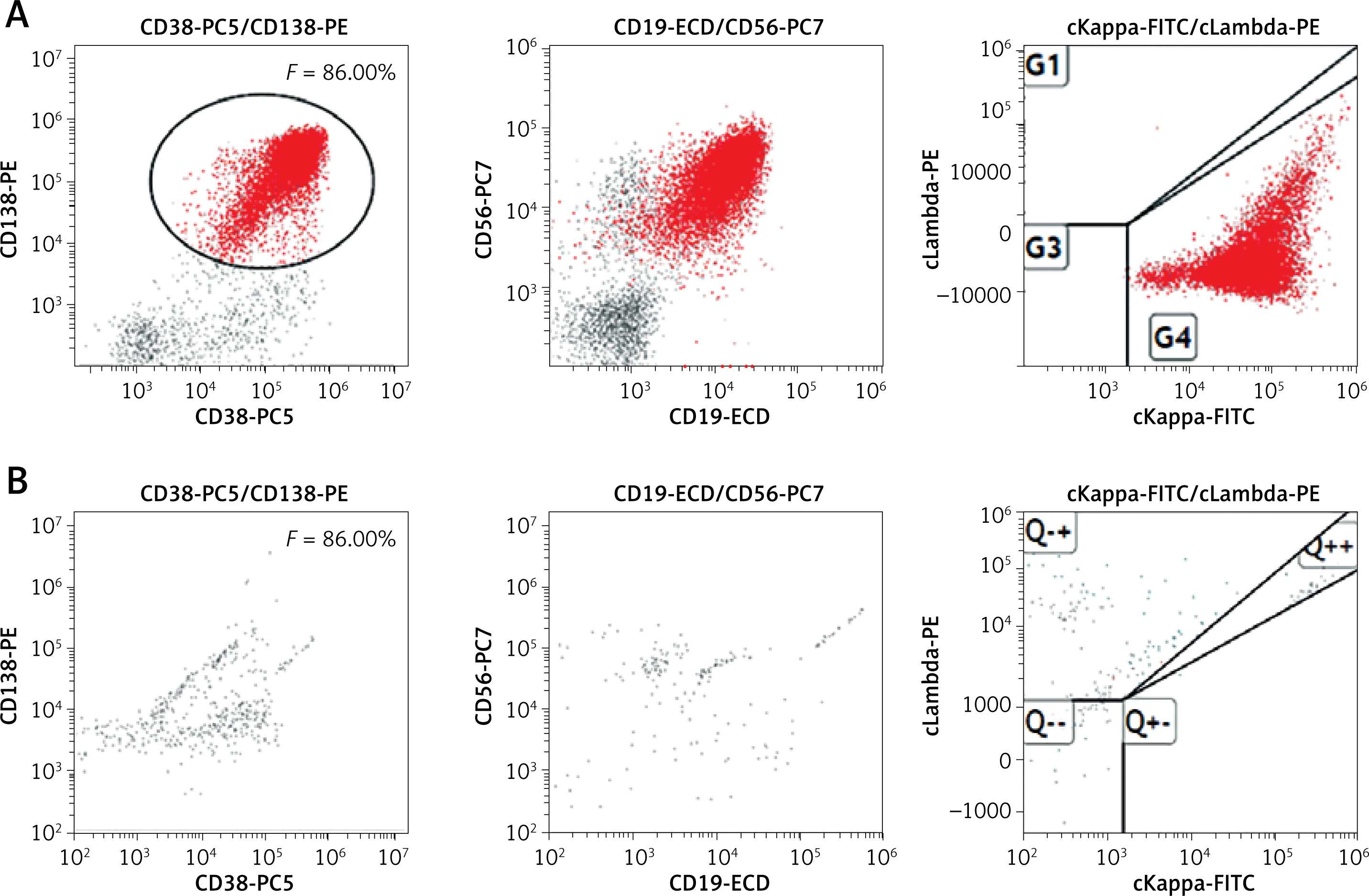
Given the refractory course and limited CNS-penetrant options, it was decided to restart teclistamab treatment, using the same weekly step-up dosing regimen as in the bridging phase. After 12 weeks of treatment, the patient’s neurological symptoms had completely resolved and remained stable thereafter, with the ECOG performance status improving from 4 to 2 and the CTCAE (version 5.0) neurotoxicity grade improving from 3 to 1. Repeat flow cytometry of the cerebrospinal fluid revealed MRD negativity (Figure 2 B), and contrast-enhanced MRI of the whole spine demonstrated a marked reduction in leptomeningeal enhancement (Figure 1 B) as well as a significantly reduced occupying lesion within the left neural foramen at the T5/6 level (Figure 1 D). Therefore, teclistamab was switched to 1.5 mg/kg once every 2 weeks, and 6 months after the initiation of CNS relapse treatment, it was further changed to once monthly. No significant cytokine release syndrome or immune effector cell-associated neurotoxicity syndrome was observed.
Isolated CNS relapse in MM represents a rare but increasingly recognized pattern, potentially reflecting the selection of resistant clones under the pressure of novel therapies [5]. Our patient harbored multiple high-risk features, notably del(17p), which portends/carries an extremely poor prognosis [6], all of which are associated with aggressive behavior and CNS tropism. The uniqueness of this case lies in its isolated CNS recurrence, the mechanism of which we hypothesize to involve two factors. First, although the short course of teclistamab bridging therapy effectively reduced systemic tumor burden, its CSF concentration – had it been measurable – would likely have been far below the IC50 required for T-cell redirection. This pharmacokinetic gradient would have rendered the CNS a pharmacological sanctuary for myeloma cells, enabling their outgrowth without synchronous systemic progression. Second, at the time of CNS relapse, this patient exhibited markedly reduced CD4+ T-cell counts with an inverted CD4/CD8 ratio, a hallmark feature of delayed immune reconstitution following ASCT. At this early post-transplant phase, thymus-dependent neogenesis of naïve T cells remained severely impaired, and the peripheral T-cell pool was largely composed of memory-type cells with limited clonal diversity and proliferative capacity. In the context of teclistamab therapy, whose efficacy relies on the formation of cytolytic synapses between T cells and BCMA+ plasma cells, the paucity of CD4+ T cells may critically constrain the magnitude and durability of T-cell redirection.
Although we cannot attribute the CNS relapse solely to teclistamab exposure, the temporal sequence strongly supports the sanctuary hypothesis. Furthermore, the dissociation between systemic MRD negativity and active CNS recurrence observed in this case challenges the existing monitoring frameworks. This case thus serves as a critical reminder that in highrisk MM receiving BiTEs as bridging therapy, rigorous CNS surveillance – including baseline MRI and CSF evaluation – must be strongly emphasized to avoid overlooking pharmacologically shielded disease reservoirs.
The optimal management of CNS-MM remains undefined. Conventional anti-myeloma agents have limited CNS penetration, and intrathecal chemotherapy or radiotherapy offers only transient responses [7]. BCMA CAR-T therapy has demonstrated activity in CNS-MM, likely due to disruption of the BBB by meningeal disease, which facilitates drug entry [8]. However, the lengthy manufacturing time of CAR-T cells limits rapid accessibility for patients.
Teclistamab is a BiTE targeting CD3 on T cells and BCMA on plasma cells, leading to T-cell-mediated cytotoxicity. Although BCMA-targeting BiTEs are large-molecule agents that may limit CNS penetration, cases of CNS efficacy have been reported, though the underlying mechanisms remain to be fully elucidated [9]. Prior small-cohort studies of BiTEs in CNS-MM have demonstrated definitive therapeutic efficacy of talquetamab [10]. However, in our patient, teclistamab was selected based on the patient’s prior favorable response to teclistamab and the patient’s preference. Indeed, the observed radiographic response to teclistamab in our patient, along with the patient’s neurological functional recovery, supports the biologic plausibility of BCMA-directed BiTE activity in the CNS. Importantly, this case does not suggest that teclistamab is superior to talquetamab, but rather that BCMA-targeted BiTE, as a class, may have activity in CNS-MM. In the patient, teclistamab also achieved rapid and durable clearance of CSF plasma cells with a manageable safety profile, underscoring its potential utility in this challenging scenario. Further studies are needed to define the optimal timing, combination regimens, and monitoring strategies for BiTEs in the setting of CNS involvement, as well as to directly compare the efficacy of different BiTEs in this rare population.