Introduction
Necrotizing enterocolitis (NEC) is the most common life-threatening intestinal surgical disease in the neonatal stage, especially in premature infants. The incidence of necrotizing enterocolitis is higher with the decrease of body weight and gestational age. National Institute of Child Health and Human Development (NICHD) reported that more than 90% of NEC infants were under 36 weeks of gestation, and 15% of the infants had birth weight below 600 g. The incidence of NEC in very low birth weight (VLBW) infants and extremely low birth weight (ELBW) infants was 5% and 10%, respectively [1–4]. With the strengthening and improvement of neonatal care measures, the survival rate of preterm infants has gradually improved, but the incidence of NEC has gradually increased, and the clinical treatment of NEC has made little progress in recent years [3]. Recent trends indicate that NEC is likely to become the leading cause of premature infant death in the United States. Preterm delivery, gastrointestinal immaturity, hypoxia, artificial feeding, bacterial transplantation and hypoperfusion are the major risk factors for NEC. Preterm birth is the single most dangerous factor, and excessive inflammation is the main part. But the NEC pathogenesis is complex, and the specific pathogenesis has been not very clear.
Phosphatase and tensin homology deleted on chromosome ten (PTEN)/phosphoinositide-3 kinase (PI3K)/serine/threonine (AKT)/glycogen synthase kinase-3β (GSK-3β) signaling pathway played important roles in energy metabolism, apoptosis, differentiation and reproduction [5–7]. Recent studies found that miRNAs could regulate cell apoptosis and proliferation by relative pathways [8–10]. miRNA-21 has an important role in the miRNA family [11, 12]. In previous studies, the results showed that PTEN was a target gene of miRNA-21 [13, 14].
In our present study, we wanted to explain the effect and mechanism of miRNA-21 in the development of NEC.
Material and methods
Clinical data
Thirty pairs of normal newborn and NEC children who were treated and born in our hospital from 2015.6 to 2016.6 were collected and the venous blood was taken.
Animals and grouping
Thirty newborn Wistar rats aged 1 day, body weight was 5.5–10 g, were purchased from Shandong University Medical School. The rats were divided into 3 groups. There were 10 rats in every group. The NC group was injected with 0.2 ml of normal saline; the Model group was injected with 0.2 ml of normal saline based on the model; the miRNA group was injected with 0.2 ml of miRNA-21 which was purchased from Gen Script (Nanjing) Co., Ltd (Nanjing, China) based on the model. All the drugs were administered by the intraperitoneal injection route. Rat pups are separated from their mothers at 24 h after birth, room temperature was maintained at about 21°C, natural lighting, with the formula Mead Johnson infant oral feeding in neonatal rats (200 kcal/kg/day), feeding 3 h/once, 0.1 ml each time, and increasing the diet with body weight increasing.
RT-PCR
The total mRNA was extracted by Trizol which was purchased from TOYOBO (Japan), and the total mRNA concentration was measured by a UV-Vis spectrophotometer, taking 4 µg of total RNA for reverse transcription into cDNA. A certain amount of cDNA was used for PCR amplification. The miRNA- 21 gene expression primer sequences were synthesized from Gen Script (Nanjing) Co., Ltd (Nanjing, China) as follows: miRNA-21 F: 5’-TCCGAAGTTGTAGTCAGACT- 3’; R: 5’-GTGCAGGGTCCGAGGT- 3’. U6 F: 5’-CTCGCTTCGGCAGCACA-3’; R: AACGCTTCACGAATTTGCGT-3’. In this experiment, we used U6 as a reference. miRNA-21 mRNA expression was measured by the 2–ΔΔCT method.
NEC animal model [15, 16]
After zeroing the portable RSS-5100 oxygen meter, the probe was connected to the top notch of the anoxic box sealed with adhesive tape, and pure nitrogen was filled into the other notch. After the oxygen concentration in the tank dropped to zero, the newborn rats were put into the tank quickly, with nitrogen continuously filled until it reached 100%, after which the environment was maintained for 10 min. Subsequently, the rats were put into an oxygen chamber with 100% oxygen for 10 min, and then immediately placed into a refrigerator at 4°C for 10 min. The treatment was repeated 3 times a day, for a total of 9 times. After the treatment, the rats were sent back to the incubator. In addition, LPS (10 mg/kg) was diluted in 0.1 ml of sterile water for intragastric administration once a day. The rats in different groups were administered according to their prescribed dosage for 3 consecutive days, and sacrificed by decollation on the 4th day.
Histopathological examination of intestinal tract
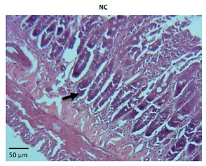
1–2 cm of the proximal ileocecal tissue was fixed immediately in paraformaldehyde, paraffin embedded, sliced, and stained with H&E staining. The pathological scores were scored by Nadler criteria: 0 points: The villi and epithelium of the intestine are intact and the tissue structure is normal; 1 point: Slight submucosal or lamina propria swelling separation; 3 point: Moderate submucosal and/or lamina propria separation, submucosal and/or muscular edema; 4 point: Intestinal villus disappearance with intestinal necrosis. The pathological score is greater than or equal to 2 points, which is considered to be the onset of NEC.
TUNEL assay
1–2 cm of the proximal ileocecal tissue was fixed immediately in paraformaldehyde, paraffin embedded, sliced, and subjected to TUNEL assay. The cell apoptosis was measured by a TUNEL kit (Roche, USA). Paraffin sections were dewaxed in xylene, followed by hydration in a gradient of alcohol; protease K (20 µg/ml) was used for digestion for 30 min at 37°C, mixed reaction solution containing terminal deoxynucleotidyl transferase and fluorescein labeled dUTP were added to culture for 1 h at 37°C, followed by adding anti-fluorescein antibodies linked to alkaline phosphatase to culture for 30 min at 37°C, hematoxylin light lining dye, dehydrated, transparent, mounting, observation under light microscope and measuring the cell apoptosis rate.
WB assay
The total protein of proximal ileocecal tissue was extracted, and we measured the concentration by the spectrophotometer method, trim as 2 g/l, electrophoresis by 10% SDS-PAGE. After the film was transferred by the wet rotating method, it was put into the closed liquid, and the 2 h was closed on the shaking table. The first antibody was added to culture at 4°C overnight. After that, the second antibody was added to culture at room temperature overnight. After exposure in a darkroom, developing and fixing. The GAPDH was considered as a reference in this study. The relative antibodies (PTEN, PI3K, p-PI3k, AKT, p-AKT, GSK-3β and GAPDH) were purchased from Abcam (USA) and p-GSK-3β was purchased from Cell Signaling (USA). PTEN (ab32199, Abcam, USA); PI3K (ab32089, Abcam, USA); p-PI3K (ab182651, Abcam, USA); AKT(ab79463, Abcam, USA); p-AKT (ab38449, Abcam, USA); GSK-3β (ab93926, Abcam, USA), p-GSK-3β (mAb #5558, Cell Signaling, USA)
Statistical analysis
The relative data from our study were analyzed by SPSS 20.0 software. The data are shown as mean ± SD (standard deviation). The significance between multiple sets of data was analyzed by one-way ANOVA with LSD test. P < 0.05 showed that the difference was statistically significant.
Results
Clinical data and analysis
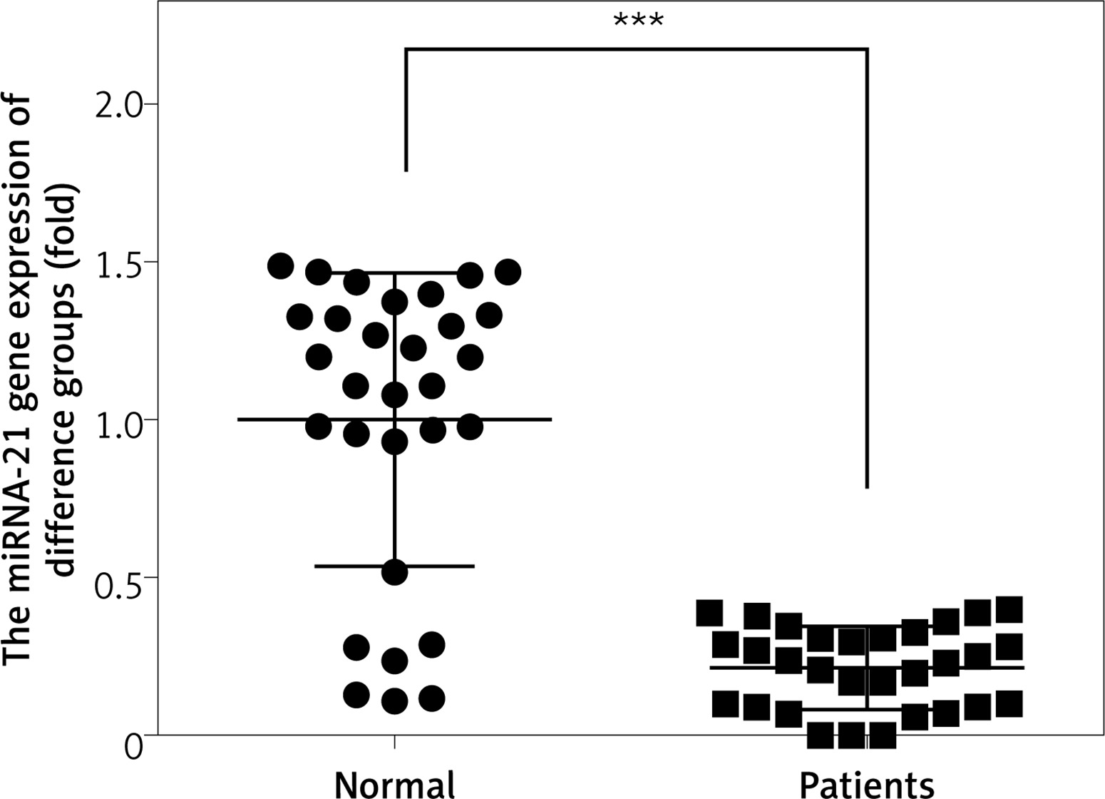
Compared with the NC group, the serum miRNA- 21 gene expression of NEC children was significantly down-regulated (p < 0.05). The relative data are shown in Figure 1.
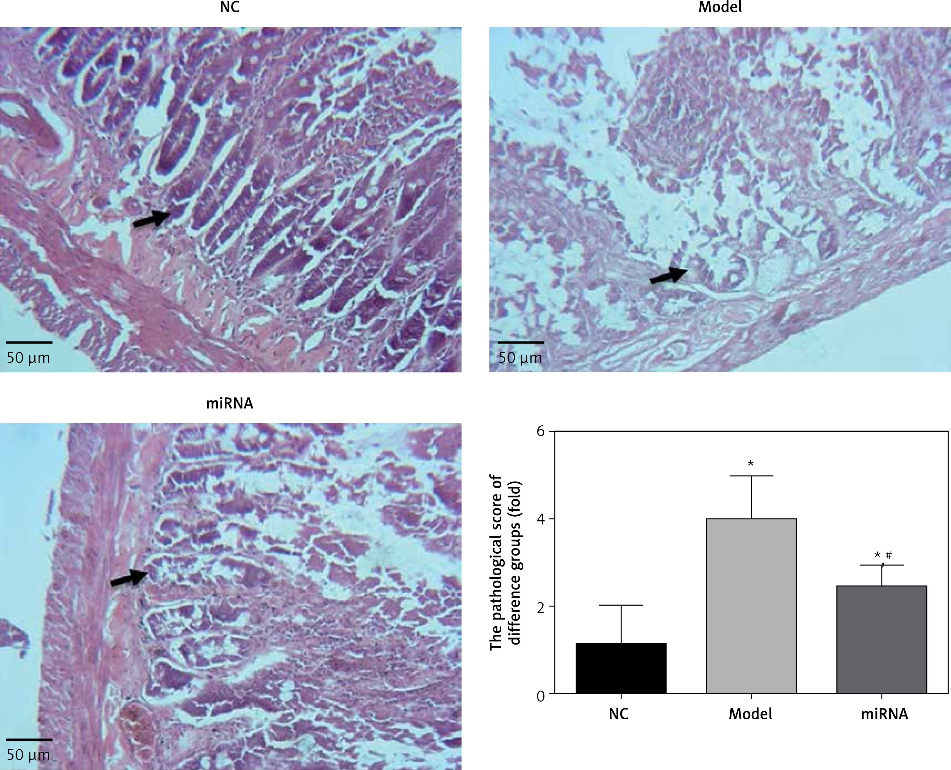
Pathology of difference groups by H + E staining
The pathological score of Model and miRNA groups was significantly higher than that of the NC group (p < 0.05, respectively). This result showed that our NEC model was successful. Meanwhile, compared with the Model group, the pathological score of the miRNA group was significantly increased compared with that of the Model group (p < 0.05) with miRNA-21 over-expression. The relative data are shown in Figure 2.
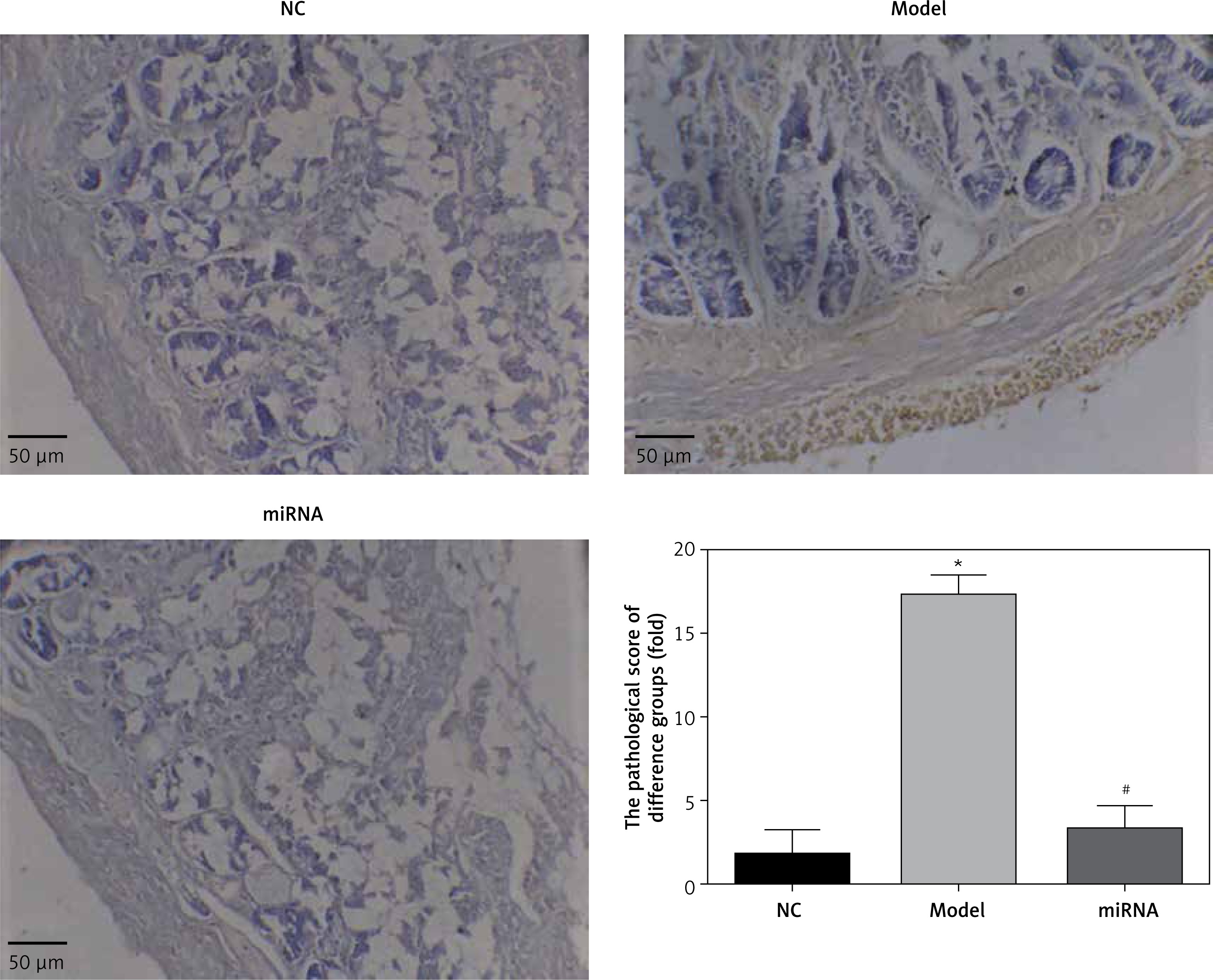
Cell apoptosis rate of different groups by TUNEL assay
Compared with the NC group, the positive apoptosis cell number of the Model group was significantly increased (p < 0.05). However, the cell apoptosis rate of the miRNA group was significantly suppressed compared with that of the Model group (p < 0.05). The relative data are shown in Figure 3.
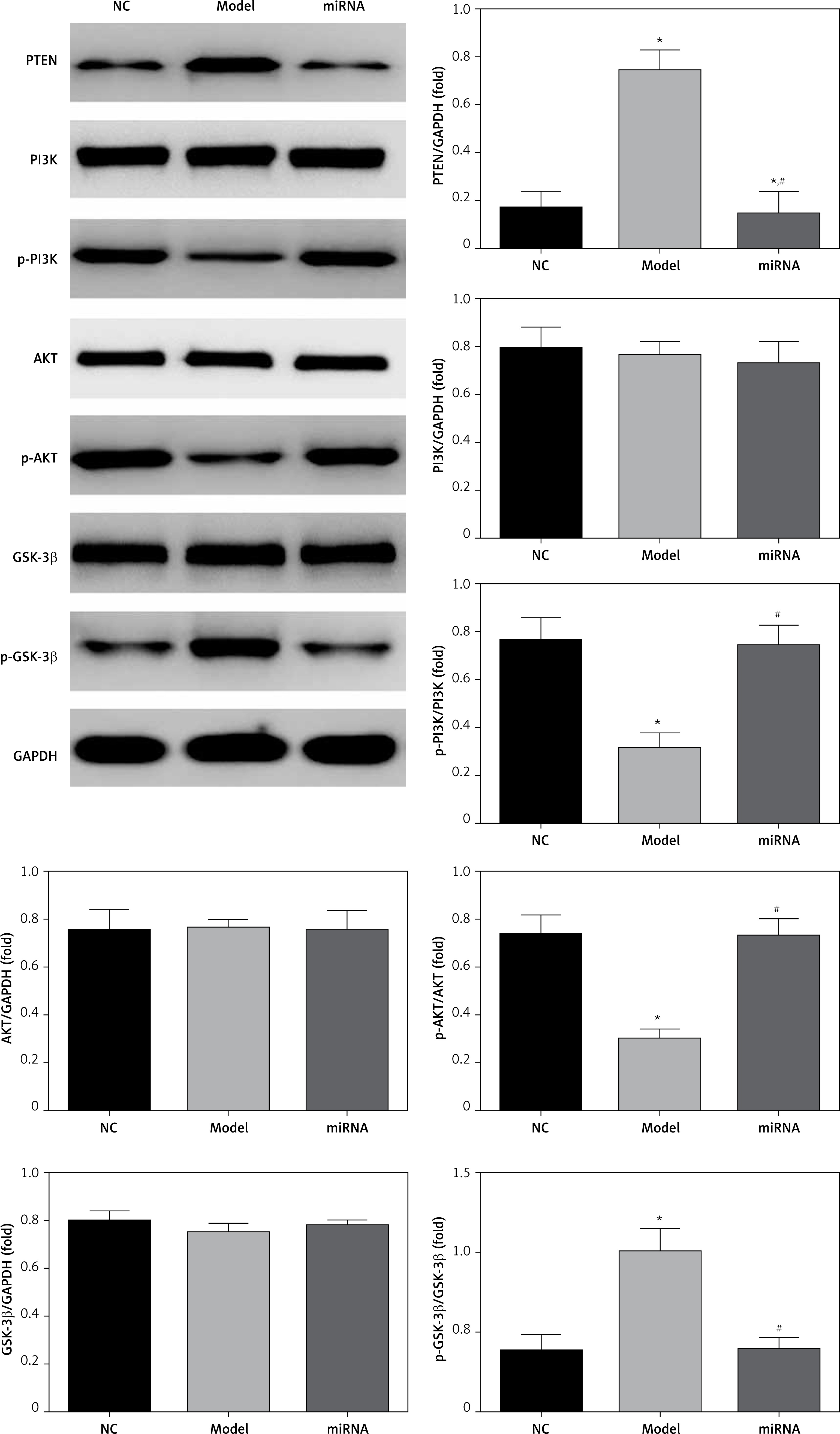
Relative protein expression by WB assay
There were no significantly differences among NC, Model and miRNA groups in PI3K, AKT and GSK-3β protein expression levels. However, compared with the NC group, the PTEN and p-GSK-3β protein expression levels of the Model group were significantly up-regulation (p < 0.05, respectively), and p-PI3K and p-AKT protein expression levels of the Model group were significantly down-regulated (p < 0.05, respectively). With miRNA-21 over-expression, the PTEN and p-GSK-3β protein expression levels of the miRNA group were significantly suppressed (p < 0.05, respectively), and p-PI3K and p-AKT protein expression levels of the miRNA group were significantly increased (p < 0.05, respectively) compared with the Model group. The relative data are shown in Figure 4.
Discussion
The NEC is the main effect of low birth weight and low survival rate of the disease and has become very challenging. Although in recent years, many clinical preventive measures such as the increment of enteral feeding and treatment measures based on the standard scheme have resulted in great progress, there is still a risk tendency of premature infants [15]. This experiment mainly simulated the following risk factors of neonatal NEC: ischemia reperfusion injury, hypoxia, hypothermia, artificial feeding, endotoxin caused by hypoxia reoxygenation. Hypoxia, on the one hand, causes diving reflex, redistributes blood and reduces gastrointestinal blood flow and oxygen delivery through adrenaline-induced vasoconstriction; on the other hand, it causes ischemia-reperfusion injury, which leads to increased intestinal mucosal permeability and eventually intestinal mucosal injury. Hypoxia, endotoxin, and ischemia-reperfusion injury stimulate intestinal Paneth cells to secrete type II phospholipase-A2 (PLA2-II), thus producing platelet-activating factor (PAF) and activating platelet-activating factor receptor (PAFR). Activation of PAFR can activate multiple cytokines including signal transduction and transcription activator of transcription 3 (STAT3) and nuclear factor NF-κB, causing pathological changes, such as vasoconstriction and/or vasodilation, activation and translocation of cytokines, activation of cell adhesion molecules, increase in capillary permeability, production of reactive oxygen species and nitric oxide, as well as increase in intestinal mucosal permeability. PAF by inducing STAT3 phosphorylation and NF-κB activation induced ileum up-regulation of TLR4. TLR4 increased Notch signaling pathway activated by reduction of goblet cell differentiation. The above pathological changes eventually lead to the occurrence of NEC [16, 17]. Sodhi et al. [18] showed that the TRL4–/– and conditional destruction of TRL4 in the knockout mice did not occur in NEC, but the NEC in the wild type mice was more serious. The PTEN/PI3K/AKT signaling pathway and ischemia reperfusion injury, cell proliferation, differentiation, migration, apoptosis, inflammation and so on participate in the process of apoptosis by regulating its downstream signal molecule GSK-3β activity, while the initial NEC pathological process is related to the apoptosis of intestinal tissue cell death [19]. Some scholars have suggested that PTEN/P13K/AKT and its downstream signal molecules may regulate the expression of GSK- 3β by a negative factor induced by hypoxia in the intestine and can have a protective effect on the intestinal tissue in NEC. GSK-3β widely exists in eukaryotic cells and is involved in transcriptional activation, cell proliferation, cell differentiation and cell movement and other physiological processes. Oxidative stress can activate GSK-3β activation of a pro-apoptotic mechanism [20].
MicroRNAs are a kind of endogenous single stranded molecule found in recent years, non-encoding RNA, size of 18–25 nucleotides, with target mRNA 3’-untranslated region combination at the post-transcriptional level degradation of mRNA or inhibiting mRNA translation, thereby affecting the expression of the target gene [21, 22]. Some previous studies reported that miRNA-21 has an important role in cell apoptosis via regulation of the relative pathway [23–25]. Meanwhile, the studies also found that PTEN was a potential target for miRNA-21 [26, 27]. In our present study, we firstly measured miRNA-21 gene expression in NEC and normal children. The results showed that miRNA-21 gene expression was down-regulated in the NEC children. In the animal experiment, the pathology and cell apoptosis rate were improved in the miRNA-21 over-expression group. This result confirmed that miRNA-21 up-regulation had improved NEC induced pathology and cell apoptosis. In molecular biology, we measured the relative proteins such as PTEN, PI3K, AKT and GSK-3β expression levels. The results showed that PTEN and GSK-3β, which were two kind roles to improve cell apoptosis [28, 29] were suppressed and the PI3K/ AKT pathway, which was negatively regulatory for cell apoptosis [30], was stimulated.
In conclusion, depending on our results, we inferred that miRNA-21 over-expression can improve NEC induced small intestine tissue necrosis via the PTEN/PI3K/AKT/GSK-3β pathway. miRNA-21 might be a potential target in diagnosis and treatment of NEC.