
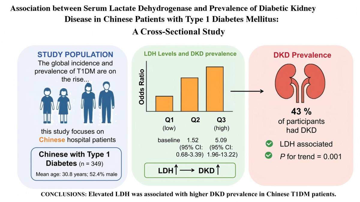
Type 1 diabetes mellitus (T1DM) is an autoimmune disease marked by irreversible loss of pancreatic β-cells, which ultimately causes insulin deficiency and hyperglycemia. As of 2022, the global prevalence reached 8.75 million cases [1]. T1DM demonstrates a rising global incidence [2] and marked autoimmune comorbidity (thyroid disease, celiac disease, Addison disease, and vitiligo), yet Mendelian randomization analysis suggests an independent causal relationship between T1DM and non-immune diseases [3]. Diabetic kidney disease (DKD) represents a prevalent and severe microvascular complication of T1DM, frequently progressing to end-stage renal disease (ESRD) [4]. ESRD associated with diabetes exerts a particularly heavy burden on low- and middle-income countries; this is especially true for China, which has the highest prevalence of diabetes globally [5]. Therefore, identifying biomarkers associated with DKD may help elucidate disease mechanisms and inform future research directions.
Lactate, the terminal glycolytic metabolite, is generated from pyruvate by lactate dehydrogenase (LDH) [6]. Beyond its crucial role in renal metabolism, lactate serves as a pathogenic factor in the progression of kidney disease. The lactate-LDH-lactation signaling axis is increasingly recognized for its significant role in disease pathogenesis [7, 8]. LDH is a frequently employed biomarker in various clinical contexts, including myocardial infarction and tissue injury [9], and has been associated with metabolic conditions such as diabetes [10]. Preclinical evidence suggests that targeting lactate metabolism may offer a potential strategy for renal protection.
Increasing evidence [11–13] suggests an association between LDH and DKD in type 2 diabetes mellitus (T2DM). Up-regulated LDH catalytic capacity and gene expression in DKD may drive disease initiation and progression. While LDH and lactate are emerging biomarkers for DKD in T2DM, their association with DKD in T1DM is unknown. We examined this relationship in Chinese hospital patients.
Methods
Study design and individuals
The present cross-sectional study included sociodemographic and laboratory data from patients who first visited the endocrinology department between January 2015 and December 2024. T1DM diagnosis followed ADA criteria [14], requiring clinical features supported by laboratory confirmation: history of diabetic ketoacidosis/ketosis, symptoms of hyperglycemia at diagnosis, fasting or stimulated C-peptide < 200 pmol/l, or positivity for T1DM-associated autoantibodies. To confirm insulin dependency and exclude misdiagnosis, participants were followed for ≥ 18 months after initial diagnosis [15]. Medical records contained pre-admission laboratory data (≥ 3 months before admission) from external or outpatient sources, including at least one urinary albumin-to-creatinine ratio (UACR) and serum creatinine measurement.
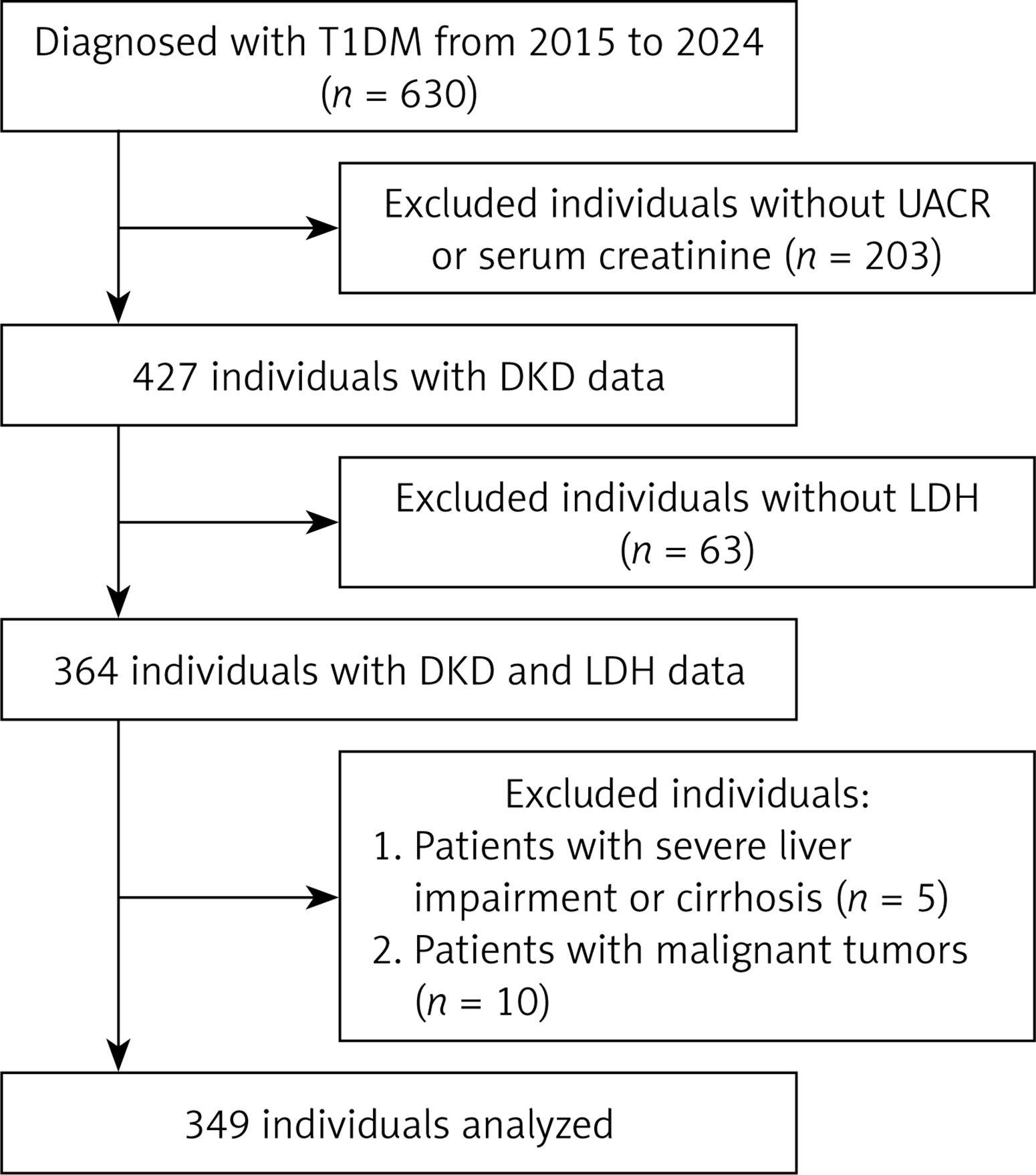
Initially, 630 participants were diagnosed with T1DM. Exclusion criteria were: (1) UACR and serum creatinine not measured within 3 months before and after admission, or discordant UACR results not confirmed by repeat testing; (2) missing LDH data; (3) history of acute myocardial infarction, myocarditis, pericarditis, heart failure, or surgery within 1 month; (4) rhabdomyolysis, myositis, muscular dystrophy, or strenuous exercise within 7 days; (5) severe infection or pulmonary embolism; (6) non-diabetic kidney disease; (7) liver impairment, cirrhosis, or malignant tumors. Finally, 349 participants with complete LDH and DKD data were included. Figure 1 shows the flowchart of participant selection.
Study variables and outcome
Demographics (sex, age, ethnicity, and marital status), clinical variables (height, weight, systolic and diastolic blood pressure [SBP and DBP]) and laboratory results were extracted from electronic medical records and manually verified to ensure completeness and accuracy. After more than 5 min of rest, seated blood pressure was measured twice (2 min interval) and averaged; hypertension (HTN) was defined as systolic (SBP) ≥ 140 mm Hg, diastolic (DBP) ≥ 90 mm Hg, or antihypertensive use. Body mass index (BMI) was calculated as weight (kg) divided by height squared (m2).
After an overnight fast of 8 to 12 h, fasting venous blood and urine samples were collected. Routine biochemical analyses, including glycated hemoglobin (HbA1c), fasting blood glucose (FBG), aspartate aminotransferase (AST), alanine aminotransferase (ALT), serum creatinine (SCr), serum albumin (ALB), uric acid (UA) and lactate dehydrogenase (LDH), were performed using the Abbott Architect ci8200. Triglycerides (TG, measured by GPO-PAP), total cholesterol (TC, measured by CHOD-PAP), low-density lipoprotein cholesterol (LDL-C, measured using surfactant), and high-density lipoprotein cholesterol (HDL-C, measured by CAT) were quantified on the Beckman Coulter AU5800. Thyroid-stimulating hormone levels were determined using chemiluminescence on the IMMULITE 2000. The UACR was calculated from morning spot urine samples, with specimens collected during menstruation, pregnancy, febrile illness, infection, or heavy exercise excluded when identified. DKD was diagnosed according to KDIGO 2022 criteria [16] when UACR ≥ 30 mg/g was documented on at least 2 occasions ≥ 3 months apart (one prior, two at admission) or a reduced estimated glomerular filtration rate (eGFR) below 60 ml/min/1.73 m2. Diagnoses were verified by integrating historical and admission laboratory data.
Statistical analysis
Normality was examined with histograms, Q-Q plots, and the Kolmogorov-Smirnov test. Continuous variables are presented as means ± standard deviations (SD) when normally distributed and as medians and interquartile ranges (IQR) otherwise; categorical variables are reported as frequencies and percentages (%). Group comparisons employed χ2 or Fisher exact tests for categorical variables, one-way ANOVA for normally distributed continuous variables, and Kruskal-Wallis tests for non-normally distributed data. Logistic regression was used to evaluate the association between LDH (per 10 U/l increment) and DKD. Results are presented as odds ratios (OR) accompanied by 95% confidence intervals (CI). Covariates were selected based on prior literature and expert judgment. Multicollinearity among covariates was quantified using variance inflation factors (VIFs). Three progressively adjusted models were constructed: Model 1 (unadjusted); Model 2 (adjusted for age, sex, duration of diabetes, and HTN status); Model 3 (additionally adjusted for BMI, hemoglobin, ALB, ALT, SCr, UA, TC, TG, CRP, and HbA1c).
To confirm consistency, LDH was analyzed both continuously and in tertiles (p for trend was calculated); restricted cubic spline (RCS) regression was used to assess linearity and dose-response with respect to DKD after adjustment for Model 3 covariates. Subgroup analyses were performed, with interactions tested using the likelihood ratio. Among 349 participants, missing data for each covariate were less than 20% (BMI, n = 38; HbA1c, n = 22; TC, n = 4; TG, n = 4; hemoglobin, n = 3; albumin, n = 1). Multiple imputation (m = 5) was performed under the missing-at-random assumption. Sensitivity analyses comparing results with complete-case analysis confirmed the robustness of our findings.
Analyses were performed with R Statistical Software (Version 4.2.2, http://www.R-project.org, The R Foundation) and the Free Statistics analysis platform (Version 2.2.2, Beijing, China, http://www.clinicalscientists.cn/freestatistics). Two-sided p < 0.05 indicated statistical significance.
Results
Baseline characteristics
A total of 349 individuals with T1DM were included in this study. The median LDH level was 179 U/l (range: 86–506 U/l) among all participants. The median age was 30.8 years, and 52.4% of the participants were male. The overall prevalence of DKD was 43%. Table I shows the baseline characteristics of the groups stratified by LDH levels. Participants in the highest LDH tertile (Q3) exhibited longer diabetes duration, higher SBP and DBP, elevated renal markers (BUN, SCr, UA, UACR), lower eGFR, and adverse lipid profiles (TC, TG, LDL-C) compared to lower tertiles (all p < 0.01). Notably, DKD prevalence increased markedly across ascending tertiles (Q1: 26.7%, Q2: 33.9%, Q3: 67.8%, p < 0.001). No significant differences were detected for sex, age, ethnicity, marital status, BMI, HDL-C, HbA1c, FBG, or TSH (all p > 0.05).
Table I
Baseline characteristics of participants
[i] Data are expressed as mean ± standard deviation or median (interquartile range) or percentage (%). BMI – body mass index, SBP – systolic blood pressure, DBP – diastolic blood pressure, Hb – hemoglobin, ALB – serum albumin, ALT – alanine aminotransferase, AST – glutamic oxaloacetic transaminase, BUN – blood urea nitrogen, SCr – serum creatinine, UA – uric acid, TC – total cholesterol, TG – triglycerides, LDL-C – low-density lipoprotein cholesterol, HDL-C – high-density lipoprotein cholesterol, HbA1c – glycated hemoglobin, FBG – fasting blood glucose, TSH – thyroid stimulating hormone, CRP – C-reactive protein, UACR – urinary albumin-to-creatinine ratio, eGFR – estimated glomerular filtration rate, RAASi – renin-angiotensin-aldosterone system inhibitors, SGLT2i – sodium-glucose linked transporter-2 inhibitors, HTN – hypertension, DKD – diabetic kidney disease.
Association between LDH and the presence of DKD
To investigate the association between LDH and DKD in patients with T1DM, we fitted logistic regression models (Table II). Elevated LDH levels were significantly associated with DKD prevalence in multivariable analyses (Table II). Each 10 U/l increase in LDH conferred 8% higher odds of DKD in the fully adjusted model (OR = 1.08, 95% CI: 1.01–1.14, p = 0.017). Tertile analysis revealed that compared with the reference tertile (< 160 U/l), the highest tertile (≥ 203 U/l) exhibited 3.49-fold higher odds of prevalent DKD (OR = 3.49, 95% CI: 1.60–7.58, p = 0.002), whereas the middle tertile (160–203 U/l) showed no significant association (p > 0.05). Significant trend associations persisted across models (p for trend = 0.002).
Table II
Association between LDH and DKD in Chinese patients with T1DM
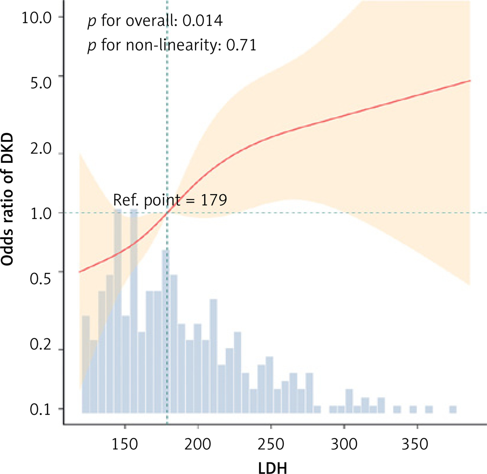
In this study, RCS analysis was used to further explore the non-linear relationship between LDH and the prevalence of DKD. As the level of LDH increased, the prevalence of DKD showed an upward trend. Within the sample population, a linear relationship was observed between LDH and DKD prevalence, with a p-value of 0.71 indicating no significant non-linearity (Figure 2).
Figure 2
The smooth curve fit for the association between LDH and DKD. Data were fit by a multivariable logistic regression model based on restricted cubic splines. LDH was entered as a continuous variable. Data were adjusted for age, sex, BMI, duration of diabetes, HTN, Hb, ALB, ALT, SCr, UA, TC, TG, CRP, and HbA1c. The background histograms (light blue color) represent the percentage of the density distribution of LDH in the study population. Solid lines signify the predicted values, while the 95% confidence intervals are marked with dashed lines. Only 95% of the data is shown
Stratified analysis
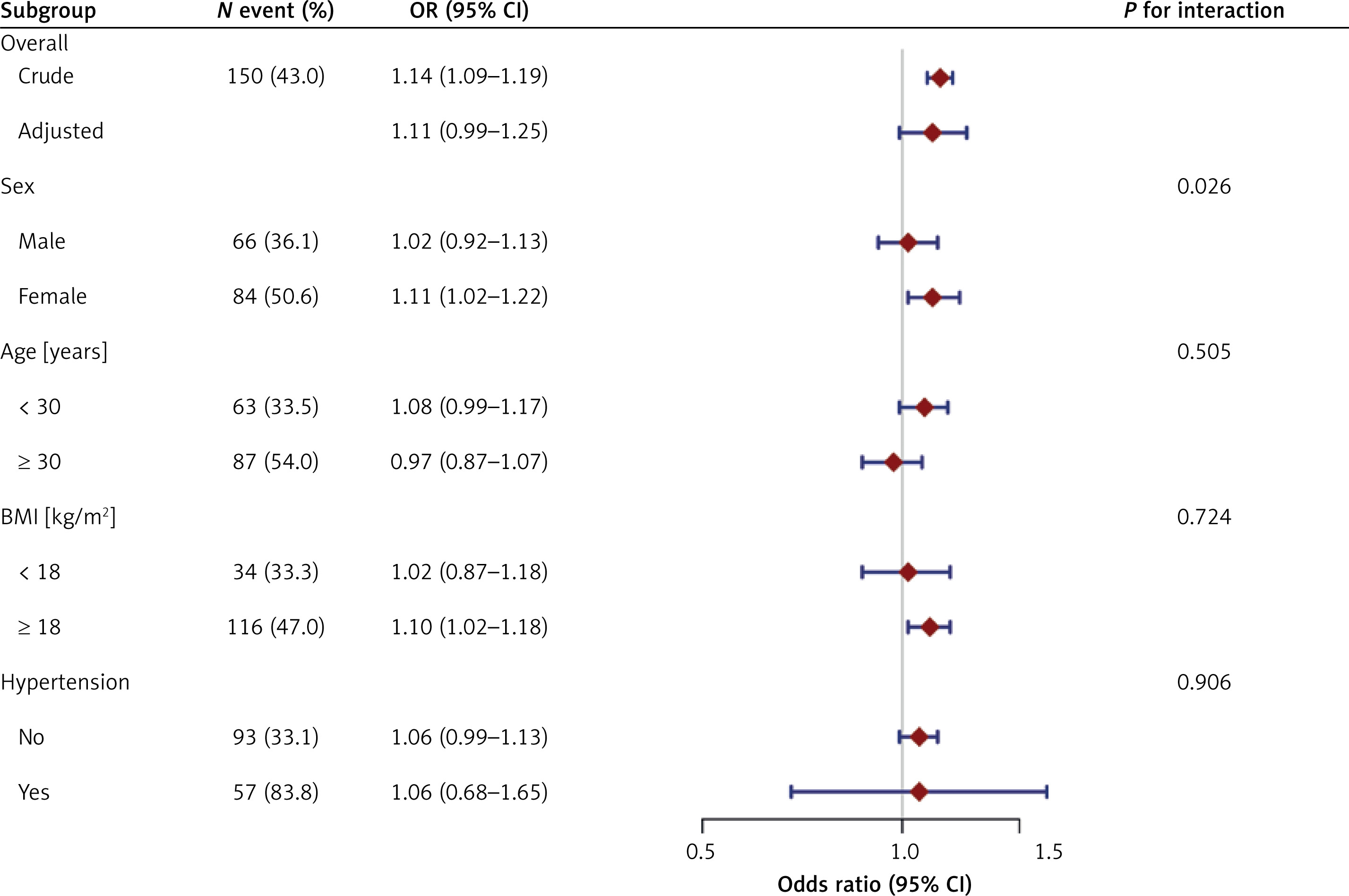
To assess the effect on the relationship between LDH and DKD, stratified analyses were conducted across predefined subgroups: sex, age, BMI, and HTN. A significant interaction by sex was observed (p for interaction = 0.026), with elevated LDH showing a stronger DKD association in females (adjusted OR = 1.11, 95% CI: 1.02–1.22) versus males (adjusted OR = 1.02, 95% CI: 0.92–1.13), whereas no additive interaction was evident (Supplementary Table SI). No interactions were observed for age, BMI, or HTN (all p for interaction > 0.05). The overall adjusted association followed a consistent trend (aOR = 1.11, 95% CI: 0.99–1.25) (Figure 3).
Figure 3
The relationship between LDH and DKD in different subgroups. This figure shows the association between LDH and DKD, stratified by age, sex, BMI, and HTN. A potential multiplicative interaction was observed for sex (p for interaction = 0.026), with a stronger association in females (OR = 1.11, 95% CI: 1.02–1.22) compared to males (OR = 1.02, 95% CI: 0.92–1.13). No significant interactions were found for age, BMI, or HTN (all p for interaction > 0.5). LDH was entered as a continuous variable per 10 U/l increase. Except for the stratification component itself, each stratification factor was adjusted for all other variables (age, sex, BMI, duration of diabetes, HTN, Hb, ALB, ALT, SCr, UA, TC, TG, CRP, and HbA1c)
Sensitivity analysis
Sensitivity analyses using the complete-case analysis yielded consistent findings for the association between LDH tertiles and DKD (Supplementary Table SII). The highest tertile (≥ 203 U/l) had higher odds of DKD compared with the lowest tertile (< 160 U/l) (OR = 4.21, 95% CI: 1.23–14.46, p = 0.022), whereas the middle tertile showed no significant association. The continuous LDH variable did not show a significant association (OR = 1.11 per 10 U/l increase, 95% CI: 0.99–1.25, p = 0.066); however, the trend test remained significant (p for trend = 0.024).
Discussion
This study demonstrated that elevated LDH levels were concurrently associated with higher DKD prevalence in Chinese patients with T1DM. Multiple imputation analysis showed that continuous LDH was significantly associated with DKD (Table II), although this association attenuated in complete case analysis (Supplementary Tables SII–SIV), likely due to selection bias from non-random missing data [17]. Notably, the tertile analysis remained consistent across both approaches, reinforcing robustness. A preliminary subgroup analysis suggested a potential multiplicative interaction by sex; however, the additive interaction was not significant, the sample was underpowered for interaction testing, and no correction for multiple comparisons was applied. This observation is therefore highly exploratory and should be interpreted with extreme caution.
LDH is released from damaged cells and participates in lactate metabolism; elevated LDH has been reported to coexist with early renal dysfunction in previous investigations [18]. Elevated lactate itself has been independently associated with acute kidney injury [19] and CKD progression [20]. In the present cross-sectional analysis, a graded increase in DKD prevalence across LDH tertiles was observed, suggesting that LDH levels are closely correlated with DKD prevalence even within the normal reference range.
The observed cross-sectional association between LDH and DKD could be consistent with concurrent glomerular injury (manifested as albuminuria, a key feature of DKD [21]) and tubular damage. Hyperglycemia-induced mitochondrial dysfunction has been proposed to elevate lactate and LDH, which are regarded as markers of cellular injury and cytotoxicity [22, 23]. LDH has also been hypothesized to reflect tubular metabolic stress under hypoxic and oxidative conditions [24, 25]. However, because LDH is released non-specifically from injured tissues, non-renal injury may attenuate its association with UACR. Whether LDH is associated with DKD in prospective settings warrants further investigation rather than inference from cross-sectional data alone.
The observed association between LDH and DKD is consistent with previous reports in T2DKD [12, 26] and hemodialysis patients [27]. Existing mechanistic studies suggest that lactate may contribute to kidney injury beyond being a nonspecific damage marker, but the present data cannot confirm whether lactate exerts direct pathogenic effects or define causal directionality in this study. As a novel epigenetic modification, lactylation influences kidney disease progression [28–30]. In DKD, elevated lactate drives histone H3K14la modification, activating KLF5 and triggering tubular epithelial-to-mesenchymal transition [31]. Podocyte-specific lactate accumulation induces global histone lactylation, suppressing nephrin and tight junction proteins while upregulating fibrotic markers [32]. These modifications collectively accelerate renal fibrosis. These mechanistic hypotheses provide biological plausibility for the observed cross-sectional association but do not establish causality or temporal direction from the present data.
This study is among the first to examine the relationship between LDH and the prevalence of DKD in Chinese populations using hospital-based data. The observed association may generate mechanistic and etiological hypotheses for future research. However, several limitations are evident. First, as a cross-sectional study, the associations identified here do not establish causality and may be subject to unmeasured confounding (e.g., insulin doses, smoking status, and physical activity). Second, this was a single-center hospital-based study, and participants were limited to patients at their first admission; such a design may not represent the overall T1DM population and may introduce selection bias, thereby limiting external validity and generalizability. Third, excluding patients with severe hepatic dysfunction or malignancies may further restrict generalizability to individuals with these comorbidities. Fourth, this study is an observational association analysis and did not evaluate model discrimination (AUC), calibration, or comparative performance against standard clinical predictors; therefore, no clinical application or translation can be supported at this stage. Future research should address these limitations through carefully designed multicenter, population-based prospective studies with formal predictive modeling, discrimination, and calibration assessments to validate these observations and explore the potential role of lactate metabolism in DKD progression.
In conclusion, elevated LDH levels are independently associated with DKD in Chinese patients with T1DM. These findings are hypothesis-generating only. Future longitudinal studies should validate these findings in diverse populations and investigate the causal role of lactate metabolism in DKD progression.