
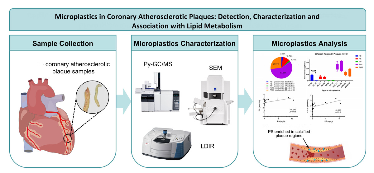
Microplastics (MPs, particles < 5 mm) are increasingly recognized as persistent environmental pollutants with severe human health implications [1, 2]. Clinical and experimental evidence indicates possible cardiovascular relevance, including inflammation, endothelial dysfunction, and detection of MPs in human blood, vascular plaques, and cardiac tissues [3–7]. In carotid atheromas, polyvinyl chloride and polyethylene have been associated with an increased risk of stroke, myocardial infarction, or all-cause death, while MPs have also been detected in carotid arteries and human coronary with atherosclerotic plaques [3, 8]. However, direct evidence from human coronary atherosclerotic plaques remains limited, because specimens of coronary plaque are difficult to obtain clinically. In this research letter, we aimed to report preliminary biological detection and characterization of MPs in human coronary atherosclerotic plaques obtained during coronary endarterectomy, using scanning electron microscopy, laser direct infrared spectroscopy, and pyrolysis-gas chromatography/mass spectrometry. This study also explored distribution of polymer between calcified and non-calcified plaque regions and assessed possible associations with circulating lipid markers.
Thirty-three specimens of coronary atherosclerotic plaque were collected from 24 patients who underwent coronary artery bypass grafting with coronary endarterectomy. After retrieval, specimens were placed immediately in sealed glass containers, anonymized, stored at –20°C, and sent to an accredited laboratory for microplastic analysis. For sample allocation, 17 individual-patient specimens were used for lipid-marker correlation analysis and quantitative polymer detection; 5 patients had plaques containing calcified and non-calcified regions, which were separated for paired regional comparison; and two representative plaques were assessed by scanning electron microscopy (SEM) and laser direct infrared spectroscopy (LDIR) for particle morphology, size, and surface characterization. Pyrolysis-gas chromatography/mass spectrometry (Py-GC/MS) was used to quantify 11 target polymers, including PS, PE, PP, PMMA, PVC, PC, PET, PA6, PA66, PLA, and PBAT, according to protocols reported in previous studies [3, 9–12]. Polymer concentrations were normalized to plaque weight and given as μg/g.
Strict contamination-control procedures were applied, including filtered reagents, plastic-free handling, procedural blanks, cleaned glassware, and recovery testing. Fasting lipid markers, including total cholesterol, triglyceride, low-density lipoprotein cholesterol, and high-density lipoprotein cholesterol were obtained before surgery. Wilcoxon signed-rank test was applied to compare paired calcified and non-calcified plaque regions. Associations between concentrations of MP and lipid markers were determined using Spearman correlation analysis with 95% confidence intervals. The study was reviewed and approved by the Medical Ethics Committee of the 2nd Xiangya Hospital, Central South University, and patient written informed consent was obtained.
The workflow of sample and particle-size distribution are given in Figure 1 A. LDIR detected multiple types of polymers, and identified that most particles were ≤ 200 μm. SEM showed heterogeneous fibrous, granular, and irregular fragment-like particles with folded or rough surfaces (Figure 1 B). In Py-GC/MS analysis of 33 coronary plaque specimens obtained from 24 patients, five different types of polymers were detected: PVC, PS, PA66, PE, and PP. The pattern of overall abundance was PVC > PA66 > PE > PS > PP, with mean concentrations of 20.78 ±7.66, 9.68 ±4.99, 3.85 ±2.15, 0.90 ±2.46, and 0.62 ±0.83 μg/g, respectively. PA66, PVC, and PE were detected in all examined specimens; PP was detected in 17, and PS in 8 out of 33 specimens (Figure 1 C). These findings demonstrate the presence of diverse MPs in human coronary plaques and are consistent with previous studies related to MPs in human vascular and cardiac tissues [3, 7, 8].
Figure 1
Detection and characterization of microplastics in human coronary atherosclerotic plaques. A – Study workflow and sample allocation. The lower graph shows particle-size distribution by laser direct infrared spectroscopy. C – Overall polymer distribution detected by pyrolysis-gas chromatography/mass spectrometry (Py-GC/MS) in 33 plaque specimens. D – Polymer concentrations in paired calcified and non-calcified plaque regions. Exploratory correlations between polystyrene (PS) concentration. E – low-density lipoprotein cholesterol (LDL-C), F – total cholesterol (TC) in 17 individual-patient specimens. p < 0.05 (LDIR). B – Representative LDIR and scanning electron microscopy (SEM) images of identified microplastic particles with corresponding infrared spectra. p < 0.05
CA – calcified area, NC – non-calcified area, PE – polyethylene, PP – polypropylene, PVC – polyvinyl chloride, PA66 – polyamide 66.
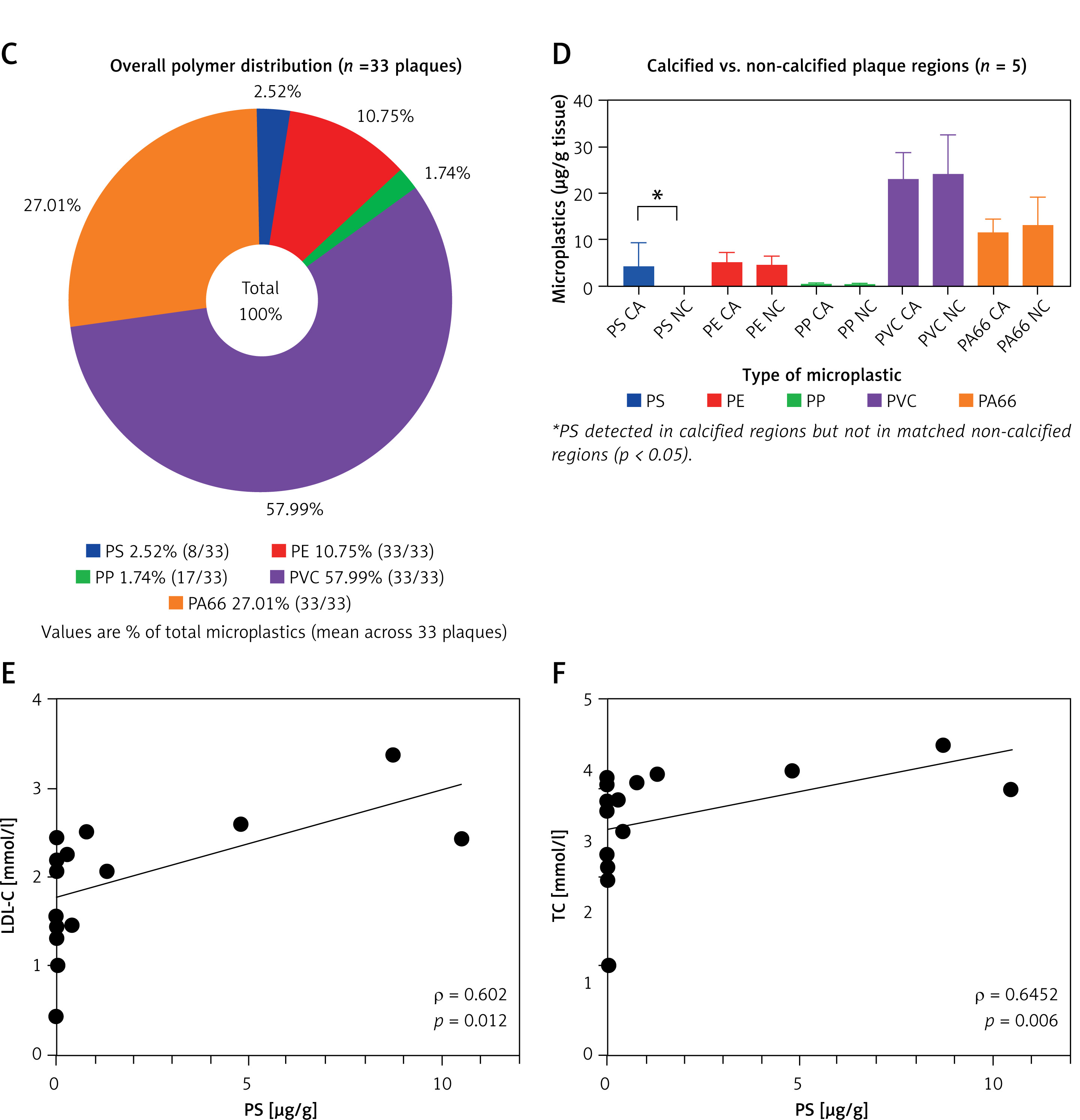
PS was detected in calcified regions but not in matched non-calcified regions (4.07 ±5.11 vs. 0.00 ±0.00 μg/g; Figure 1 D), in five paired plaques containing adjacent calcified and non-calcified regions. However, this region-specific pattern was not observed with other polymers. Although coronary calcification is a marker of advanced atherosclerosis [13], it is unclear from the present cross-sectional data whether PS contributes to calcification, accumulates preferentially in calcified tissue, or represents another feature of plaque biology.
In exploratory analyses of 17 individual-patient specimens, PE, PA66, and PVC were positively correlated with one another: PE-PA66, ρ = 0.853, 95% CI: 0.63–0.95; PE-PVC, ρ = 0.824, 95% CI: 0.57–0.93; and PVC-PA66, ρ = 0.801, 95% CI: 0.52–0.93. These findings may represent co-retention patterns or shared exposure. PS showed exploratory positive correlations with LDL-C (ρ = 0.602, 95% CI: 0.17–0.84, p = 0.012; Figure 1 E) and total cholesterol, (ρ = 0.645, 95% CI: 0.24–0.86, p = 0.006; Figure 1 F), whereas other polymers showed no consistent positive relationship with lipid markers. Given the small sample size and multiple unadjusted comparisons, these association findings should be considered an exploratory interpretation.
This research has several limitations, including the small sample size, cross-sectional design, selected surgical population, limited number of specimens examined by LDIR/SEM, and limited number of paired calcified/non-calcified samples. Therefore, the findings of this study do not establish causality or prove an initiating role for MPs in coronary plaque calcification. Larger studies with rigorous contamination control, prespecified polymers and outcomes, and longitudinal clinical follow-up are needed.