

Introduction
Pressure ulcers, also known as decubitus ulcers, are a type of wound that initially develops in the superficial layers of the skin as a result of prolonged external pressure, and subsequently extends both radially and into deeper tissue layers [1]. In pressure ulcers, continuous external pressure on the skin leads to circulatory impairment in the skin and underlying tissues, resulting in hypoxic tissue injury and necrosis [1]. Prolonged external pressure on the body surface increases capillary pressure, leading to reduced blood flow and tissue ischemia, which in turn causes pressure ulcers, particularly in areas such as the sacrum, buttocks, heels, ankles, and elbows [1]. The pathogenesis of pressure ulcers involves multiple contributing factors, including immobility-induced tissue pressure, as well as physical factors such as friction at the skin surface and moisture [1]. Additional risk factors include malnutrition, inadequate fluid intake, motor and neurological disorders, mental illness, cardiovascular conditions such as peripheral arterial occlusive disease and congestive heart failure, and sensory impairments [1]. More than 60% of patients with pressure ulcers are elderly [1]. Aging is associated with thinning of the dermis and subcutaneous fat tissue, along with a reduction in collagen content and hair follicle density, which collectively contribute to delayed wound healing [2–4].
Wound healing is a complex biological process involving regulated cellular events and the participation of multiple cytokines and growth factors, including fibroblast growth factor (FGF), epidermal growth factor, transforming growth factor, platelet-derived growth factor, insulin-like growth factor-1 (IGF-1), interleukins (IL-1, IL-2, IL-6), and tumor necrosis factor-α (TNF-α) [2, 3]. In addition, a variety of cell types, such as keratinocytes, fibroblasts, endothelial cells, macrophages, and platelets, interact in a coordinated and regulated manner to support the complex process of wound healing [4, 5]. FGFs, initially isolated from bovine pituitary glands, constitute a family of 22 members classified into seven subfamilies [3, 6, 7]. Among these, the FGF1 subfamily includes FGF1 and FGF2 [5, 6]. FGF2, also known as basic FGF (bFGF), not only exerts mitogenic effects but also regulates cellular migration, differentiation, and vascular angiogenesis [4–10]. Consequently, FGF2 is considered critical for optimal wound healing [4, 8, 9].
The Klotho protein, identified in 1997 and described as an anti-aging factor, comprises three isoforms: α-Klotho, β-Klotho, and γ-Klotho [6, 7, 11–14]. α-Klotho is the most abundantly expressed isoform in human tissues, while γ-Klotho is expressed at relatively low levels [12, 15, 16]. Also known as human lactase-like protein (LCTL) or Klotho-LPH-associated protein (KLPH), γ-Klotho remains the only Klotho isoform confirmed to be present in the skin, although its precise mechanisms of action and tissue targets have not yet been fully elucidated [7, 11, 12, 15, 17]. Decreased Klotho levels in the skin result in cutaneous atrophy, similar to aging-related changes [2]. Although it is hypothesized that Klotho proteins may influence tissue repair, studies investigating this relationship are limited in number [2, 3]. The mechanisms underlying delayed wound healing remain incompletely understood [2]. Klotho protein expression and circulating levels decline with advancing age, and it is possible that this reduction may contribute to impaired wound healing [6, 17].
The aim of this study was to evaluate serum levels of γ-Klotho and FGF2 in patients with pressure ulcers.
Material and methods
This study included patients aged 18 years and older who were followed by the Home Health Care Services (HHCS) of the Sultangazi Haseki Training and Research Hospital between December 1, 2024, and March 1, 2025. Patients with stage 2, 3, or 4 pressure ulcers were included in the case group. Pressure ulcers are classified into four stages: stage 2 involves partial-thickness loss of the epidermis and/or dermis; stage 3 includes full-thickness skin loss with damage extending into the subcutaneous tissue, potentially reaching the fascia; and stage 4 involves extensive tissue loss including muscles, bones, tendons, and joint capsules [1]. Open wounds are present in stage 2 and higher, resulting in the loss of the normal barrier function of the intact skin [1]. The control group consisted of patients matched 1 : 1 with the case group for age and sex, who did not have pressure ulcers.
Sociodemographic and clinical data, including age, sex, comorbidities, smoking status, duration of HHCS utilization, Mini Nutritional Assessment (MNA) scores, use of nutritional supplementation products, and use of medical devices such as urinary catheters, endotracheal tubes, nasogastric tubes, and percutaneous endoscopic gastrostomy (PEG), were obtained from medical records. In the case group, wound characteristics were further evaluated, including wound location, duration, stage, dimensions (length, width, and depth), total number of wounds, presence of exudate, odor, signs of infection, and periwound tissue appearance.
Blood samples were collected from all patients in the morning after an overnight fast. Samples were drawn into Vacusera vacuum blood collection tubes (Disera, Izmir, Turkey) and processed in accordance with the CLSI PRE04-ED1:2023 guidelines (Handling, Transport, Processing, and Storage of Blood Specimens for Routine Laboratory Examinations, 1st Edition). Samples were centrifuged according to the manufacturer’s recommendations and serum samples were stored at –80°C until analysis.
The following laboratory parameters were measured: creatinine, C-reactive protein (CRP), albumin, total protein, hemoglobin, and hematocrit. CRP, albumin, and total protein levels were measured using a Siemens Atellica CH Clinical Chemistry Analyzer (Siemens Healthcare GmbH, Erlangen, Germany) with manufacturer-provided commercial kits. Hemoglobin and hematocrit were measured using a Mindray BC 6800 Plus automated complete blood count analyzer (Shenzhen, China) using corresponding proprietary reagents.
Serum levels of γ-Klotho and FGF2 were measured using enzyme-linked immunosorbent assay (ELISA). γ-Klotho concentrations were determined using a LCTL ELISA Kit (Shanghai Yehua Biological Technology Co. Ltd., Shanghai, China), with a sensitivity of 10.0 pg/ml, a standard curve range of 20–2000 pg/ml, and intra- and inter-assay coefficients of variation below 7% and 10%, respectively. FGF2 levels were measured using a Heparin-Binding Growth Factor 2 (FGF2) Human ELISA Kit from the same manufacturer, with a sensitivity of 10.0 pg/ml, a standard curve range of 15–800 pg/ml, and intra- and inter-assay coefficients of variation also below 7% and 10%.
Statistical analysis
Statistical analysis were performed using IBM SPSS Statistics version 22. The normality of data distribution was assessed using the Kolmogorov–Smirnov and Shapiro–Wilk tests. Descriptive statistics were presented as frequency, percentage, mean, median, standard deviation, and interquartile range, as appropriate. For comparison of continuous variables between groups, Student’s t-test was used for normally distributed data, and the Mann–Whitney U test was used for non-normally distributed data. Spearman correlation analysis was employed to assess relationships between continuous variables, while categorical variables were analyzed using the χ2 test. A p-value of < 0.05 was considered statistically significant for all analyses.
Results
A total of 80 patients were included in the study, comprising 40 (50.0%) patients in the case group and 40 (50.0%) in the control group. The demographic and laboratory characteristics of the patients according to their group are summarized in Table I.
Table I
Demographic and laboratory characteristics of the patients according to their group
| Parameter | Total (n = 80) | Case group (n = 40) | Control group (n = 40) | P-value |
|---|---|---|---|---|
| Age [years] | 78.0 [24.8] | 74.3 ±14.1 | 74.3 ±14.1 | 0.994a |
| Duration of HHCS [months] | 24.0 [35.8] | 30.5 [33.0] | 15.0 [36.0] | 0.012b |
| Sex | 0.813c | |||
| Female | 53 (66.3) | 26 (65.0) | 27 (67.5) | |
| Male | 27 (33.7) | 14 (35.0) | 13 (32.5) | |
| Comorbidities | ||||
| Neurological disorders | 46 (57.5) | 24 (60.0) | 22 (55.0) | 0.651c |
| Cardiovascular diseases | 57 (71.3) | 27 (67.5) | 30 (75.0) | 0.459c |
| Diabetes mellitus | 24 (30.0) | 15 (37.5) | 9 (22.5) | 0.143c |
| Thyroid disorders | 12 (15.0) | 5 (12.5) | 7 (17.5) | 0.531c |
| Hyperlipidemia | 9 (11.3) | 3 (7.5) | 6 (15.0) | 0.288c |
| Malignancies | 8 (10.0) | 1 (2.5) | 7 (17.5) | 0.025c |
| Other | 24 (30.0) | 8 (20.0) | 16 (40.0) | 0.051c |
| Tobacco use | 0.013c | |||
| No | 61 (76.2) | 35 (87.5) | 26 (65.0) | |
| Yes | 5 (6.3) | 3 (7.5) | 2 (5.0) | |
| Former smokers | 14 (17.5) | 2 (5.0) | 12 (30.0) | |
| Urinary catheter | 22 (27.5) | 16 (40.0) | 6 (15.0) | 0.012c |
| Endotracheal tube | 4 (5.0) | 3 (7.5) | 1 (2.5) | 0.305c |
| Nasogastric tube | 3 (3.8) | 3 (7.5) | 0 (0.0) | 0.077c |
| PEG | 13 (16.3) | 11 (27.5) | 2 (5.0) | 0.006c |
| Presence of oral feeding | 64 (79.9) | 26 (65.0) | 38 (95.0) | 0.001c |
| Utilization of nutritional supplementation products | 28 (35.0) | 22 (55.0) | 6 (15.0) | < 0.001c |
| MNA scores | 9.0 [4.0] | 7.0 [4.0] | 10.0 [2.0] | < 0.001c |
| Albumin [g/l] | 37.3 ±4.9 | 39.7 ±4.2 | 35.0 ±4.6 | < 0.001a |
| Total protein [g/l] | 65.1 [8.9] | 67.1 [8.3] | 62.7 [6.3] | 0.001b |
| Hemoglobin [g/l] | 11.2 ±1.9 | 11.7 ±2.0 | 10.6 ±1.7 | 0.012a |
| Hematocrit [%] | 35.5 ±5.8 | 37.0 ±6.2 | 34.0 ±5.1 | 0.023a |
| Creatinine [mg/dl] | 0.8 [0.5] | 0.9 [0.53] | 0.7 [0.34] | < 0.001b |
| CRP [mg/l] | 13.1 [17.5] | 8.6 [14.6] | 18.9 [23.4] | 0.006b |
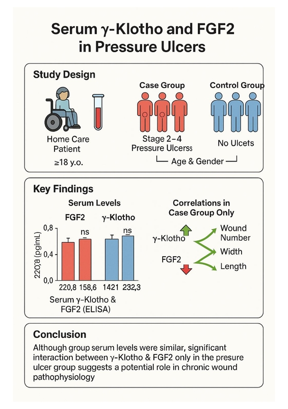
| FGF2 [pg/ml] | 220.0 [27.7] | 220.8 [26.8] | 219.1 [29.1] | 0.577b |
| γ-Klotho [pg/ml] | 1441.8 [183.2] | 1448.4 [158.6] | 1421.3 [232.8] | 0.453b |
In the case group, the median wound duration was 12.0 (9.8) months, and the median number of wounds per patient was 1.0 (1.0). Wound-related characteristics of the case group are detailed in Table II.
Table II
Wound-related characteristics of the case group
| Parameter | Case group (n = 40) | |
|---|---|---|
| Wound locationsa | ||
| Sacrum | 27 (67.5) | |
| Gluteal | 8 (20.0) | |
| Hip | 9 (22.5) | |
| Lower extremities | 9 (22.5) | |
| Back | 3 (7.5) | |
| Wound stagesa | ||
| Stage 2 | 14 (35.0) | |
| Stage 3 | 26 (65.0) | |
| Stage 4 | 12 (30.0) | |
| Wound dimensions | ||
| Length | 3.0 [2.0] | |
| Width | 4.0 [4.75] | |
| Depth | 2.0 [2.0] | |
| Presence of exudate | 13 (32.5) | |
| Type of exudate | ||
| Serous discharge | 35 (87.5) | |
| Purulent or hemorrhagic discharge | 5 (12.5) | |
| Signs of infection | 5 (12.5) | |
| Presence of odor | 4 (10.0) | |
| Periwound tissue appearance | ||
| Healthy | 25 (62.5) | |
| Unhealthy | 15 (37.5) | |
When the relationship between FGF2 and γ-Klotho was evaluated, a weak positive correlation was observed in the case group (r = 0.314, p = 0.048), whereas no such association was found in the control group (p = 0.164).
In the case group, FGF2 levels were positively correlated with the number of wounds (r = 0.454, p = 0.003), wound width (r = 0.422, p = 0.007), and wound length (r = 0.396, p = 0.012). However, no correlation was found between FGF2 levels and wound duration (p = 0.469) or wound depth (p = 0.319). Additionally, γ-Klotho levels in the case group were negatively correlated with wound duration (r = –0.352, p = 0.026), whereas no significant correlations were identified between γ-Klotho levels and number of wounds (p = 0.946), wound width (p = 0.298), wound length (p = 0.257), or wound depth (p = 0.559). FGF2 and γ-Klotho levels according to wound characteristics in the case group are summarized in Table III.
Table III
FGF2 and γ-Klotho levels according to wound characteristics in the case group
No significant correlations were found between FGF2 levels and age, duration of HHCS utilization, or MNA scores in either group (p > 0.05). Similarly, no significant associations were found between γ-Klotho levels and age, duration of HHCS utilization, or MNA scores in both the case and control groups (p > 0.05). FGF2 and γ-Klotho levels based on demographic characteristics of both groups are presented in Table IV.
Table IV
FGF2 and γ-Klotho levels based on demographic characteristics of both groups
| Parameter | Case group (n = 40) | Control group (n = 40) | ||||||||
|---|---|---|---|---|---|---|---|---|---|---|
| n (%) | FGF 2 [pg/ml] | P-value | γ-Klotho [pg/ml] | P-value | n (%) | FGF 2 [pg/ml] | P-value | γ-Klotho [pg/ml] | P-value | |
| Sex | 0.604a | 0.664a | 0.909a | 0.345a | ||||||
| Female | 26 (65.0) | 216.9 [25.1] | 1430.3 [229.9] | 27 (67.5) | 220.5 [32.8] | 1478.7 [180.3] | ||||
| Male | 14 (35.0) | 224.1 [45.1] | 1397.5 [484.4] | 13 (32.5) | 221.1 [22.3] | 1416.4 [147.5] | ||||
| Comorbidities | ||||||||||
| Neurological disorders | 0.713a | 0.613a | 0.861a | 0.262a | ||||||
| No | 16 (40.0) | 216.9 [33.9] | 1393.5 [180.3] | 18 (45.0) | 223.8 [34.7] | 1423.8 [132.4] | ||||
| Yes | 24 (60.0) | 219.7 [24.7] | 1430.3 [247.1] | 22 (55.0) | 220.2 [18.8] | 1470.5 [167.6] | ||||
| Cardiovascular diseases | 0.177a | 0.798a | 0.221a | 0.590a | ||||||
| No | 13 (32.5) | 224.7 [28.6] | 1431.1 [214.8] | 10 (25.0) | 217.3 [25.5] | 1479.5 [256.9] | ||||
| Yes | 27 (67.5) | 215.6 [27.4] | 1380.3 [265.6] | 30 (75.0) | 221.4 [27.8] | 1442.6 [143.9] | ||||
| Diabetes mellitus | 0.233a | 0.699a | 0.924a | 0.975a | ||||||
| No | 25 (62.5) | 220.2 [37.3] | 1450.8 [231.1] | 31 (77.5) | 221.1 [31.3] | 1436.1 [168.9] | ||||
| Yes | 15 (37.5) | 213.5 [26.2] | 1372.1 [244.3] | 9 (22.5) | 220.5 [16.5] | 1478.7 [164.8] | ||||
| Thyroid disorders | 0.551a | 1.000a | 0.807a | 0.781a | ||||||
| No | 35 (87.5) | 220.2 [30.1] | 1411.5 [240.9] | 33 (82.5) | 220.5 [25.9] | 1436.1 [180.3] | ||||
| Yes | 5 (12.5) | 215.6 [32.8] | 1485.3 [259.8] | 7 (17.5) | 223.5 [44.8] | 1449.2 [75.4] | ||||
| Hyperlipidemia | 0.072a | 0.144a | 0.985a | 0.019a | ||||||
| No | 37 (92.5) | 220.2 [27.8] | 1411.5 [206.6] | 34 (85.0) | 220.8 [28.4] | 1430.3 [149.2] | ||||
| Yes | 3 (7.5) | 193.8 [–] | 1613.1 [–] | 6 (15.0) | 222.6 [23.2] | 1561.5 [184.0] | ||||
| Malignancies | 0.150a | 0.900a | 0.363a | 0.577a | ||||||
| No | 39 (97.5) | 218.9 [29.2] | 1411.5 [242.6] | 33 (82.5) | 219.9 [29.0] | 1447.5 [159.0] | ||||
| Yes | 1 (2.5) | – | – | 7 (17.5) | 224.7 [19.9] | 1449.2 [167.2] | ||||
| Other | 0.070a | 0.009a | 0.774a | 0.389a | ||||||
| No | 32 (80.0) | 216.5 [27.3] | 1368.0 [230.7] | 24 (60.0) | 220.2 [15.9] | 1482.8 [148.4] | ||||
| Yes | 8 (20.0) | 226.8 [28.9] | 1533.6 [583.6] | 16 (40.0) | 225.7 [47.3] | 1423.8 [198.8] | ||||
| Tobacco use | 0.340b | 0.271b | 0.270b | 0.762b | ||||||
| No | 35 (87.5) | 219.3 [31.9] | 1431.2 [257.4] | 26 (65.0) | 221.1 [28.7] | 1474.6 [179.1] | ||||
| Yes | 3 (7.5) | 227.9 [–] | 1347.5 [–] | 2 (5.0) | 242.9 [–] | 1463.9 [–] | ||||
| Former smokers | 2 (5.0) | 199.5 [–] | 1727.1 [–] | 12 (30.0) | 216.9 [22.4] | 1425.4 [137.7] | ||||
| Urinary catheter | 0.255a | 0.795a | 0.324a | 0.019a | ||||||
| No | 24 (60.0) | 215.2 [27.3] | 1414.8 [184.8] | 34 (85.0) | 222.6 [26.1] | 1482.8 [168.0] | ||||
| Yes | 16 (40.0) | 221.8 [32.0] | 1421.3 [289.3] | 6 (15.0) | 213.8 [33.5] | 1383.6 [406.1] | ||||
| Endotracheal tube | 1.000a | 0.064a | – | – | ||||||
| No | 37 (92.5) | 219.3 [31.3] | 1380.3 [206.6] | 39 (97.5) | 220.5 [26.8] | 1449.2 [163.9] | ||||
| Yes | 3 (7.5) | 218.9 [–] | 1565.6 [–] | 1 (2.5) | – | – | ||||
| Nasogastric tube | 0.557a | 1.000a | – | – | ||||||
| No | 37 (92.5) | 218.9 [29.9] | 1431.2 [235.2] | 40 (100.0) | 220.8 [26.8] | 1448.4 [158.6] | ||||
| Yes | 3 (7.5) | 224.7 [–] | 1363.9 [–] | 0 (0.0) | – | – | ||||
| PEG | 0.492a | 0.530a | 0.400a | 0.831a | ||||||
| No | 29 (72.5) | 219.3 [30.2] | 1450.8 [233.6] | 38 (95.0) | 221.4 [28.4] | 1448.4 [148.4] | ||||
| Yes | 11 (27.5) | 218.9 [18.9] | 1372.1 [185.2] | 2 (5.0) | 213.7 [–] | 1485.3 [–] | ||||
| Presence of oral feeding | 0.900a | 0.834a | 0.400a | 0.831a | ||||||
| No | 14 (35.0) | 218.2 [17.8] | 1368.0 [258.2] | 2 (5.0) | 213.7 [–] | 1485.3 [–] | ||||
| Yes | 26 (65.0) | 219.7 [31.9] | 1450.0 [228.3] | 38 (95.0) | 221.4 [28.4] | 1448.4 [148.4] | ||||
| Utilization of nutritional supplementation products | 0.325a | 0.717a | 0.839a | 0.470a | ||||||
| No | 18 (45.0) | 217.9 [27.6] | 1454.1 [197.5] | 34 (85.0) | 221.1 [31.7] | 1448.4 [144.3] | ||||
| Yes | 22 (55.0) | 221.2 [26.8] | 1391.8 [270.5] | 6 (15.0) | 217.9 [17.3] | 1488.5 [300.0] | ||||
FGF2 levels were positively correlated with CRP in the case group (r = 0.398, p = 0.011). However, no significant associations were found between either FGF2 or γ-Klotho and levels of albumin, total protein, creatinine, hemoglobin, or hematocrit in either the case or control group (p > 0.05).
Discussion
Wound healing is a complex physiological process involving various cell types such as fibroblasts, endothelial cells, keratinocytes, macrophages, and platelets [6]. This process is regulated by multiple cytokines and growth factors, including FGFs [2, 5]. FGFs constitute a family of cell signaling proteins that mediate angiogenesis, wound healing, metabolic regulation, and embryonic development by binding to FGF receptors (FGFRs) [5, 6, 10, 11]. In wound healing, FGFs stimulate fibroblast proliferation and strongly activate other mesoderm-derived cells such as vascular endothelial and smooth muscle cells, osteoblasts, and chondrocytes [4–6]. As a key regulator of wound healing, angiogenesis facilitates the delivery of immune cells, clearance of cellular debris, and transport of oxygen and nutrients for tissue regeneration, with FGF2 playing a stimulatory role in this process [6]. Among the FGFs, FGF1, FGF2, FGF4, FGF7, FGF16, FGF21, and FGF23 have been shown to be effective in the treatment of diabetic ulcers [5, 6]. Previous studies have demonstrated improved healing of pressure ulcers following FGF2-based treatments [4, 6, 8, 9]. In animal models, impaired wound healing in aged mice has been associated with decreased FGF2 levels, and FGF2-null mice exhibit reduced collagen deposition, delayed re-epithelialization, and impaired wound closure [8]. In this study, no significant difference in FGF2 levels was observed between patients with and without pressure ulcers. A potential explanation for this finding is that, unlike previous studies which primarily assessed FGF2 expression in wound tissue, the present study evaluated circulating FGF2 levels. Nonetheless, positive correlations were observed between FGF2 levels and the number, width, and length of wounds in patients with pressure ulcers. As the size and number of wounds increase, the extent of skin tissue damage is likely to become greater, which may contribute to elevated FGF2 levels due to its pivotal role in tissue repair.
Klotho protein expression and circulating levels decline with aging and have been associated with reduced lifespan, pulmonary atrophy, atherosclerosis, osteopenia, and infertility [2, 12, 14]. Among the three subtypes of Klotho, α-Klotho is primarily secreted by the kidneys and is notably reduced in chronic kidney disease [12, 14, 15]. β-Klotho is synthesized mainly in adipose tissue, as well as the lungs and pancreas, and plays a role in metabolic regulation [11, 12, 15]. γ-Klotho, the least extensively studied isoform, is expressed in the skin, testes, and kidneys [5, 7, 11, 12, 15]. Although elevated γ-Klotho expression has been observed in cancers such as breast, bladder, and prostate, its specific target tissues and underlying mechanisms of action remain poorly understood [7, 11, 17]. Aging, which may delay wound healing, is also associated with a physiological decline in Klotho levels [12, 16], suggesting a potential role for Klotho proteins in wound repair. However, only a limited number of studies have explored this relationship [2, 3]. An animal study demonstrated that the skin of Klotho-deficient mice resembled that of aged humans; however, delayed wound healing was not directly associated with α-Klotho deficiency [2]. Activation of the β-Klotho gene in adipose-derived stem cells has been shown to enhance basal membrane regeneration, angiogenesis, and epithelialization, thereby promoting cell proliferation [3]. Although γ-Klotho is reported to be expressed in skin tissue [11, 17], to the best of our knowledge, no previous studies have directly assessed its role in wound healing. In the current study, no significant difference was found in γ-Klotho levels between patients with and without pressure ulcers. This may be due to the evaluation of serum rather than tissue-specific levels of γ-Klotho. Klotho proteins exist in both membrane-bound and soluble forms, with the soluble form being primarily derived from α-Klotho [11]. In this context, the levels of γ-Klotho, which has been reported to be expressed in skin tissue, may not differ between patients with pressure ulcers and those without. Furthermore, a negative correlation was observed between γ-Klotho levels and wound duration in this study. A reduction in circulating Klotho protein levels associated with aging has been correlated with delayed wound healing in the skin [6, 17], ultimately leading to an extended wound duration.
Klotho proteins are considered cofactors for various FGFs [7, 13, 18]. The FGF family can be classified into intracrine, endocrine, and paracrine factors based on their respective functions [6]. In particular, endocrine FGFs (e.g., FGF15, FGF19, FGF21, and FGF23) require α- or β-Klotho as cofactors for their activity [5, 10, 11]. For instance, α-Klotho functions as a cofactor for FGF23, thereby influencing kidney function and bone metabolism [7, 11, 12, 15]. Additionally, β-Klotho mediates insulin signaling in conjunction with FGF15 and FGF19, and regulates cholesterol, bile acid, and lipid metabolism in hepatic and adipose tissues [11, 15, 18]. However, the specific FGF subtype for which γ-Klotho functions as a cofactor has not yet been identified [11]. γ-Klotho has been shown to enhance FGF15/19 signaling in human embryonic kidney 293 cells. Interestingly, depletion of γ-Klotho results in constitutive activation of extracellular signal-regulated kinase (ERK) and a reduced ERK response to canonical FGF2 stimulation. Furthermore, γ-Klotho has been reported to directly interact with FGF23 and FGFRs, potentially contributing to an FGFR-mediated immunosuppressive function [7]. In addition, γ-Klotho is also proposed to act as a co-receptor for FGF19 in cultured cells by forming complexes with FGFR1b, FGFR1c, FGFR2c, and FGFR4 [15]. While endocrine FGFs require Klotho proteins as cofactors to exert their biological functions, paracrine FGFs such as FGF2 do not depend on Klotho proteins for their activity [5, 10, 11]. Nonetheless, in vivo studies have reported that Klotho induction may suppress FGF2 expression [17], and to date, no Klotho subtype has been identified as a direct cofactor for FGF2. FGF2, a key mediator of wound healing, may potentially interact with γ-Klotho, the only Klotho isoform known to be expressed in the skin. In support of this hypothesis, the present study demonstrated a positive correlation between serum γ-Klotho and FGF2 levels in patients with pressure ulcers, whereas no such association was observed in the control group.
One limitation of this study is the relatively small sample size. Additionally, evaluating FGF2 and γ-Klotho levels in blood samples rather than directly in wound tissue may represent another constraint.
In conclusion, wound healing is a complex process that relies on the coordinated regulation of multiple cellular mechanisms. FGF2 has been identified as an important regulator of wound repair. The Klotho protein family, often characterized as anti-aging factors, has also been implicated in wound healing; however, its role remains controversial. Notably, γ-Klotho is reported to be expressed in skin tissue; however, its role in wound healing remains unclear. In the present study, no significant differences were observed in the serum levels of γ-Klotho and FGF2 between patients with and without pressure ulcers. However, a weak positive correlation between γ-Klotho and FGF2 was identified in the pressure ulcer group, whereas no such association was found in the control group. These results highlight the complexity of the biological processes involved in wound healing and suggest that further research is needed to better elucidate the specific roles of γ-Klotho and FGF2 in this process.

