

Introduction
Pneumothorax is a common clinical emergency characterized by the abnormal accumulation of air in the pleural cavity between the lungs and chest wall, which prevents lung expansion and can lead to lung atrophy through mechanisms such as alveolar collapse and impaired ventilation [1]. Pneumothorax is mainly classified into spontaneous and traumatic types [2]. Spontaneous pneumothorax includes primary and secondary types. Primary pneumothorax occurs when the thin walls of the alveoli, the tiny air sacs in the lungs responsible for gas exchange, rupture and allow air to enter the pleural cavity, causing lung collapse. Secondary pneumothorax occurs in patients with existing lung diseases such as chronic obstructive pulmonary disease (COPD), asthma, or tuberculosis [3–5]. Traumatic pneumothorax results from blunt, penetrating, or explosive chest injuries. Clinically, pneumothorax often presents with sudden chest pain and dyspnea, and severe cases may progress to hypoxia, hypotension, or even death [6–11]. Epidemiological data reveal significant gender and age differences in pneumothorax incidence. Men are more susceptible than women, and this gender difference can be attributed to several factors, including higher rates of smoking in men, differences in body size (e.g., larger and taller body frames increase alveolar wall tension), and more [3]. Incidence peaks vary across age groups, reflecting complex underlying factors. Known risk factors include smoking, body mass index (BMI), height, exposure to toxic metals, and environmental influences [11–16].
Physical activity, defined as any bodily movement produced by skeletal muscles requiring energy expenditure, ranges from low-intensity walking to vigorous exercise [17, 18]. According to the World Health Organization (WHO), nearly one third of adults globally lack sufficient physical activity, which is linked to increased risks of cardiovascular disease, diabetes, certain cancers, and mental health disorders [19–23]. Emerging evidence suggests that both insufficient and excessive physical activity may influence lung health and potentially increase pneumothorax risk, especially in individuals with underlying pulmonary conditions [24–26]. Additionally, factors such as smoking and body habitus associated with pneumothorax risk may correlate with physical activity patterns [27–30].
Frailty is a geriatric syndrome characterized by weight loss, fatigue, muscle weakness, and reduced mobility, leading to diminished quality of life and increased risks of falls, hospitalization, and mortality [31, 32]. In epidemiological studies, risk factors for frailty include age, gender, chronic disease, poor nutrition and insufficient physical activity [33, 34]. Although the relationship between frailty and pneumothorax is not well studied, preliminary data indicate that frail individuals, particularly the elderly, may have compromised lung function, often demonstrating weakened respiratory muscles and reduced lung ventilation, which results in fragile alveoli that are more susceptible to spontaneous pneumothorax in the event of stress or external injury [35, 36]. Frailty is also linked to respiratory diseases such as COPD, which are established pneumothorax risk factors [37, 38]. This combination of diminished lung function and chronic comorbidities likely amplifies the vulnerability of frail patients to pneumothorax. In adolescents and young adults, frailty has also been identified as a potential risk factor for primary spontaneous pneumothorax [14, 16, 39].
Although studies suggest physical activity and frailty are linked to pneumothorax, these associations are often influenced by factors such as smoking, age, and sex. We hypothesized that more physical activity would be potentially protective against pneumothorax and that frailty would increase the risk of pneumothorax. Traditional studies struggle to prove this because of confounding and reverse causation. To address this, we used Mendelian randomization (MR), a genetic method that helps identify causal effects by using genetic variants as tools, reducing bias [40, 41]. We applied two-sample MR (TSMR) and multivariable MR (MVMR) to examine how physical activity and frailty independently affect pneumothorax risk. Our goal is to provide strong evidence on these causal links to support better prevention and treatment strategies.
Material and methods
Study design
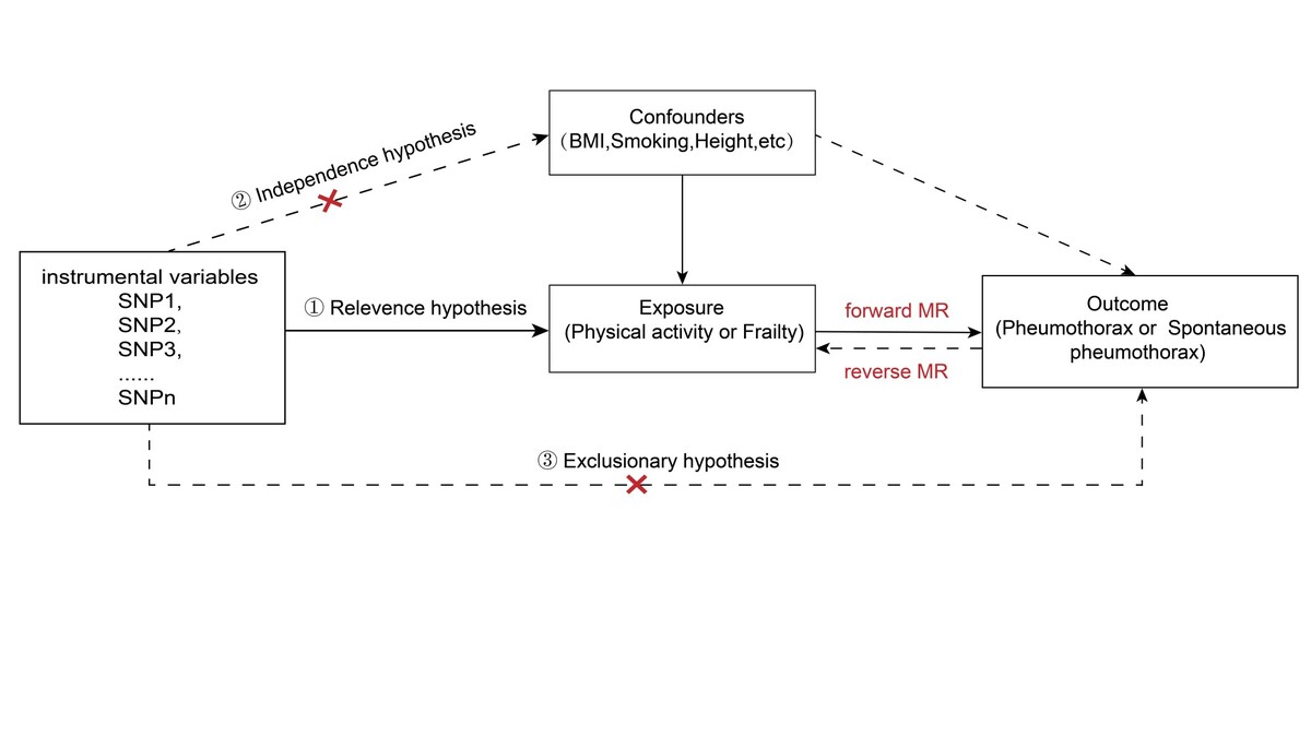
The overall design of the MR analysis in this study is illustrated in Figure 1. We conducted a TSMR analysis to investigate the causal effects of physical activity or frailty on pneumothorax and spontaneous pneumothorax using genome-wide association study (GWAS) data. We applied two-sample MR leverages genetic variants as instrumental variables (IVs) derived from two independent GWAS datasets, one for the exposure and one for the outcome, to estimate causal relationships. This approach helps to minimize bias from confounding factors and reverse causation, as genetic variants are randomly assorted at conception and fixed throughout life [42]. The core assumptions for valid IVs are: (1) strong association with the exposure; (2) no association with confounders; (3) influence on the outcome only through the exposure. All GWAS datasets included in this study derive from large-scale European cohorts, primarily the UK Biobank and Integrative Epidemiology Unit (IEU) studies. Detailed information on sample sizes, case-control numbers, and population characteristics are summarized in Table I. The study populations represent diverse localities across Europe, ensuring broad applicability within this ancestry group.
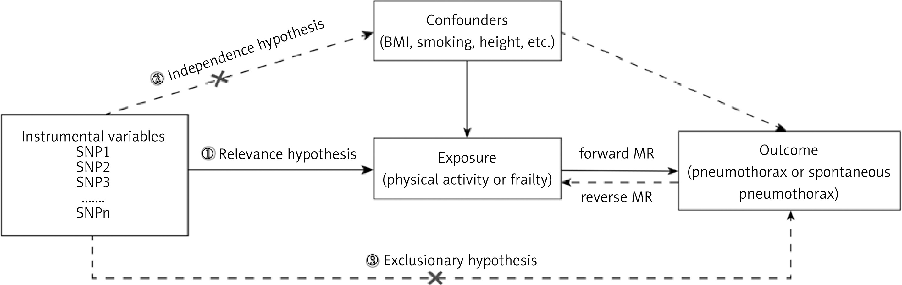
Table I
Detailed information of the GWAS in our analysis
Data sources for exposure and outcome
Exposure data include physical activity (four categories) [43], frailty (Frailty Index (FI) [44, 45] and Frailty Phenotype (FP) [32, 46]), and body mass index (BMI) [47] as a confounder. Outcome data comprise pneumothorax [47] and spontaneous pneumothorax [48] GWAS summary statistics. Detailed dataset information and GWAS catalog IDs are listed in Table I. All datasets are publicly accessible via the IEU OpenGWAS project (https://gwas.mrcieu.ac.uk/). Due to their availability in public databases, no additional ethical approval is required, allowing researchers unrestricted access. Further details regarding the data are provided in Supplementary Table SI.
Selection of instrumental variables
Single nucleotide polymorphisms (SNPs) were selected as IVs based on genome-wide significance (p < 5e-08), with relaxed thresholds (p < 5e-06) for exposures with fewer loci [49]. Linkage disequilibrium was controlled by clumping (r2 = 0.001, distance > 10,000 kb). Single nucleotide polymorphisms with weak instrument bias (F-statistic < 10) or pleiotropic effects related to confounders (BMI, smoking, alcohol use, height, lung function, respiratory diseases) were excluded. By querying the LDtrait module in the LDlink database (https://ldlink.nih.gov/) [50], the system retrieved published GWAS results for each SNP associated with potential confounders. SNPs that reached the genomic significance level (p < 5e-08, r2 = 0.001) in the associated confounding GWAS were excluded to reduce the effect of confounding bias. SNPs with palindromic structures were removed. Outlier SNPs identified by MR-PRESSO were removed [51, 52].
Analysis of Mendelian randomization
Five MR methods were applied: MR-Egger regression [53], inverse variance weighted (IVW) method [54], weighted median [55], weighted mode, and simple mode [56], with the IVW method being the primary approach [57]. Univariable MR (UVMR) assessed causal effects of physical activity and frailty on pneumothorax outcomes. In addition, to explore the possibility of reverse causality, we also performed a bidirectional MR analysis [58] with pneumothorax (including spontaneous pneumothorax) as the exposure variable and light DIY or FI as the outcome. This reverse MR used the same workflow as the forward MR analysis and was designed to test for potential reverse causality. High BMI is associated with chronic diseases such as cardiovascular disease, diabetes, and certain cancers, and studies have shown that people with lower BMI may be at increased risk for spontaneous pneumothorax due to less chest wall fat and increased mechanical stress on lung tissue [59]. Moreover, BMI is a shared risk factor for pneumothorax, physical activity, and frailty. Here, BMI was used as a confounder or mediator variable in MVMR analyses to distinguish the independent effects of physical activity and debility on pneumothorax. The MVMR analysis adhered to the same IV selection criteria, with F- statistics of 10.3, 26.5, and 30.3 for BMI, light DIY, and FI respectively.
Sensitivity analysis
To ensure the reliability and robustness of our results, we performed sensitivity analyses including heterogeneity tests using Cochran’s Q, horizontal pleiotropy assessment with MR-Egger regression, and leave-one-out analysis [49, 51, 60]. Random-effects IVW models were used when heterogeneity was significant (p < 0.05); otherwise, fixed-effects models were applied. A significant MR-Egger intercept (p < 0.05) indicated directional pleiotropy [52]. MR-PRESSO was used to detect and correct for outlying SNPs. Leave-one-out analysis was used to check whether any individual SNPs had a disproportionate effect on the results.
Results
Causal association between physical activity and pneumothorax
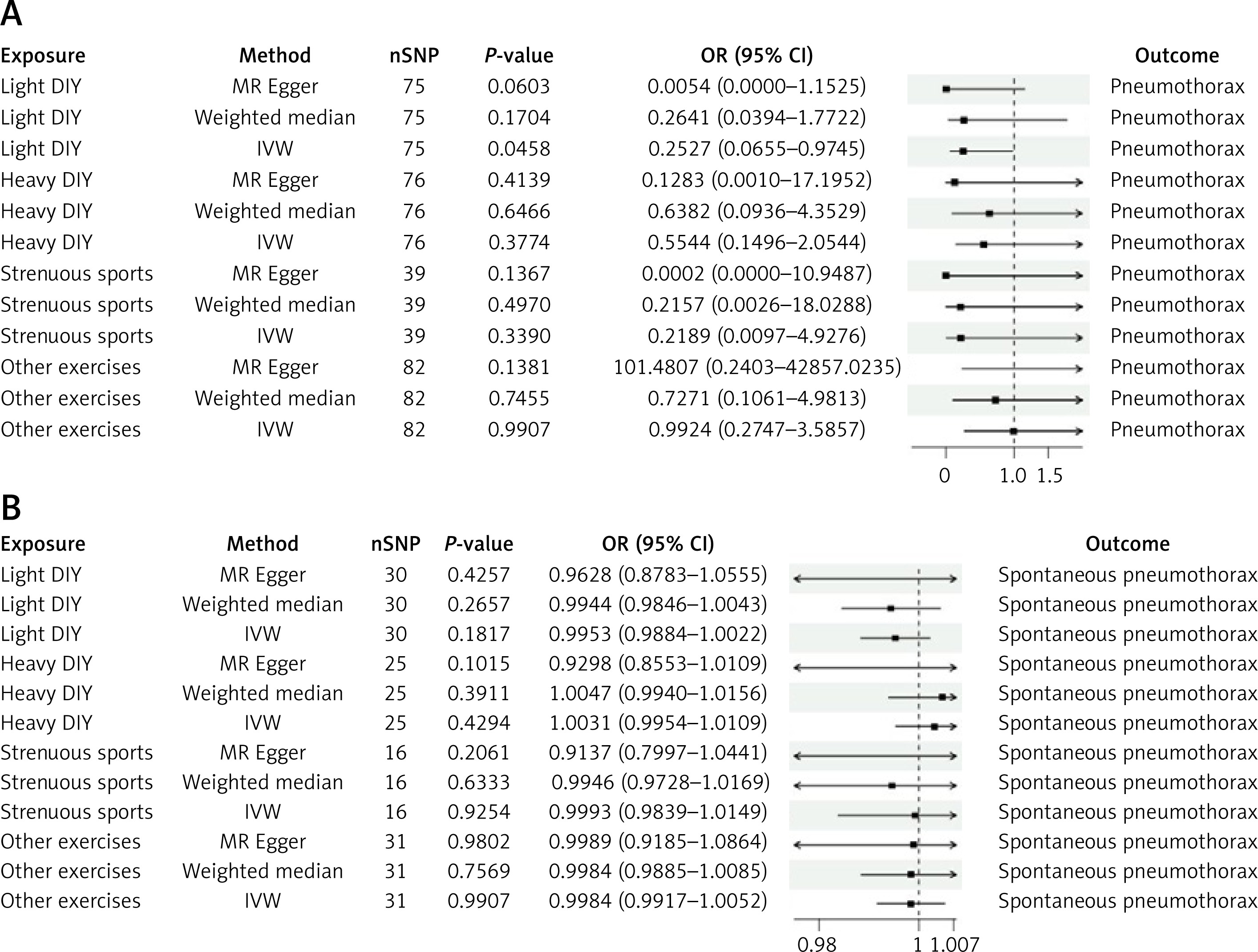
We selected SNPs closely associated with four different intensities of physical activity through conditional filtering and conducted MR analyses to investigate the potential causal relationship between physical activity and pneumothorax. The MR analyses primarily relied on the IVW method. Results from supplementary analyses are presented in Supplementary Table SII. As shown in Figure 2 A, the results indicated a significant causal relationship between light DIY and pneumothorax (IVW: odds ratio (OR) = 0.2527, 95% CI: 0.0655–0.9745, p = 0.0458). No causal associations were observed between any physical activity and spontaneous pneumothorax (Figure 2 B). Our findings suggest that higher light DIY was associated with a lower risk of pneumothorax, serving as a protective factor.
Figure 2
Causal relationships between physical activity and the risk of pneumothorax and spontaneous pneumothorax. A – MR analysis of physical activity and pneumothorax. B – MR analysis of physical activity and spontaneous pneumothorax. Four different intensities of physical activity were used as exposure factors, with pneumothorax/ spontaneous pneumothorax as the outcome. The inverse variance weighted (IVW) results were used as the primary reference, with p < 0.05 indicating statistically significant causal relationships
OR – odds ratio, per genetically predicted 1 standard deviation increase in exposure.
Causal association between frailty and pneumothorax
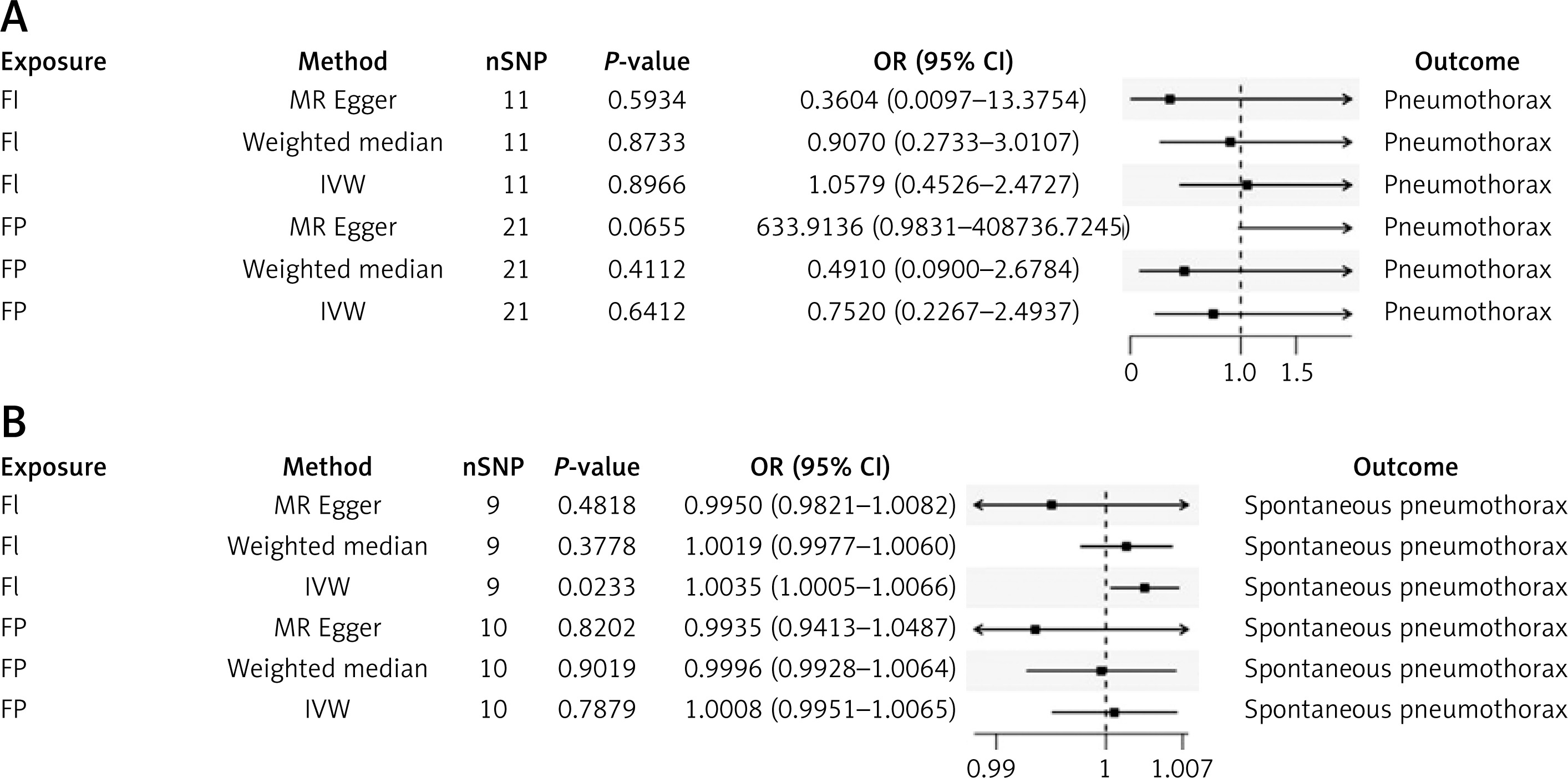
To explore whether frailty has a potential impact on the development of pneumothorax, we selected the FI and FP as exposures for MR analysis. The primary results of the MR analysis were derived from the IVW method, with additional results presented in Supplementary Table SII. Our findings indicated that although no causal relationship was observed between frailty and pneumothorax (Figure 3 A), there was a significant causal relationship between the FI and spontaneous pneumothorax, suggesting that the FI may be a risk factor for spontaneous pneumothorax (IVW: OR = 1.0035, 95% CI: 1.0005–1.0066, p = 0.0233, Figure 3 B).
Figure 3
Causal relationships between frailty and the risk of pneumothorax and spontaneous pneumothorax. A – MR analysis of frailty and pneumothorax. B – MR analysis of frailty and spontaneous pneumothorax. Two frailty assessment metrics, FI and FP, were used as exposure factors, with pneumothorax/spontaneous pneumothorax as the outcome. The IVW results were used as the primary reference, with p < 0.05 indicating statistically significant causal relationships
FI – Frailty Index, FP – frailty phenotype, OR – odds ratio, per genetically predicted 1 standard deviation increase in exposure.
Results of inverse Mendelian randomization analysis
To investigate whether there are reverse causal effects between physical activity, frailty, and pneumothorax, we conducted reverse MR analyses on the positive findings obtained earlier. In these analyses, pneumothorax and spontaneous pneumothorax were used as exposures, while light DIY and the FI were used as outcomes. The results showed that a weak inverse association between light DIY and pneumothorax, with an effect size very close to no effect (IVW: OR = 0.9951, 95% CI: 0.9912–0.9989, p = 0.0124, Figure 4), but no reverse causal relationship was observed between the FI and spontaneous pneumothorax.
Figure 4
Results of reverse MR analyses for physical activity and frailty with pneumothorax. Pneumothorax and spontaneous pneumothorax were used as exposure factors, while light DIY and FI were used as outcomes. The IVW results were used as the primary reference, with p < 0.05 indicating statistically significant causal relationships
FI – Frailty Index, OR – odds ratio, per genetically predicted 1 standard deviation increase in exposure.

This suggests that a higher genetic liability to pneumothorax was associated with lower light DIY.
Results of multivariate Mendelian randomization analyses
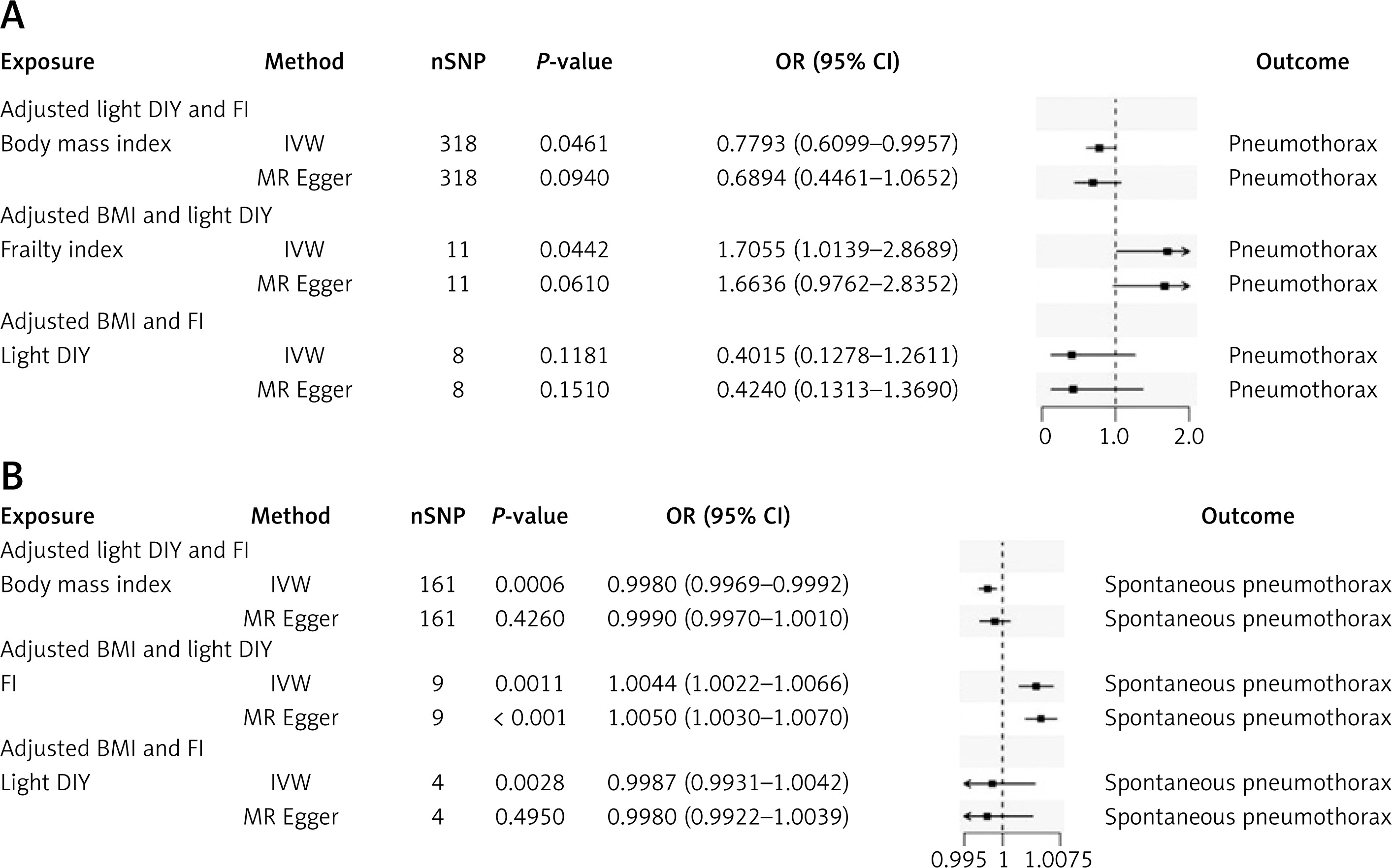
To further investigate whether physical activity and frailty influence the development of pneumothorax after adjusting for BMI, physical activity or frailty, we conducted a MVMR analysis. The IVW results showed that light DIY, which originally had a causal effect on pneumothorax, no longer had a causal effect after adjusting for BMI and the FI (Figure 5 A). This suggests that BMI may play a mediating or confounding role in the association between light DIY and pneumothorax. However, after adjusting for BMI and light DIY activity, the FI still had a causal effect on spontaneous pneumothorax (IVW: OR = 1.0044, 95% CI: 1.0022–1.0066, p = 0.0011, Figure 5 B).
Figure 5
Results of MVMR analyses for physical activity and frailty with pneumothorax. A – MVMR analysis of physical activity and frailty with pneumothorax. B – MVMR analysis of physical activity and frailty with spontaneous pneumothorax. BMI, FI, and light DIY were used as exposure factors, with pneumothorax/spontaneous pneumothorax as the outcome. The IVW results were used as the primary reference, with p < 0.05 indicating statistically significant causal relationships
BMI – body mass index, FI – Frailty Index, OR – odds ratio, per genetically predicted 1 standard deviation increase in exposure.
Sensitivity analysis results
To enhance the robustness and reliability of our findings, we employed multiple sensitivity analyses, including Cochran’s Q tests, MR-Egger intercept tests, and MR-PRESSO global tests. For most exposures, the Cochran’s Q test for heterogeneity yielded p-values greater than 0.05, indicating no significant heterogeneity observed. Although heterogeneity was detected in some results, it did not invalidate the MR estimates in this study, as the random-effects IVW model could balance the aggregated heterogeneity. Horizontal pleiotropy occurs when genetic variants related to the exposures of interest directly affect the outcomes through multiple pathways beyond the hypothesized exposure. Therefore, we further used MR-Egger intercept tests and MR-PRESSO global tests to detect pleiotropy and assess the robustness of our results. The results showed p-values greater than 0.05, indicating no significant pleiotropy observed. Detailed results are presented in Tables II, III, and Supplementary Table SIII. Additionally, the results of reverse MR analyses and MVMR analyses are provided in Supplementary Table SIII. Leave-one-out analyses, forest plot, scatter plots, and funnel plots further validated the stability of our findings (Supplementary Figures S1–S3). Overall, the sensitivity analyses ensured the robustness and reliability of our results.
Table II
Sensitivity analysis of the causal relationship between physical activity and pneumothorax or spontaneous pneumothorax
Table III
Sensitivity analysis of the causal relationship between frailty and pneumothorax or spontaneous pneumothorax
Discussion
Pneumothorax remains a significant global health challenge, with treatment strategies ranging from conservative management to surgical interventions depending on disease severity and clinical presentation [61–63]. Despite advances in minimally invasive surgical techniques, the complex etiology and high recurrence rate of pneumothorax highlight the need to better understand modifiable risk factors to improve prevention [64, 65]. There have been no large-scale observational or MR studies directly linking physical activity and frailty to pneumothorax in previous studies. However, observational studies have shown that physical activity is strongly associated with lung function [66]. The positive effects of physical activity on lung function have been demonstrated in a variety of respiratory diseases [67–69]. In addition, a part of the literature has highlighted the negative association between frailty and lung diseases and their complications [70, 71]. Our study found a causal association between light DIY and frailty with pneumothorax, suggesting that frailty may be an important risk factor for the development of pneumothorax. In particular, light DIY had an inverse causal association with pneumothorax, but this inverse association was very weak, suggesting that the inverse association may be influenced by shared pleiotropy or confounding factors (e.g., BMI). Further MVMR analyses showed that the independent effect of light DIY on pneumothorax disappeared after adjusting for BMI, suggesting that BMI may play a mediating or confounding role in the association between physical activity and pneumothorax. In contrast, the effect of FI on pneumothorax remained significant after adjustment, supporting its independent causal role. These findings contribute to a more accurate understanding of the mechanisms of physical activity and frailty in the development of pneumothorax.
Our study found that light DIY activities, such as pruning and watering lawns, may be potentially protective against pneumothorax. Light DIY is defined as a type of low-intensity physical activity over the past 4 weeks. The impact of physical activity on pneumothorax may be related to changes in lung pressure during exercise [72, 73]. Regular physical activity can improve lung ventilation and gas exchange efficiency, and enhance alveolar stability, reducing the fragility of alveoli, thereby reducing the likelihood of pneumothorax [74, 75]. In addition, regular physical activity helps to improve systemic blood circulation, enhance oxygen supply, maintain lung tissue health, and reduce lung injury triggered by hypoxia [75]. Some studies suggest that insufficient physical activity may lead to decreased lung function, thereby increasing the risk of pneumothorax [76]. Of note, our analyses showed that not all types of physical activity had a significant effect on pneumothorax risk. With the exception of light DIY activities, other intensities of physical activity did not show significant causal associations. This result may partly stem from the nature of light DIY activities, which are typically low-impact, moderate exercises that improve lung ventilation and respiratory muscle function without exerting excessive mechanical stress on lung tissue. In contrast, high-intensity activities may cause rapid changes in intrathoracic pressure and increase the risk of alveolar rupture, which may explain the lack of protective effects seen with activities of other intensity levels. In addition, sample size differences in physical activity intensity and variable definitions may affect the efficacy of statistical tests. Some studies have also pointed out that the higher incidence of pneumothorax after strenuous or high-intensity physical activity may be due to increased mechanical stress on the lungs [77–82]. In conclusion, moderate physical activity has a potential protective effect against pneumothorax.
In addition, our study identified that frailty is an independent risk factor for the development of pneumothorax. This causal relationship remained significant even after adjusting for BMI and light DIY activity, suggesting the importance of frailty in pneumothorax risk assessment. The frailty syndrome is characterized by muscle weakness, weight loss, and reduced mobility, which not only reflects the deterioration of the patient’s overall health, but also increases his or her susceptibility to lung disease [83]. From the point of view of physiological mechanisms, the impact of frailty on the risk of pneumothorax may be realized mainly through pulmonary dysfunction [84, 85]. Frail patients are often associated with fragility of alveolar structures and reduced respiratory muscle function, which makes lung tissue more susceptible to injury and rupture in response to mechanical stress or inflammatory states, which in turn induces pneumothorax [86]. In addition, frailty often coexists with chronic respiratory diseases such as COPD, further weakening the integrity of lung tissue and respiratory function through airway inflammation, airway remodeling and alveolar destruction, which superimposed on each other significantly increases the vulnerability of the lungs and the likelihood of pneumothorax occurrence [87–94]. In summary, frailty not only exists as an independent risk factor, but it contributes to the occurrence of pneumothorax by affecting lung tissue structure and respiratory muscle function, as well as interacting with chronic lung diseases. In future clinical management, emphasis should be placed on the identification and intervention of frailty status to reduce the incidence of pneumothorax.
Our findings reinforce the importance of tailored interventions promoting appropriate physical activity to strengthen pulmonary health, especially among frail and elderly populations. However, several limitations warrant consideration. Firstly, our study focused on individuals of European origin and there were some limitations in the sample size, which limits the generalisability of the study; future studies should include larger sample sizes and more diverse populations. Second, some potential residual confounders, such as smoking and other incompletely controlled confounders, may affect the accuracy of causal inferences. In addition, the p-values reported in this study were not corrected for multiple testing. For example, the association between light DIY physical activity and pneumothorax (p = 0.0458) was borderline significant and no longer statistically significant after Bonferroni multiple correction, suggesting a risk of false positives for this result, which needs to be further validated in larger samples and independent cohorts. Finally, while we provide evidence for causal links between physical activity, frailty, and pneumothorax, mechanistic pathways remain to be elucidated. In conclusion, this study highlights the dual roles of physical activity and frailty in pneumothorax risk, providing a foundation for developing preventive and therapeutic strategies aimed at improving patient outcomes.
Our results highlight the crucial roles of physical activity and frailty in pneumothorax risk. These findings highlight the importance of targeted prevention strategies for at-risk populations and provide a basis for future pneumothorax management. Future research should validate these results in diverse populations and explore the protective effects of physical activity through intervention studies.

