

Introduction
Breast cancer is the most common cancer in women and a commonly seen cause of death among women worldwide. In China, it has been reported that deaths due to breast cancer have accounted for 6.9% of all cancer deaths [1, 2]. It has been reported that breast cancer is associated with increased health burden, reduced life quality, and decreased life expectancy [3, 4]. The early detection and diagnosis of breast cancer is essential to the prognosis of the patients. Currently, despite the significant benefits of using conventional chemotherapy and monoclonal antibodies such as anti-PD-L1 antibody in the treatment of breast cancer patients, the development of effective treatments for breast cancer remains a huge challenge for health care providers [5–7].
The tumour microenvironment (TME) plays a very important role in the occurrence and development of breast cancers [8, 9]. Recent studies [10–12] have shown that extracellular ATP is likely to be an important invasive factor in TME. The ATP-gated receptor P2X7 is expressed in multiple malignant tumours, including breast cancers [13]. Furthermore, it has been reported that P2X7R is the key mediator of ATP to promote the invasion of cancer growths [14]. P2X7R is the most unique subtype of the purine P2 receptor family, and so research on the structure, physiological characteristics, and functions of P2X7R has progressed rapidly in recent years. Multiple studies [15–17] have shown that P2X7R plays an important role in the proliferation and apoptosis of breast cancer cells and has different expression levels in normal breast tissue, precancerous lesions, and breast cancer tissues. P2X7R is a potent stimulant of inflammation and immunity and a promoter of cancer cell growth [18]. This makes P2X7R an appealing target for anti-inflammatory and anti-cancer therapy.
It has been reported [19] that the P2X7/NLRP3/caspase 1 pathways are closely linked to Alzheimer’s disease (AD) via neuroinflammation. And previous studies [20–22] have reported that P2X7 can activate NOD-like receptor pyrin-domain-containing 3 (NLRP3), thereby activating inflammatory factors and inducing inflammation, leading to a TME that is more favourable to the growth of cancers. Based on a literature review, to date no reports on the role of P2X7-NLRP3 pathway in the breast cancer have been found.
Therefore, we attempted to investigate the hypothesis that the P2X7/NLRP3/caspase 1 signalling pathway may be relevant in the occurrence and growth of breast cancer.
Material and methods
Ethical considerations
All the animal experiment protocols were certified and approved by the Animal Care and Use Committee of our hospital (20170912) and complied with the guidelines of Animal Research: Reporting of In Vivo Experiments (ARRIVE). Our study was approved by the Ethics Committee of our Hospital (20170912).
Animals
Female Balb/C mice with an age of 5–6 weeks were used in this study (Animal Centre of Chinese Academy of Sciences, China). During the period of the experiment, all animals were housed three per cage and in a temperature-controlled (23–26°C) environment with a 12-hour light/day cycle and free access to food and water. For the development of the breast cancer-bearing model, the related mice were injected orthotopically with 1 × 10⁴ 4T1 murine breast cancer cells into one right thoracic mammary gland [23]. Thirty days after injection the mice were used for further intervention. Subsequently, all the animals were euthanised by i.p. injection of ketamine and xylazine (100 and 10 mg/kg, respectively).
Experimental design
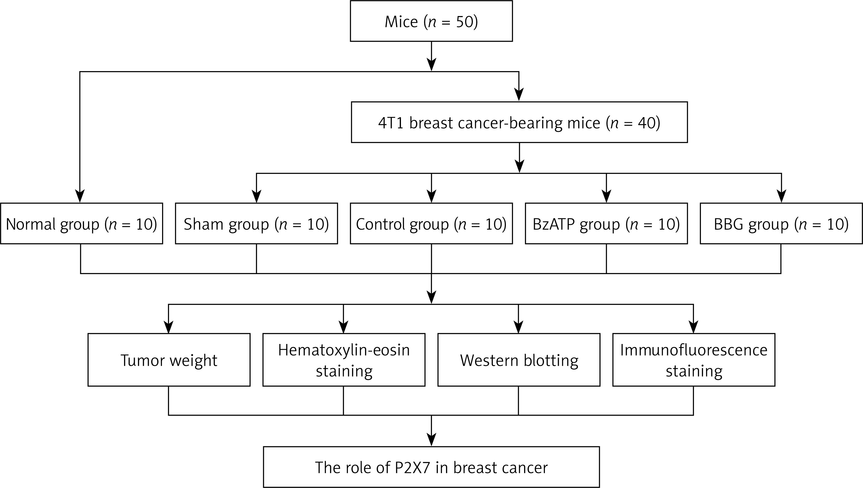
The experimental design is shown in Figure 1. Fifty Balb/C mice were randomly distributed to the normal group, sham group, control group, BzATP (P2X7 agonist) group, and BBG (P2X7 antagonist) group, with 10 mice in each group. The sham, control, BzATP, and BBG groups all comprised 4T1 breast cancer-bearing mice; the mice in the sham group received no intervention, the mice in the control group received 2 ml demineralised water injected intraperitoneally, and mice in the BzATP and BBG groups received BzATP and BBG intervention, respectively. BzATP (soluble in demineralised water, 50 µg/kg, 2 ml, Sigma-Aldrich, Shanghai, China) was intraperitoneally administered 1 h after 4T1 murine breast cancer cell injections, and BBG (soluble in demineralised water, 30 mg/kg, 2 ml, Sigma-Aldrich, Shanghai, China) was intraperitoneally administered [24]. All of the P2X7 agonist and antagonist were used in one dose, and animals were sacrificed 7 days after drug injection,
Tumour weight calculation
The food was inaccessible to mice 6 h before sacrifice. The mice were sacrificed by the method of cervical dislocation, then the tumour was carefully peeled off with delicate equipment in sterile conditions, and filter paper was used to absorb the related blood around the tumour whenever necessary. Finally, the tumour was weighed with an electric balance (S3080, Xinghe, Nanjing), and the related weight was recorded accordingly.
Haematoxylin and eosin (HE) staining
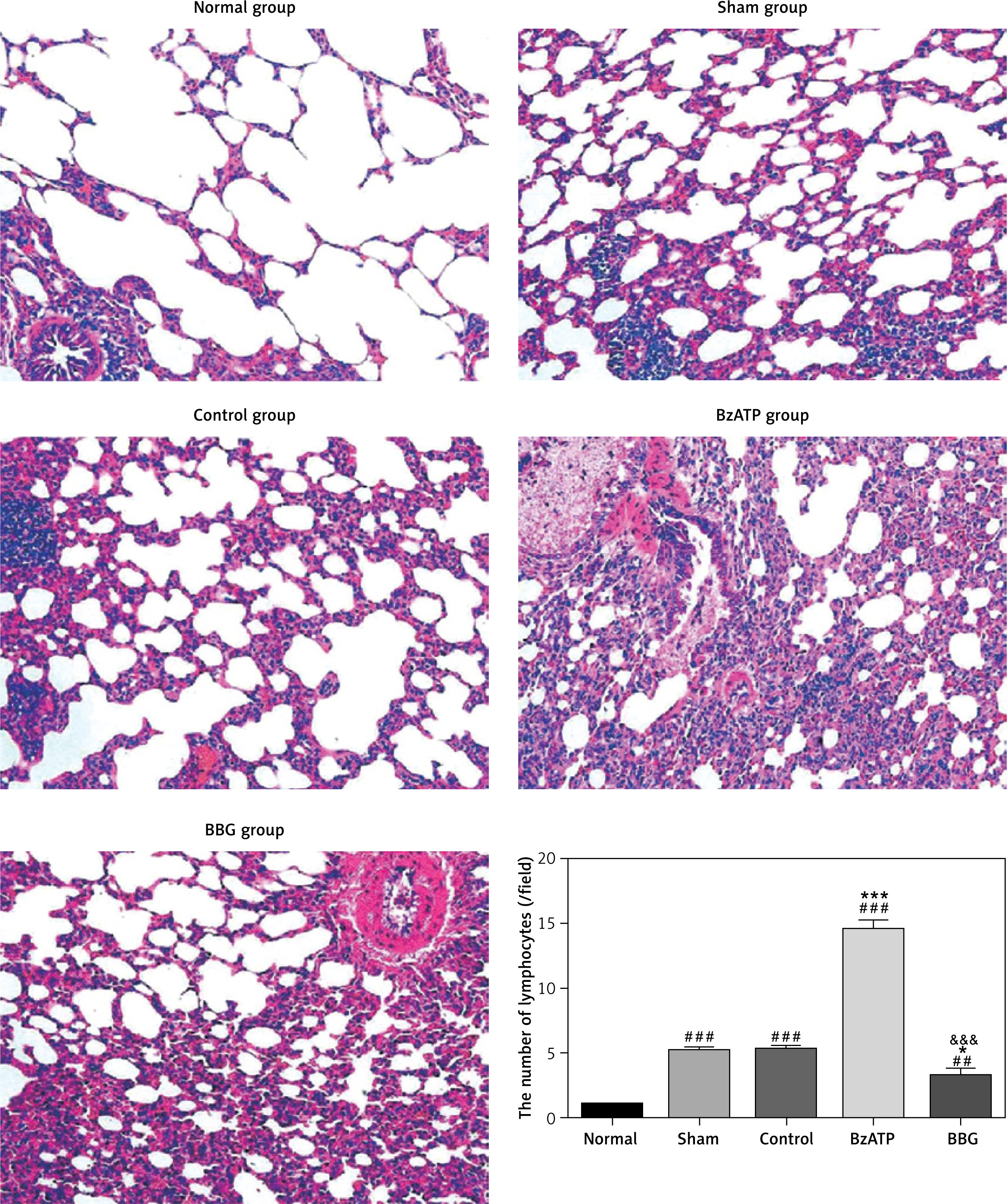
The cancer tissues were weighed and dehydrated, and then embedded in paraffin. Furthermore, the transverse section of collected tissues was discreetly sliced for HE staining, as previously reported [25]. The specimens then were photographed and analysed with a microscope (Olympus S100, Japan) at 40× magnification. For data analysis, we calculated and compared the number of lymphocytes at up to 10 pictures for each group.
Western blotting
The frozen tissue samples were lysed mechanically in cell lysis buffer for total protein extraction. Protein extraction kits (P0033, Beyotime, China) were used. The lysates were centrifuged at 12,000 rpm for 15 min at 4°C, and the concentration was measured by the bicinchoninic acid (BCA) method. The samples were further separated using related SDS-PAGE and were electro-transferred onto nitrocellulose membrane. Then, the membranes were blocked with 5% non-fat milk for 1 h at room temperature and were further incubated with primary antibodies at 4°C. In this present study, β-tublin (ab179513, Abcam, USA) was used as a loading control. The membranes were then washed three times for 5 min each in TBS + 0.1%Tween 20 and were then incubated in the appropriate secondary antibodies for 2 h at room temperature. Finally, the protein bands were visualised with the method of enhanced chemiluminescence, and the relative expression level of detected proteins was processed and analysed with Image J software. The antibodies used in this study included the following: rabbit anti-P2X7 (ab48871, Abcam, USA), rabbit anti-NLRP3 (ab214185, Abcam, USA), and rabbit anti-caspase 1 (ab238979, Abcam, USA).
Immunofluorescence staining
The immunofluorescence staining was conducted in concordance with the related guidelines [26]. Generally, the tumour sections were incubated with primary antibodies overnight at 4°C and were further covered with secondary antibodies (Jackson Immunoresearch, West Grove, PA) for 2 h at room temperature accordingly. We observed the subcellular localisation of proteins with a confocal laser scanning microscope (Olympus Z600, Japan).
Statistical analysis
We used SPSS22.0 software (SPSS Chicago, USA) for the statistical processing and analysis. All the collected data were expressed as mean ± standard deviation (SD) in the present study. We compared the differences between groups with one-way ANOVA method followed by a post hoc test (LSD). P < 0.05 was considered as being statistically significant in this study.
Results
Tumour weights
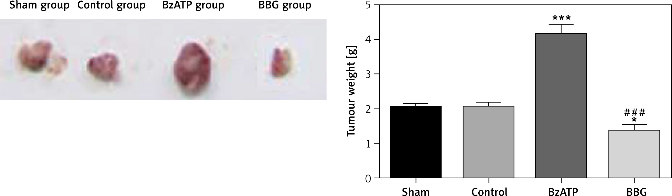
As can be seen in Figure 2, the tumour weight in the BzATP group was significantly higher than that of the sham and control groups, but the tumour weight in the BBG group was significantly lower than that of the sham, control, and BzATP groups.
HE staining analysis
As can be seen in Figure 3, for the normal group, the structure of breast tissue was complete and relatively independent. Meanwhile, for the other breast cancer group, the hyperplasia of mammary glands was obvious, and the structure was not clear. Furthermore, the medullary lymphocytes in the BzATP group were significantly higher than those of the other groups, but the medullary lymphocytes in the BBG group were significantly lower than those of the sham, control, and BzATP groups.
Immunofluorescence staining analysis
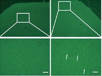
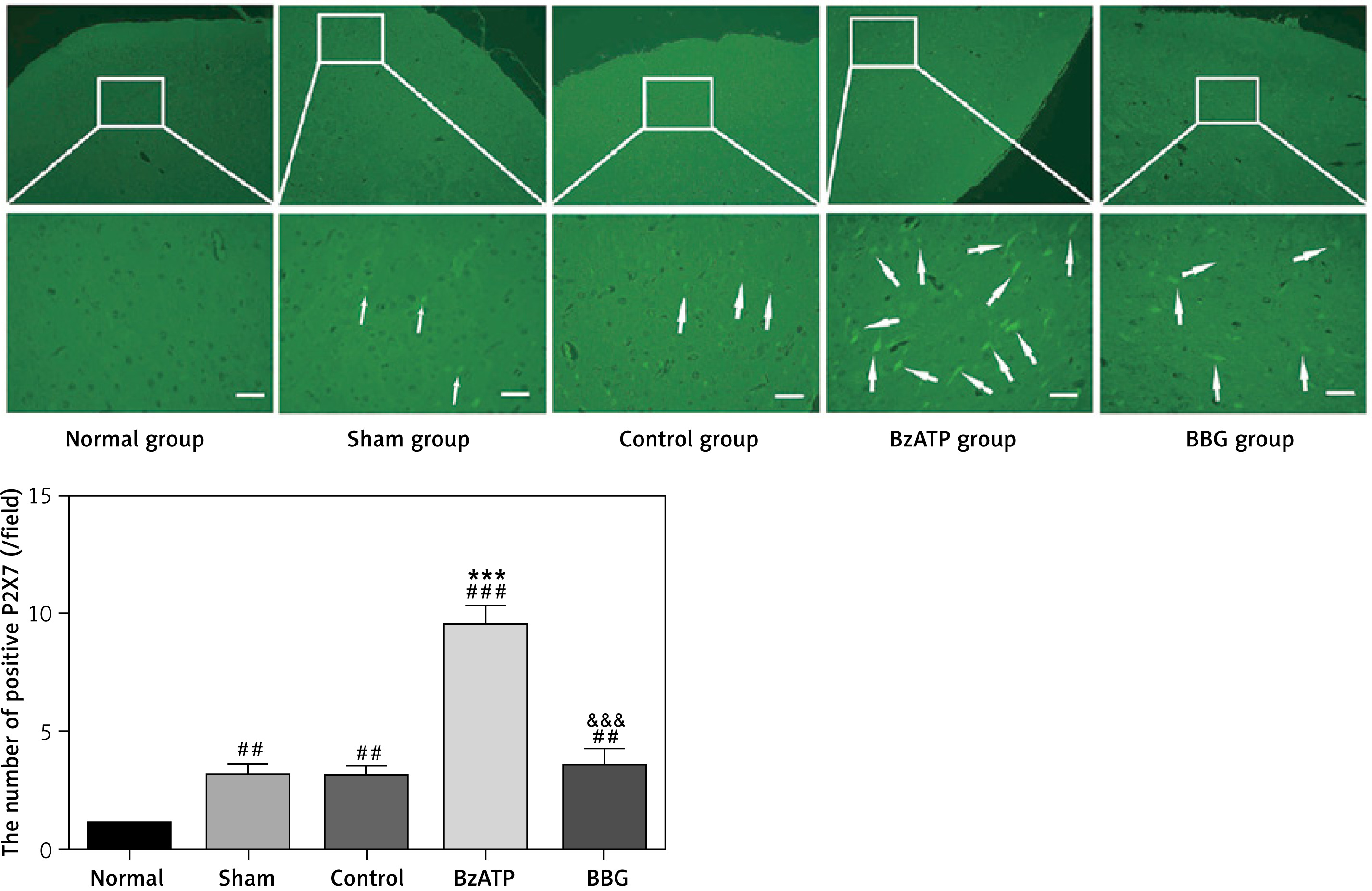
As can be seen in Figure 4, the immunofluorescence staining indicated that the number of positive P2X7 was significantly increased for breast cancer-bearing groups, and the number of positive P2X7 in the BzATP group was significantly higher than that of the other groups, but the BBG significantly reduced the number of P2X7.
Western blot analysis of expression of P2X7
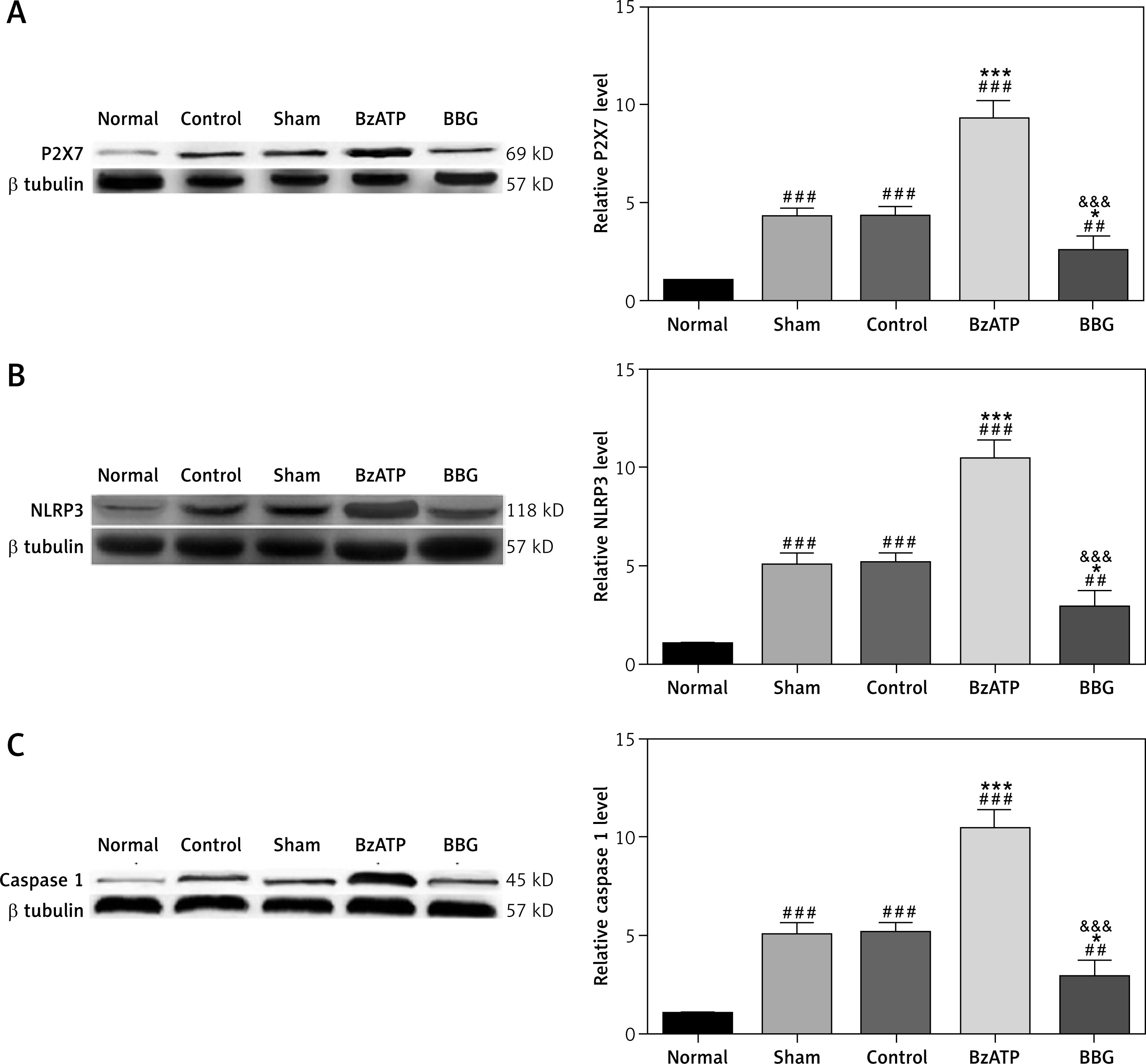
As can be seen in Figure 5 A, the relative expression level of P2X7 was significantly increased for breast cancer-bearing groups, and the relative expression level of P2X7 in the BzATP group was significantly higher than that of other groups, yet the relative expression level of P2X7 in the BBG group was significantly lower than that of other breast cancer-bearing groups.
Western blot analysis of the expression of NLRP3 and caspase 1
As can be seen in Figures 5 B and C, the relative expression level of NLRP3 and caspase 1 were significantly increased for breast cancer-bearing groups, and the relative expression levels of NLRP3 and caspase 1 in the BzATP group were significantly higher than those of other groups, but the relative expression levels of NLRP3 and caspase 1 in the BBG group were significantly lower than those of other breast cancer-bearing groups.
Discussion
With the progression of research, it has been found that abnormal expression of P2X7 is related to the occurrence and development of various cancers, and it may even become a new target for cancer treatment and prognosis [14, 27]. Therefore, it is necessary to conduct more related studies to elucidate the mechanism of P2X7 on the development and treatment of breast cancer. The results of the present study confirm that P2X7 plays an important role in the development of breast cancer, downregulating P2X7 can inhibit the growth of breast cancer, and that the P2X7/NLRP3/caspase-1 signalling pathway is closely involved in this process.
The P2X7 receptor is a member of the P2X family, with unique structure and function. Its biological functions include participating in cell signal transduction, cytokine secretion, and mediating cell survival and growth [28, 29]. The production of high concentrations of ATP after tissue damage activates the P2X7, causing the transient opening of cell membrane pathways and leading to an influx of calcium ions. Subsequently it catalyses the hydrolysis of phosphatidylcholine to produce a second messenger phospholipid (PA) [30]. It further activates the Src kinase family (SFKs), hydrolyses protein precursors, and forms ligands, which then bind to VEGFR, which in turn promotes the growth of tissues [31]. Also, the binding of ATP to the P2X7 receptor induces the formation of membrane pores of lysed cells, which can lead to cellular Ca2+-independent cell necrosis [24, 32]. On the other hand, continuous ATP stimulation activates P2X7 to trigger excessive Ca2+ to enter cells and induce apoptosis [33]. Therefore, P2X7 is closely associated with the development of cancers.
Breast cancer is a malignant cancer with high incidence. Previous studies [12, 34] found that the expression of P2X7 in normal, mildly proliferative breast epithelium is almost absent, while P2X7 receptors are overexpressed in the ductal or lobular carcinoma. Also, it has been reported that the ATP concentration in the tumour stroma is significantly higher than that of normal tissues [35]. Therefore, P2X7 is likely to have a pro-proliferative effect in the development of breast cancer. At the same time, the expression of P2X7 and oestrogen receptor (OR) in breast cancer tissues has a positive correlation [17]. The expression of P2X7R is abnormal in human MDA-MB-435s breast cancer cells and A549 lung adenocarcinoma cells, and the application of the P2X7R inhibitor KN-62 can block ATP-induced tumour cell migration and invasion [36], which is consistent with our findings.
Inflammatory response is a key factor in tumourigenesis and development, and its secreted multiple inflammatory factors can mediate the emergence of the tumour microenvironment. Also, it plays an important role in initiating, maintaining, and promoting tumour growth [37]. Of these, NLRP3 influences the pathogenesis of cancer by modulating innate and adaptive immune responses, cell death, proliferation, and gut microbiota [38]. NLRP3 can be assembled with the adaptor protein ASC and caspase 1 to form a multi-protein complex called an inflammasome. The assembly of inflammasome leads to the activation of caspase 1 and the maturation and secretion of pro-inflammatory cytokines such as IL-18, thus exerting a variety of immunological effects [39, 40]. The results of our studies confirm that NLRP3 plays an important role in the progression of breast cancers.
Several limitations in this study must be considered. Firstly, we only performed a primary investigation on the role of P2X7/NLRP3/caspase 1 pathway in breast cancers. The ERK and JNK pathways should be further investigated in more detail because they are also a part of this signalling pathway [41]. The underlying mechanisms of how these synergistic effects work remain unclear, which also warrants further investigation. Secondly, activation of P2X7 leads to calcium mobilisation, which may be the mechanism link of P2X7 response to cells. We did not measure intracellular calcium mobilisation in this study; future studies focusing on calcium mobilisation measurement will help us confirm the acting mechanism of P2X7 in breast cancer. Thirdly, studies should be conducted to confirm the role of P2X7 in breast cancers; we will verify this in our future studies.
In conclusion, P2X7 plays an important role in the development of breast cancer; blocking P2X7 can inhibit the growth of breast cancer, and it is associated with NLRP3/caspase 1 pathway. It is worth noting that P2X7 activation involves the activation of multiple different intracellular effector pathways, resulting in complex biological effects, and its application to clinical treatment has a long way to go – further explorations on the mechanisms of the antitumour effect of P2X7 are needed.

