

Introduction
Acute coronary syndrome (ACS) is reported to be one of the most important causes of mortality and morbidity worldwide [1]. It is widely accepted that plaque rupture or erosion followed by obstructive thrombus formation is the main feature of ACS. Based on quantitative analysis of coronary angiography, it is evident that diverse parameters, including the presence of coronary plaque and the degree of luminal stenosis, carry incremental predictive value and improve risk stratification of ACS [2]. On the other hand, intravascular optical coherence tomography (OCT) is considered to generate high-resolution cross-sectional images of plaque morphology, allowing physicians to visualize the development of the distinct vulnerable plaques characterized by a rupture-prone thin fibrous cap and a lipid-rich necrotic core [3]. In addition, thin cap fibrous atheroma (TCFA) is more likely to have higher numbers of pro-inflammatory macrophages, with punctate signal-rich spots exceeding the background noise on OCT images [4].
Suppression of tumorigenicity 2 (ST2), a member of the interleukin 1 receptor (IL-1R) family, constitutes two primary isoforms regulated by IL-33 [5, 6]. The transmembrane ST2 receptor (ST2L) consists of a membrane-bound isoform with 3 extracellular IgG domains, while a soluble ST2 receptor (sST2), lacking transmembrane and intracellular domains, acts as an IL-33 decoy receptor and could be detected in circulating serum [7]. Growing evidence has expanded our knowledge about the biology function of IL-33/ST2 signaling and the pathogenesis of IL-33/ST2 in tumor development and cardiovascular abnormality [8, 9]. Depletion of ST2 in a mouse pressure overload model results in excessive ventricular hypertrophy, chamber dilation, and cardiac fibrosis [10]. Moreover, increased serum sST2 predicts poor outcomes in patients with ACS and chronic heart failure [6, 11–14]. By contrast, ST2L, predominantly expressed in macrophages, activates Th2 cells and mast cells, retards atherosclerotic development and alleviates cardiac remodeling through interaction of IL-33 and enhancement of its intracellular signaling [15]. However, the association of ST2L, especially ST2L expression on monocyte subsets, with plaque characteristics remains unknown. Therefore, we aimed to determine the association of sST2 and ST2L expression on monocyte subsets with the severity and vulnerability of coronary plaques in this study.
Material and methods
The data that support the findings of this study are available from the corresponding author upon reasonable request.
Study population
Forty-eight patients with unstable angina pectoris (UAP) and 31 patients with ST-elevation myocardial infarction (STEMI) admitted to Shanghai Tenth People’s Hospital between April 2013 and July 2014 were recruited as previously described [16]. UAP was diagnosed based on the manifestation of angina at rest, accelerated chest pain, or new-onset chest pain without any abnormality of cardiac markers. STEMI was diagnosed according to the criteria of the Fourth Universal Definition of Myocardial Infarction [17] and further confirmed by coronary angiography, with the presence of acute thrombosis secondary to plaque rupture. Thirty-three age- and sex-matched subjects with suspected angina but free of luminal diameter narrowing ≥ 50% confirmed by angiography served as controls. Patients with severe liver and kidney failure, acute infectious disease, and malignancies were excluded. All patients provided written informed consent before enrollment. The study was approved by Shanghai Tenth People’s Hospital and followed the guidelines of the Declaration of Helsinki.
Quantification of plaque characteristics by angiography and optical coherence tomography
Coronary angiography and optical coherence tomography (OCT) were performed as previously described [16]. We defined the severity of ACS by evaluating the severity of coronary plaques obtained from coronary angiography. The severity of coronary plaques was evaluated by the length of plaque (> 20 mm as long lesions), the degree of stenosis (moderate [from 50% to 69%]; severe [from 70% to 99%], or occlusion [100%]) with or without calcification, and the number of coronary vessels with significant stenosis. Left main trunk stenosis was considered as a two-vessel disease. TCFA was defined as a plaque presenting lipid content for > 90°, and with thinnest part of the fibrous cap measuring < 65 μm obtained from OCT [17, 18].
Analysis of serum sST2
Approximately 5 ml of peripheral venous blood was collected from controls and UAP patients on the day after fasting for over 8 h. Peripheral venous blood from patients with STEMI was collected and placed in EDTA-coated tubes on the day of admission. After centrifugation, serum was stored at –80°C for further analysis in batches. Serum levels of sST2 were measured by sandwich enzyme immunoassay using Human ST2/IL-33R Quantikine ELISA kits (catalog #DST200, R&D System, USA) according to the manufacturer’s protocol. The coefficients of variation (CV) for intra-assay precision and inter-assay precision were less than 5.2% and 6.8%, respectively.
Quantification of ST2L on monocyte subsets by flow cytometry
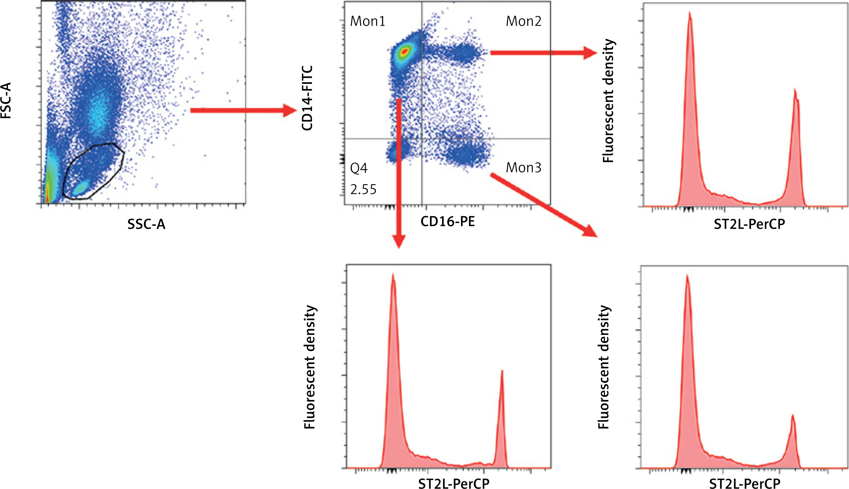
For measurement of surface ST2L expression on monocytes, 100 μl of peripheral blood was blocked with 1% bovine serum albumin (BSA, Gibco, USA) solution for 15 min at 4°C and stained with a cocktail of monocyte antibodies for 30 min at 4°C. The information and concentrations of each monocyte subset are shown in Supplementary Table SI. On the basis of flow cytometry, circulating monocytes were divided into CD14++CD16- monocytes (Mon1), CD14++CD16+ monocytes (Mon2), and CD14+CD16+ monocytes (Mon3). PerCP-eFluor710-conjugated antibodies against IL-33R(ST2) were used to analyze the expression of ST2 receptors on monocytes (Figure 1). The flow cytometry for identifying surface ST2L expression and monocyte subsets was performed by one expert technician who was blinded to the coronary angiography and OCT results [16].
Figure 1
Gating strategy for circulating monocyte subsets and ST2L expression. Whole peripheral blood cells were prepared and labeled with various antibodies. CD14 was used in conjunction with CD16 to identify monocyte subsets (Mon). Mon1 was identified as CD14++CD16-, Mon2 as CD14++CD16+, and Mon3 as CD14+CD16+. ST2L antibodies were further used to stratify the percentage of ST2L positive monocyte subsets
Induction of macrophage polarization
Animal studies were conducted in accordance with the Guide for the Care and Use of Laboratory Animals (U.S. National Institutes, MD, USA), and all the procedures were approved by the Animal Care and Use Committee of Shanghai Tenth People’s Hospital. Bone marrow cells were isolated from femurs of C57BL/6 mice following standard protocol in Dulbecco’s modified Eagle’s medium (DMEM, Gibco, USA) containing 10% fetal bovine serum (FBS, Gibco, USA) and 1% penicillin and streptomycin [19]. Following separation, bone marrow-derived macrophages (BMDMs) were cultured in 10% FBS/DMEM with recombinant M-CSF at a concentration of 100 ng/ml (Peprotech Incorporation, USA) for 3 days (M0 phenotype). BMDMs were washed and treated for 12 h with DMEM + 5% FBS alone, LPS at a concentration of 100 ng/ml, and recombinant interferon gamma (IFN-γ, Peprotech Incorporation, USA) at a concentration of 20 ng/ml for polarization to the M1 phenotype, or recombinant IL-4 at a concentration of 20 ng/ml (Peprotech Incorporation, USA) for polarization to the M2 phenotype. Likewise, human circulating monocytes were collected from healthy volunteers, differentiated into macrophages and maintained RPMI 1640 medium (Gibco, USA) supplemented with 10% FBS according to previous protocols [20].
Quantitative RT-PCR
Total cellular RNA was extracted and purified using TRIzol reagent (Invitrogen, USA). Reverse transcription was performed using the Transcriptor First Strand cDNA Synthesis Kit (Roche, Germany). Quantitative RT-PCR for ST2L, TNF-α, IL-6, CD206, and Arg1 was performed using FastStart Universal SYBR Green Master Mix (Roche, Germany) on a Roche LightCycler instrument. The primer sequences used for quantitative RT-PCR are tabulated in Supplementary Table SII.
Immunoblotting
Protein lysates were performed on 8% SDS-PAGE gels and transferred to membranes as our previously reported [21]. The membranes were probed using antibody directed against ST2L (Abcam, USA) and normalized to GAPDH (Santa Cruz, USA).
Statistical analysis
Continuous variables were presented as mean ± standard deviation (SD). Categorical variables were presented as absolute number and proportion. Differences between two groups were analyzed with Student’s t test. Differences between multiple groups were initially compared using oneway ANOVA, and posthoc tests were then performed with Bonferroni correction. Receiver-operating characteristic (ROC) curve analysis was conducted to determine the performance of serum sST2 and ST2L expression on monocytes for assessing plaque vulnerability. Youden’s index was applied to derive the best cut-off value of ST2L expression on monocytes. Based on the cut-off, we then performed backward stepwise multivariate logistic regression that included variables with p < 0.1 in univariate analysis to identify independent predictors of plaque vulnerability. Values of p < 0.05 were considered significant.
Results
Comparison of serum sST2 and ST2L on monocytes among patients with different severity of ACS
The patient characteristics, angiographic findings, and results of OCT imaging were summarized in our previous study [16]. Of the whole measurements, the proportions and the absolute number of ST2L on monocytes and Mon2 subsets in STEMI patients were significantly lower than those in UAP patients and controls (Table I). Consistent with other research, serum levels of sST2 showed a steady increase from controls to STEMI patients (control 1.35 ±0.65 ng/ml, UAP 2.42 ±0.61 ng/ml and STEMI 3.88 ±1.61 ng/ml, p < 0.001).
Table I
Comparison of serum sST2 levels and ST2L expression on monocytes among the three groups
[i] Data are presented as mean ± SD. P (group 1 vs. 2) indicates the difference between Control and UAP groups after post-hoc Bonferroni comparison. P (group 2 vs. 3) indicates the difference between UAP and STEMI groups after post-hoc Bonferroni comparison. UAP – unstable angina pectoris, STEMI – ST-elevation myocardial infarction, ST2 – suppression of tumorigenicity 2, ST2L – transmembrane ST2 isoform, Mon – monocytes.
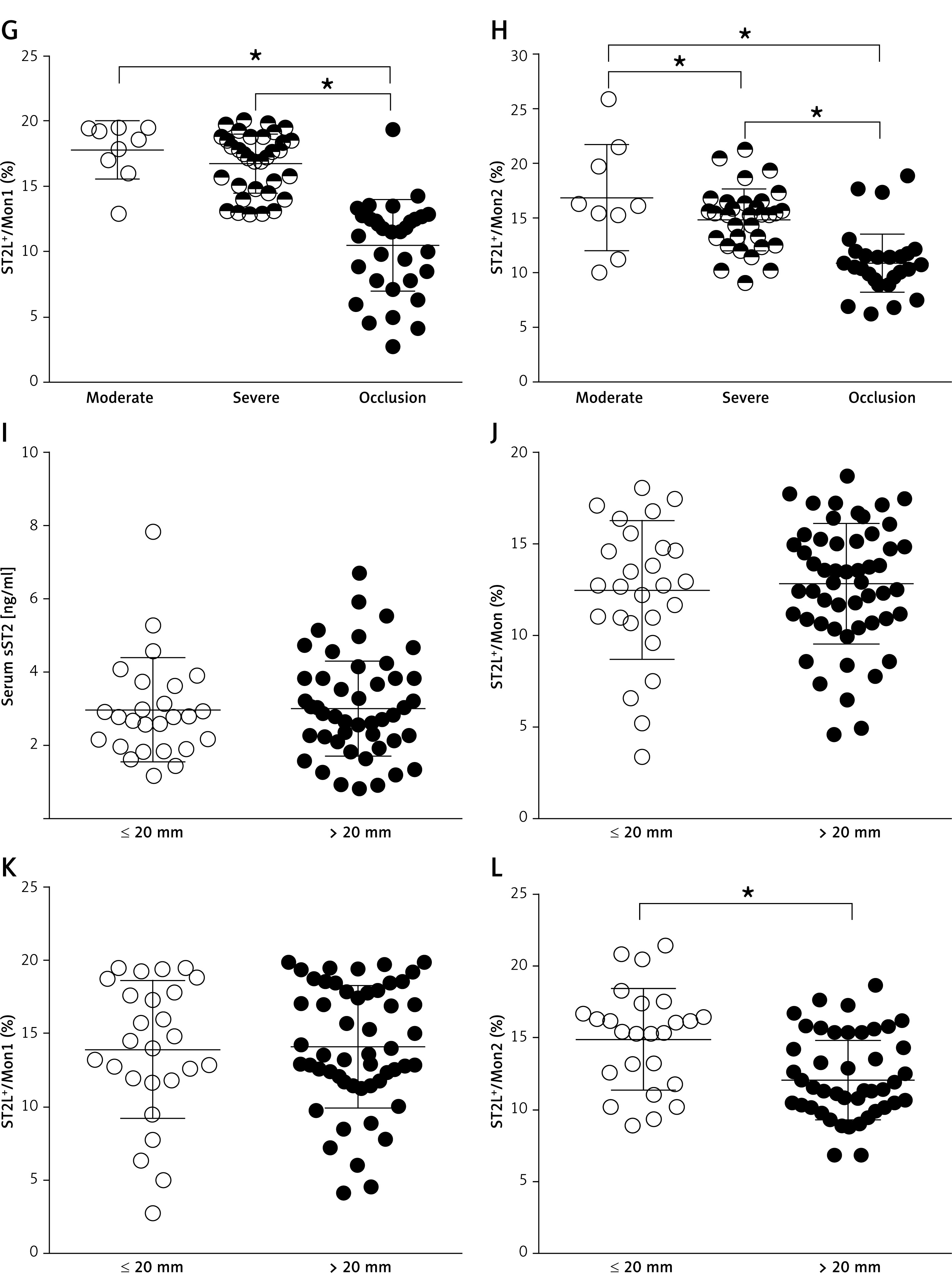
Based on quantitative analysis of coronary angiography, it is evident that diverse parameters, including the presence of coronary plaque and the degree of luminal stenosis, carry incremental predictive value and improve risk stratification of ACS [2]. We next assessed the association of serum sST2 and ST2L expression on monocyte subsets with the severity of ACS as detected by angiography. No significant association was found between serum sST2 and the atherosclerotic severity of ACS (Figures 2 A, E, I). ACS patients with lower proportions of ST2L+/Mon and ST2L+/Mon1 were more likely than those with higher proportions to have obstructive stenosis (Figures 2 B, F, J). It should be noted that the percentage of ST2L+/Mon2 was significantly inversely correlated with all three angiographic parameters (Figures 2 D, H, L).
Figure 2
Comparison of serum sST2 and the percentage of ST2L expression on monocytes in patients with different severities of coronary plaques detected by angiography. A–D – Comparison of serum sST2 and the percentage of ST2L expression on monocytes according to the number of stenotic vessels. E–F – Comparison of serum ST2 and the percentage of ST2L expression on monocytes according to the degree of stenosis. G–H – Comparison of serum ST2 and the percentage of ST2L expression on monocytes according to the degree of stenosis. I–L – Comparison of serum ST2 and the percentage of ST2L expression on monocytes according to the lesion length
ST2 = suppression of tumorigenicity 2, ST2L = transmembrane ST2 isoform, Mon = monocyte. *P < 0.05.
Relation of serum sST2 and ST2L expression on monocyte subsets to plaque stability
Plaque characteristics determined by OCT were summarized in our previous study. Of the OCT subgroup, patients with TCFA had a lower absolute number (non-TCFA 1.26 × 10⁴ ±0.63 × 10⁴/ml vs. TCFA 0.92 × 10⁴ ±0.37 × 10⁴/ml, p = 0.034) and lower percentage (non-TCFA 17.65% ±3.10% vs. TCFA 13.17% ±2.29%, p < 0.001) of ST2L+/Mon2 than those without TCFA (Table II). However, there was no difference in serum levels of sST2 between groups (Table II). We then analyzed the association of serum sST2 and ST2L expression on monocytes with plaque morphology, and found that serum sST2 was positively correlated with fibrous cap thickness (Pearson r = 0.306, p = 0.034), while no differences in serum sST2 levels according to lipid core arc and minimum lumen area were observed (Table III). In contrast, the percentage of ST2L+/Mon2 gradually increased with increasing fibrous cap thickness (Pearson r = 0.523, p < 0.001), whilst there was an inverse correlation between percentage of ST2L+/Mon2 and lipid core arc (Pearson r = –0.473, p = 0.001).
Table II
Comparison of serum sST2 and ST2L expression on monocytes in patients with and without TCFA
[i] Data are presented as mean ± SD. TCFA – thin cap fibrous atheroma. Other abbreviations listed as in Table I.
Table III
Correlation of serum sST2 levels and ST2L expression on monocytes with plaque morphology
[i] MLA – minimum lumen area. Other abbreviations listed as in Table I.
ST2L on Mon2 as determinants of plaque vulnerability
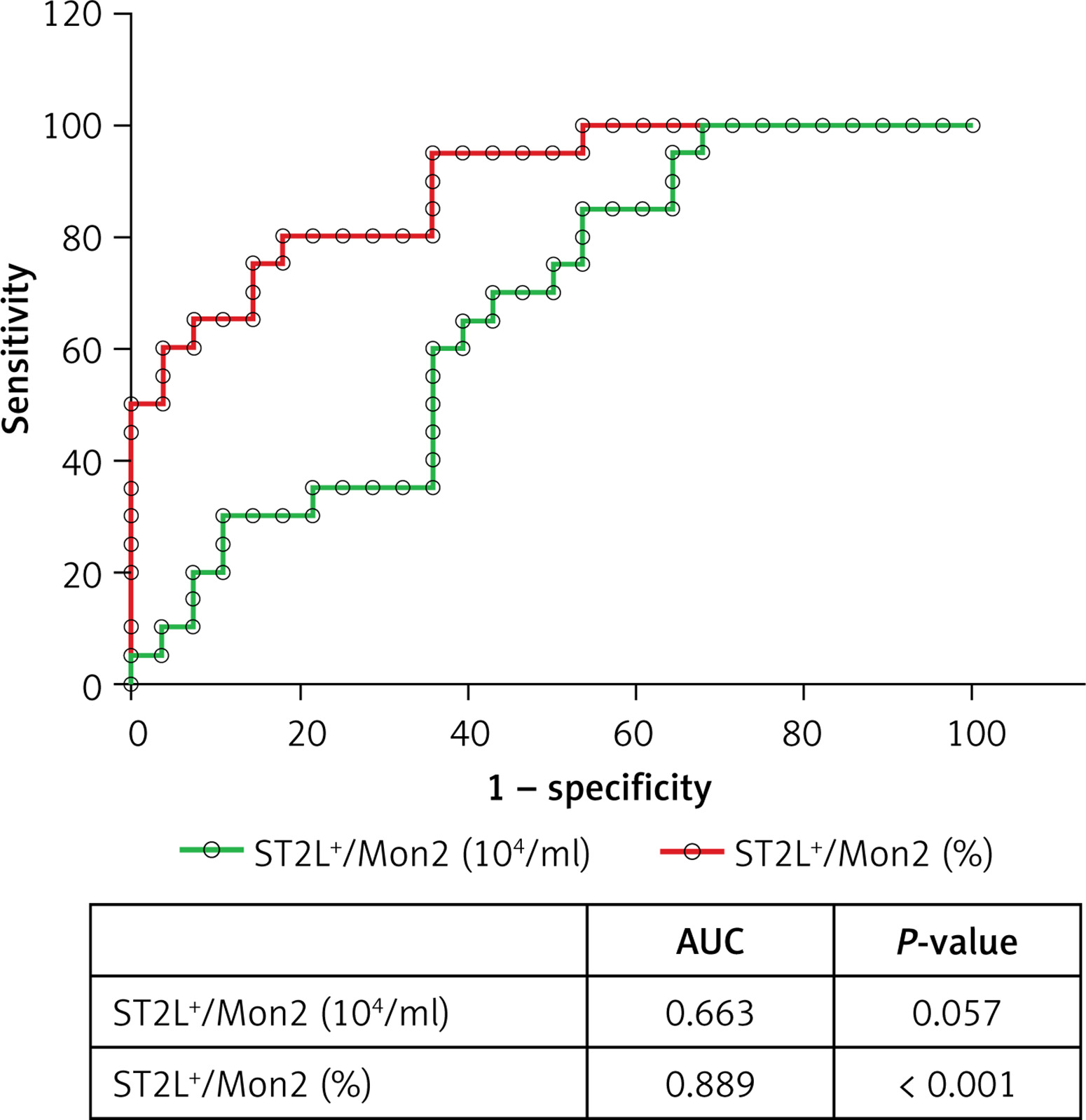
The area under the ROC curve of percentage and absolute number of ST2L+/Mon2 for determining TCFA was 0.889 and 0.663, respectively (Figure 3). The optical percentage of ST2L+/Mon2 cutoff to predict TCFA was 15.31%, which showed sensitivity of 82.41 and specificity of 85.71 (Figure 3).
Figure 3
Receiver-operating characteristics (ROC) curve analysis showing the discriminative ability of the absolute number and percentage of ST2L+/ Mon2 for thin cap fibrous atheroma (TCFA)
AUC – area under the curve.
After correction for known clinical risk factors including age, sex, smoking, hypertension, diabetes mellitus, white blood cells (WBC), low-density lipoprotein cholesterol (LDL-C), and the percentage of circulating Mon2 subsets, decreased levels of percentage of ST2L+/Mon2 remained an independent predictor of TCFA (OR = 0.07, 95% CI: 0.02–0.29, p < 0.001, Table IV).
Table IV
Multivariate logistic regression analysis for predictors of TCFA
[i] WBC, LDL-C, HDL-C were defined as continuous variables. P-values were calculated by Wald type test. WBC – white blood cells, HDL-C – high-density lipoprotein cholesterol, LDL-C – low-density lipoprotein cholesterol, OR – odds ratio, other abbreviations listed as in Table I.
ST2L was primarily expressed in M2 macrophages
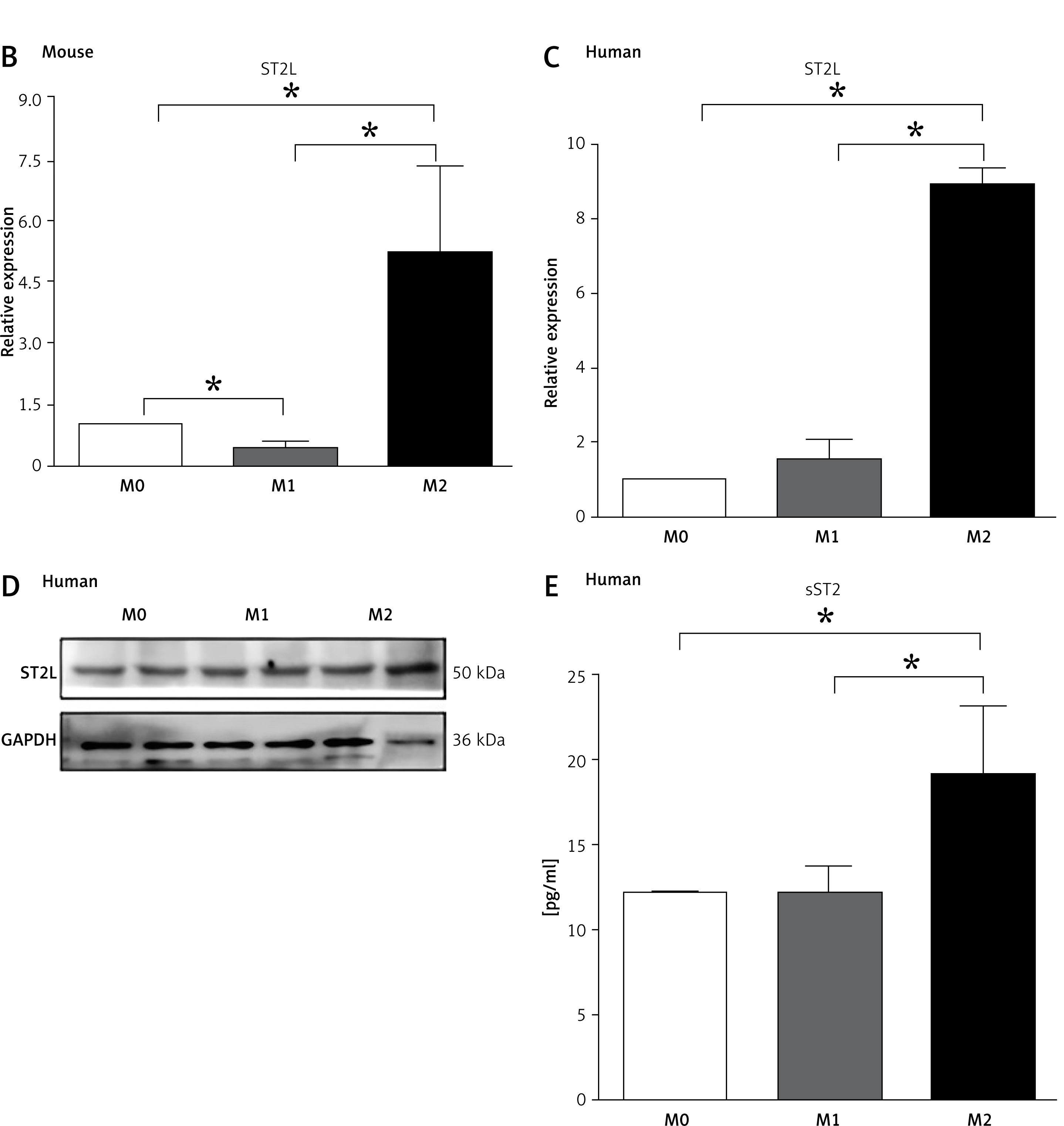
It is well known that with the development of atherosclerosis, BM-derived circulating monocytes possess the capacity to differentiate into two macrophage subsets based on their pathologic function and expressions of divergent cytokines [22]. To examine the abundance of ST2L on the macrophage subsets, we induced BMDMs and human macrophages by different stimuli, and confirmed the results by quantitative PCR and immunoblots (Figure 4 A). As compared to M1 macrophages, both ST2L mRNA and protein expression levels were higher in M2 polarized macrophages, suggesting the potential involvement of ST2L in regulating plaque instability (Figures 4 B–D). Interestingly, we found that the concentration of sST2 in the supernatant of M2 macrophages was higher than that of M1 macrophages differentiated from human circulating monocytes (Figure 4 E).
Figure 4
ST2L accumulates in M2 macrophages. A – Quantification of M1 and M2 markers in BM-derived macrophages. Macrophages were induced by LPS (100 ng/ml) and IFN-γ (20 ng/ml) for M1 polarization or by IL-4 (20 ng/ml) for M2 polarization. B, C – Quantitative PCR indicated that ST2L was upregulated in M2 polarized phenotype of mouse bone marrow-derived macrophages (B) and human macrophages (C). D – Immunoblotting showed the protein expression of ST2L in M0, M1, and M2 macrophages differentiated from human circulating monocytes. E – Comparison of the sST2 levels between the supernatant of M1 and M2 macrophages differentiated from human circulating monocytes. *P < 0.05
Discussion
To the best of our knowledge, we demonstrated for the first time that ST2L expression on Mon2, particularly the percentage of ST2L+/Mon2 among circulating leukocytes, was closely associated with the incidence and severity of ACS. The percentage of ST2L+/Mon2, rather than serum sST2, appeared to be a more suitable biomarker reflecting the signature of plaque destabilization detected by OCT.
It has been reported that IL-33 was predominantly produced by vascular smooth muscle cells and endothelial cells in response to atherogenic stimuli [8]. Likewise, cardiac hypertrophy and mechanical strain substantially induced IL-33 expression, which was predominantly observed in cardiac fibroblasts [10]. Mechanistically, through neutralization of IL-33, sST2 abrogated the cardioprotective effect of IL-33/ST2 signaling in cardiac fibrosis and hypertrophy [11]. While sST2 acted as a circulating isoform and was ubiquitously expressed in various tissues, the transmembrane isoform ST2L was inducible and primarily located in hematopoietic cells, especially Th2 cells, mast cells, monocytes, and macrophages [23]. In the present study, we found that ST2L was highly expressed in M2 macrophages, suggesting that ST2L might be involved in M2 macrophage activation, which contributes to angiogenesis and anti-inflammation. Consistently, a previous research observed that depletion of ST2L disrupts macrophage polarization to M2 phenotype with decreased M2 macrophage markers [15]. In parallel, the supernatant of M2 macrophages had a higher concentration of sST2 than that of M0 and M1 macrophages.
Weinberg et al. [6], for the first time, revealed that elevated serum sST2 predicted higher likelihood of 30-day mortality and heart failure in patients with myocardial infarction. Likewise, while the no-reflow phenomenon reflects impaired myocardial perfusion and poor prognosis [24], including mortality and heart failure, in ACS patients after percutaneous coronary intervention, Somuncu et al. [25] identified serum sST2 as an independent predictor of the no-reflow phenomenon in ACS patients. A recent meta-analysis concluded that ACS patients with higher baseline levels of serum sST2 predicted the increased possibility of all-cause mortality and major adverse cardiac events during follow-up [13]. In parallel, our study confirmed that serum sST2 level was higher in ACS patients than in controls. Nevertheless, there was no significant difference in serum sST2 between UAP patients with and without TCFA, suggesting that serum sST2 may not be considered as a predictor of plaque destabilization.
Nonetheless, little is understood about the association of ST2L with the incidence of ACS and plaque vulnerability. Circulating monocytes extravasate through the endothelium and differentiate into two opposite macrophage subsets, initiating atherosclerosis and regulating plaque stabilization [26]. To be more specific, CD14++CD16- Mon1 subsets and CD14++CD16+ Mon2 subsets more readily invade the subendothelium and differentiate into active M1 macrophages, which subsequently contribute to necrotic core formation, matrix degradation, and plaque rupture. Others’ and our previous studies have shown that CD14++CD16+ Mon2 subsets were closely associated with the incidence of ACS [16, 27]. Furthermore, higher proportions of CD14++CD16+ Mon2 subsets were more often found in patients with TCFA [16]. Therefore, together with the involvement of ST2L in macrophage polarization, these data prompted us to further investigate whether ST2L on monocyte subsets could be considered as a potential biomarker for ACS and plaque instability. In this regard, we first observed that ST2L was highly expressed in Mon1 and Mon2 subsets and lower in Mon3 subsets, suggesting that ST2L mainly functioned in pro-inflammatory monocytes which subsequently polarized to M1 macrophages and contributed to plaque development and rupture. Our study further provides evidence that a higher percentage of ST2L+/Mon2 is associated with ACS. On the other hand, numerous clinical trials have proposed that integrated measurements of plaque composition, location, and extent based on coronary computed tomography angiography or invasive coronary angiography are capable of accurate risk stratification and prognosis for ACS patients [2, 28]. In this regard, only the proportion of ST2L+/Mon2 was prevalently lower in patients with high-risk plaque features, indicating that the proportion of ST2L+/Mon2 might represent a promising biomarker for risk stratification of ACS.
Given the high image resolution with OCT, it allows physicians to identify high-risk plaques in vivo. We found that, in ACS patients, a higher percentage of ST2L+/Mon2 indicated a thinner fibrous cap accompanied by a larger extent of lipid core, both of which serve as early features of plaque instability [29]. Combined with ROC curve and multivariate analysis, we further highlighted the superiority of the percentage of ST2L+/Mon2 over other clinical factors in predicting vulnerable plaques. Based on the results of our previous study, the circulating Mon2 subset was considered as an independent predictor of the incidence of definite TCFA. It is therefore plausible to assume that ST2L, especially ST2L expression on the Mon2 subset, through its diverse pathogenic roles and clinical significances in atherosclerosis and myocardial infarction, may further improve the prediction of plaque vulnerability.
Finally, we acknowledge some potential limitations in our research. In the current study, we did not investigate the distribution of ST2L on Th2 in ACS patients. It has been demonstrated that accumulation of pro-inflammatory macrophages in infarcted myocardium and phagocytosis of necrotic cardiomyocytes by macrophages are hallmarks of the early phase of myocardial infarction [30]. While Th1 subsets seem more likely to affect the evolution of ACS, the absolute number of circulating Th2 subsets is relatively low, and there is no prominent difference in circulating Th2 subsets between patients with stable angina and ACS [31]. Despite this, a comprehensive assessment of ST2L on Th2 cells may boost the understanding of IL-33/ST2 signaling in atherosclerotic development. Particularly, a larger number of patients in another external validation cohort is required to strengthen our findings. In multivariate logistic regression analysis for predictors of TCFA, several well-known risk factors including diabetes, WBC, and LDL-C did not show significant effects on TCFA, as reported in previous studies [32–34], which may due to the small size of our study. Moreover, accumulating evidence in the functional characterization of macrophages has revealed that the phenotypes are not limited to the M1 and M2 extremes but rather represent a continuous spectrum of phenotypes associated with differential cytokine production and functional characteristics [35]. Given the difficulty in simulating the environment of macrophages in plaques in vitro, we adopted the concepts of M1 and M2 in this study, with M1 primarily representing macrophages showing a proinflammatory phenotype and M2 representing macrophages with an anti-inflammatory phenotype.
In conclusion, an enhanced proportion of ST2L+/Mon2 was associated with the severity of ACS and plaque vulnerability in coronary arteries. ST2L+/Mon2 might represent a promising biomarker for the early diagnosis of vulnerable plaques.

