
Introduction
Despite rapid advances in breast cancer diagnosis and prognosis, it is still the most frequent cancer among women globally [1]. The incidence of breast cancer continues to grow across the globe. This makes it important to search for novel therapeutic approaches. The treatment of breast cancer is multidisciplinary [2], and radiation therapy is still one of the widely used cancer treatments [3]. Radiation therapy is carried out on approximately 60% of patients as a stand-alone treatment for cancer or in addition to chemotherapy and/or surgery [4]. However, the main barrier to radiation therapy’s optimal effectiveness is still radioresistance of breast cancer cells [5], which leads to metastasis, cancer recurrence, and a poor prognosis [6]. The primary mechanism of radiation-induced cell death is assumed to be apoptosis, and radiotherapy resistance results from disruption of apoptosis signaling [7]. Activation of anti-apoptotic mechanisms is one of the strategies to avoid apoptosis, causing uncontrolled growth, tumor survival, treatment resistance, and cancer recurrence [8]. Normally, radiation-resistant cancer cells control the BCL family interaction network to prevent apoptosis [6]. The BCL-2 protein family is crucial for regulating the intrinsic apoptotic pathway. It consists of pro- and anti-apoptotic proteins with one to four BCL-2 homology domains (BH1–BH4). BCL-2 and BCL-XL are the most well-known anti-apoptotic multidomain (BH1–BH4) members. They work to inhibit the pro-apoptotic multidomain proteins BAX and BAK’s ability to generate pores, which permeabilize the outer membrane of mitochondria. Thus, overexpression of anti-apoptotic proteins is one method cancer cells use to avoid apoptosis [9].
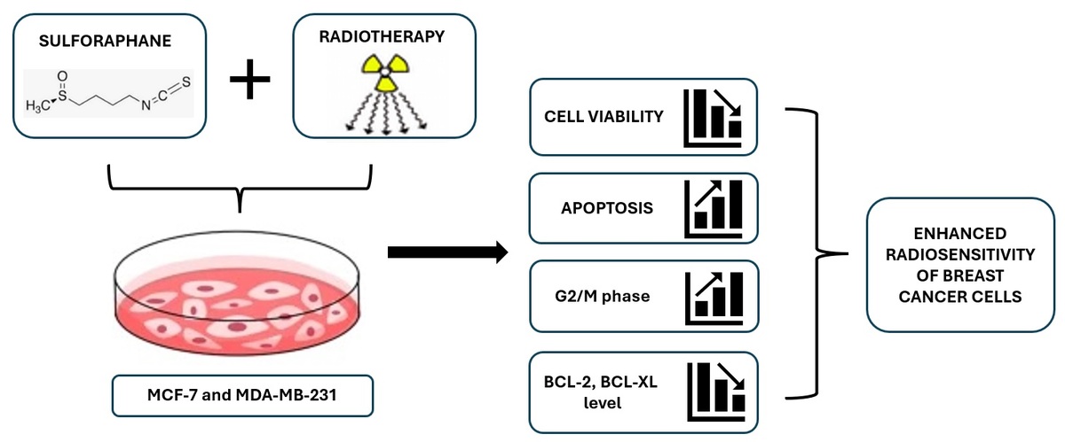
Using substances that intensify the effect of radiation is an option to increase the radiosensitivity of cancer cells [10]. Numerous studies have demonstrated the potential of phytochemicals, which are substances derived from plants, as radioprotectors and radiosensitizers [11]. One of these phytochemicals is sulforaphane (SFN). Sulforaphane is an isothiocyanate that is present naturally in widely consumed cruciferous vegetables, including broccoli, cabbage, and cauliflower [1]. This phytochemical is a natural bioactive component that has many biological properties, such as anti-inflammatory, anticancer, cardioprotective, antioxidative, cytoprotective, DNA protective, and antimicrobial properties, and functions as a strong detoxifier and immune system enhancer. While SFN is characterized by several features, its ability to prevent cancer is by far its most important property [12]. Through a variety of experimental cancer models, SFN has been extensively researched in many cancer types, including breast cancer [13]. Although SFN is widely studied as an anticancer agent, its properties as a radiosensitizer remain unclear. Several studies have demonstrated the potential of SFN to overcome radioresistance in cancer cells. SFN as a radiosensitizer has been described in studies with head and neck cancer cells [14], cervical cancer cells [15], hepatocellular carcinoma [16], prostate cancer [17], and the cervix epithelial carcinoma cell line HeLa [18]. Despite the recognized properties of SFN concerning cancers, little information is available on the radiosensitivity effect of SFN in breast cancer cells. Thus, the purpose of this study was to assess the impact of SFN on MCF-7 and MDA-MB-231 breast cancer cell lines and to determine whether this agent has a radiosensitizing effect.
Material and methods
Cell lines
The human breast cancer cell lines MCF-7 and MDA-MB-231 were obtained from CLS Cell Line Service (Eppelheim, Germany). Cells were grown as monolayers in Dulbecco’s Modified Eagle’s medium (DMEM) supplemented with 10% fetal bovine serum and 100 U/mL penicillin with 100 μg/ml streptomycin and 2 mM L-glutamine. All cell lines were maintained at 37°C in a humidified 5% CO2 incubator.
Preparation of drug solution
DL-Sulforaphane (≥ 90% (HPLC), synthetic, liquid) was purchased from Sigma. A stock solution of 1.1 mM SFN was dissolved in DMSO and frozen at –20°C. Before each experiment, working solutions were prepared, and dilutions in complete media were made to the necessary concentrations of 5–80 μM. The final concentration of the solvents in the media was 0.1% or less.
Irradiation
High-energy photon beam (X-rays 6 MeV) irradiation was performed using Varian TrueBeam STx unity. The imposed dose was 2 Gy or 4 Gy, and the source-surface distance (SSD) was set to 100 cm by using a 30 × 30 cm square 2D exposure field. The average dose rate was 300 MU (monitor units) at room temperature.
MTT assay
The cytotoxic effects of SFN on cell lines were assessed using the MTT (3-(4,5-dimethylthiazol-2-yl)-2,5-diphenyltetrazolium bromide) tetrazolium reduction test. The cells were seeded on 96-well plates and left to adhere overnight. After that, SFN was added at appropriate concentrations (0–80 μM), and the incubation continued for 24, 48, or 72 h. For the combination of SFN and IR, cells were pretreated with SFN (0–50 μM) for 48 h prior to exposure to IR. After this time, the medium was removed and replaced with a fresh SFN-free culture medium. Cells were irradiated with 2 or 4 Gy using high-energy X-rays and incubated for 72 h. Afterward, 15 μl of MTT solution was added to each well and incubated for 3–4 h before the addition of 10% SDS-0.01M HCl and left overnight at 37°C, 5% CO2. The optical density (OD) was determined at 570 nm using a microplate spectrophotometer with reference at 650 nm.
Apoptosis analysis
MCF-7 and MDA-MB-231 cell apoptosis was assayed using the multifunctional Muse Annexin V and Dead Cell kit (Millipore, USA) according to the manufacturer’s instructions. Briefly, cells were seeded in 6-well plates, incubated overnight, and treated with different concentrations of SFN (0, 10, 25, and 50 μM). After 48 h, cells were irradiated with 2 Gy and 4 Gy doses and incubated for 72 h. Next, cells were harvested, and 100 μl of Muse Annexin V and Dead Cell reagent were added. The Muse Cell Analyzer system was used for flow cytometry analysis, and the software program Muse 1.4 was used to examine the results.
Cell cycle analysis
Following the manufacturer’s recommendations, the Muse Cell Cycle Kit (Millipore, USA) was used to evaluate the cell cycle of both cancer cells. Cells were seeded, treated with SFN, and irradiated as described above in the Apoptosis analysis section. The cell pellet was reconstituted in 200 μl of Muse Cell Cycle Reagent and incubated at room temperature for 30 min, protected from light. The cell cycle was evaluated by the Muse Cell Analyzer, and the software program Muse 1.4 was used to examine the results.
RNA extraction and quantitative real-time PCR
We used reverse transcription-quantitative PCR (RT-qPCR) to determine the expression of anti-apoptotic BCL-2 and BCL-XL genes. Cells were treated with SFN (0, 25, 50 μM), after 48 h were irradiated (0, 2, 4 Gy), and after 72 h were harvested. Total RNA was extracted from cancer cells using the RNeasy Mini Kit (Qiagen, Germany) according to the manufacturer’s protocol. One microgram of total RNA was reverse transcribed to cDNA using the High-Capacity RNA-to-cDNA Kit (Applied Biosystems, USA). The expression of anti-apoptotic genes was evaluated with TaqMan Expression assays: BCL-2 (Hs00608023_m1), BCL-XL (Hs00236329_m1) (Invitrogen, Waltham, MA, USA). RT-qPCR analysis was performed on the QuantStudio3 Real-Time PCR System. The expression level for each gene was normalized to ACTB (Hs99999903_m1).
Western blot analysis
To assess the levels of BCL-2 and BCL-XL proteins in both cell lines, cells were seeded, incubated overnight, and afterward exposed to DMSO, SFN, and/or IR as described above. After that, cells were harvested gently and incubated in RIPA lysis buffer (Abcam, UK) with protease and phosphatase inhibitor cocktails (Sigma-Aldrich, USA) for 20 min on ice. Protein lysates (40 μg) were loaded into each lane and separated by electrophoresis on a 4–12% Bis-Tris Plus Gel (Invitrogen, USA). The proteins were transferred from the gel to a PVDF membrane (Invitrogen, USA) using the semi-wet transfer unit Mini Blot module. Membranes were blocked using a blocking buffer for 60 min and incubated with a primary antibody for an entire night at +4°C. These were directed against BCL-2 (product #13-8800; 1 : 1000 dilution; Invitrogen, USA), BCL-XL (product #MA5-15142; 1 : 1000 dilution; Invitrogen, USA), and GAPDH (product #TAB1001; 1 : 1000 dilution; Invitrogen, USA). After washing three times with a 1xTris-buffered saline with Tween 20 (TBST) (Thermo Fisher Scientific, USA), the blots were incubated with the relevant anti-rabbit HRP-conjugated (for BCL-XL, GAPDH), anti-mouse HRP-conjugated (for BCL-2) secondary antibody solution (Invitrogen, USA) for 1 h. The visualization of chemiluminescent imaging was done using the Azure 280 system and analyzed with AzureSpot PRO software. The GAPDH protein was used as the internal control to normalize the target protein expression level.
Statistical analysis
The statistical analysis was performed using IBM SPSS Statistics 29.0 (IBM Corp., USA). The significance of the differences between the experimental groups was evaluated using the Mann-Whitney and Student’s t-tests. All data are presented as the mean with standard deviation (S.D.), and at p < 0.05, the results were considered statistically significant. All experiments were performed at least in triplicate.
Results
SFN treatment reduces the viability of breast cancer cells
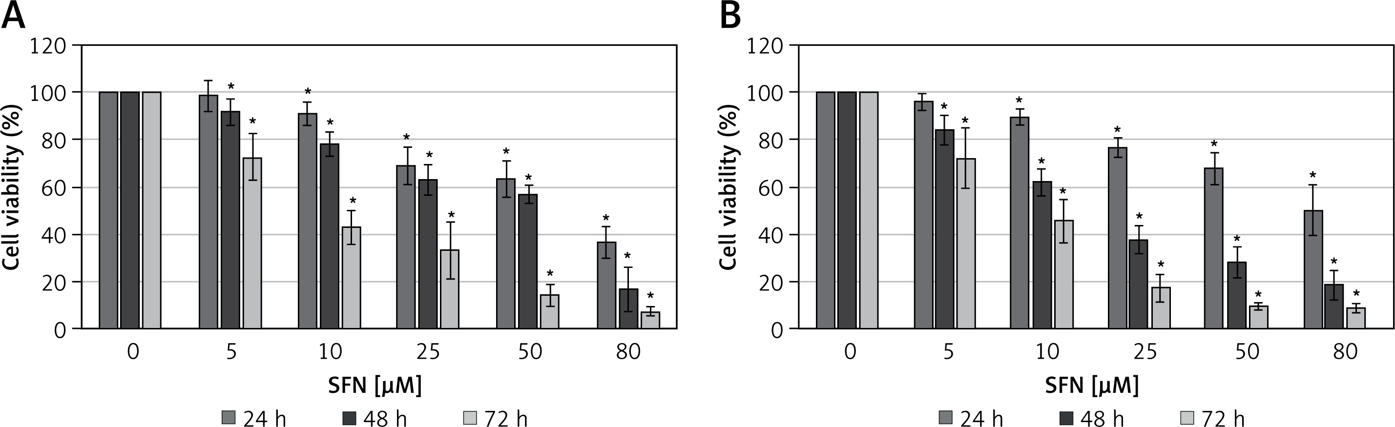
The MTT assay showed that SFN treatment at varying concentrations (5–80 μM) results basically in dose- and time-dependent growth inhibition of cells at 24, 48, and 72 h time intervals. SFN at a dose of 62.5 μM for 24 h, 46.3 μM at 48 h, and 19.3 μM at 72 h induced approximately a 50% decrease in cell viability (IC50) in MCF-7 cells. In MDA-MB-231, the IC50 value was found to be 66.9 μM, 30.8 μM, and 13.2 μM for 24, 48, and 72 h, respectively. For both cell lines, SFN caused a similar cytotoxic effect (Figure 1).
SFN enhances radiosensitivity in breast cancer cells
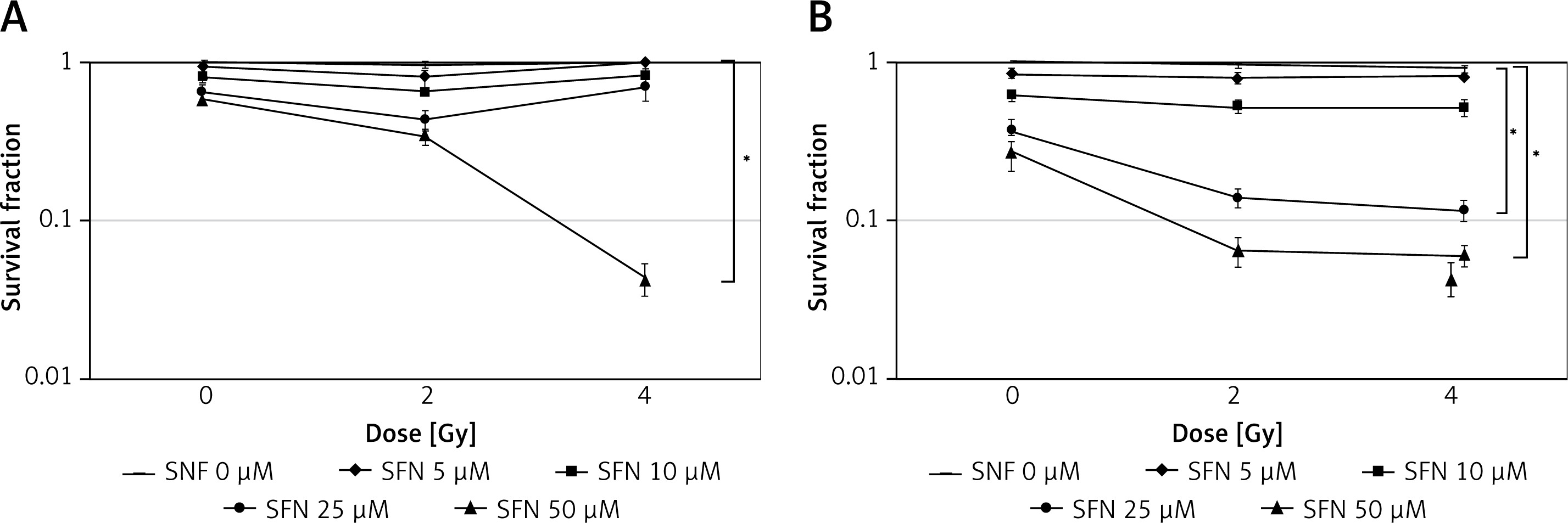
The radiosensitizing effect of SFN at different concentrations in 48 h cell culture irradiated at 2 Gy and 4 Gy was investigated. Our results revealed that only a 50 μM SFN concentration enhanced MCF-7 cell sensitivity to radiation with 2 Gy and 4 Gy doses (Figure 2 A). In the MDA-MB-231 cell line, we found that SFN treatment enhanced cell sensitivity to radiation when 25 μM and 50 μM SFN concentrations were applied with 2 Gy and 4 Gy doses (Figure 2 B). In other SFN concentrations, the effect of combinations was not statistically significant and was similar to treatment with sulforaphane alone, demonstrating no improvement in outcome when using combinations.
Apoptosis is caused by SFN and IR in breast cancer cells
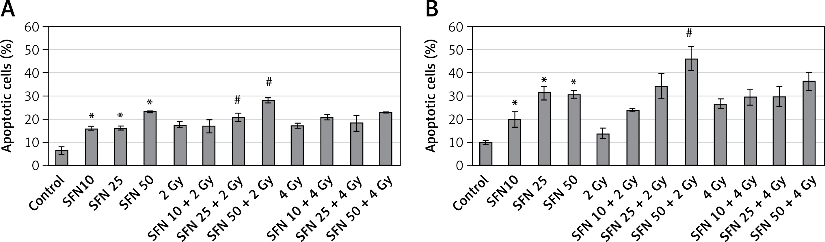
To determine whether the SFN- and IR-induced suppression of breast cancer cell growth was due to apoptosis, cells were stained with annexin V. The flow cytometry analysis showed that the apoptotic cell population increased when using all SFN concentrations in both cell lines. The MDA-MB-231 cell line contained a higher percentage of apoptotic cells than MCF-7. The impact of combination SFN+IR treatment on cell apoptosis was next examined. In MCF-7 cells, both combinations of SFN 25 μM + 2 Gy and SFN 50 μM + 2 Gy increased apoptotic cell death. In MDA-MB-231 cells, the results suggest that only the combination of a 50 μM SFN concentration with a 2 Gy radiation dose led to a statistically significant increase in the apoptotic cell population. The other treatment combinations were not statistically significantly different from the SFN-only or IR-only treatments (Figure 3).
Figure 3
Evaluation of apoptosis by flow cytometry in MCF-7 (A) and MDA-MB-231 (B) cells after SFN and/or IR exposure. Cell viability data are shown as the mean with standard deviation (SD). The mean difference was compared with the control (*) and with both groups (#) (IR-only and SFN-only) (p < 0.05)
SFN arrests the progression of the G2/M phase in the cell cycle, particularly when combined with radiation
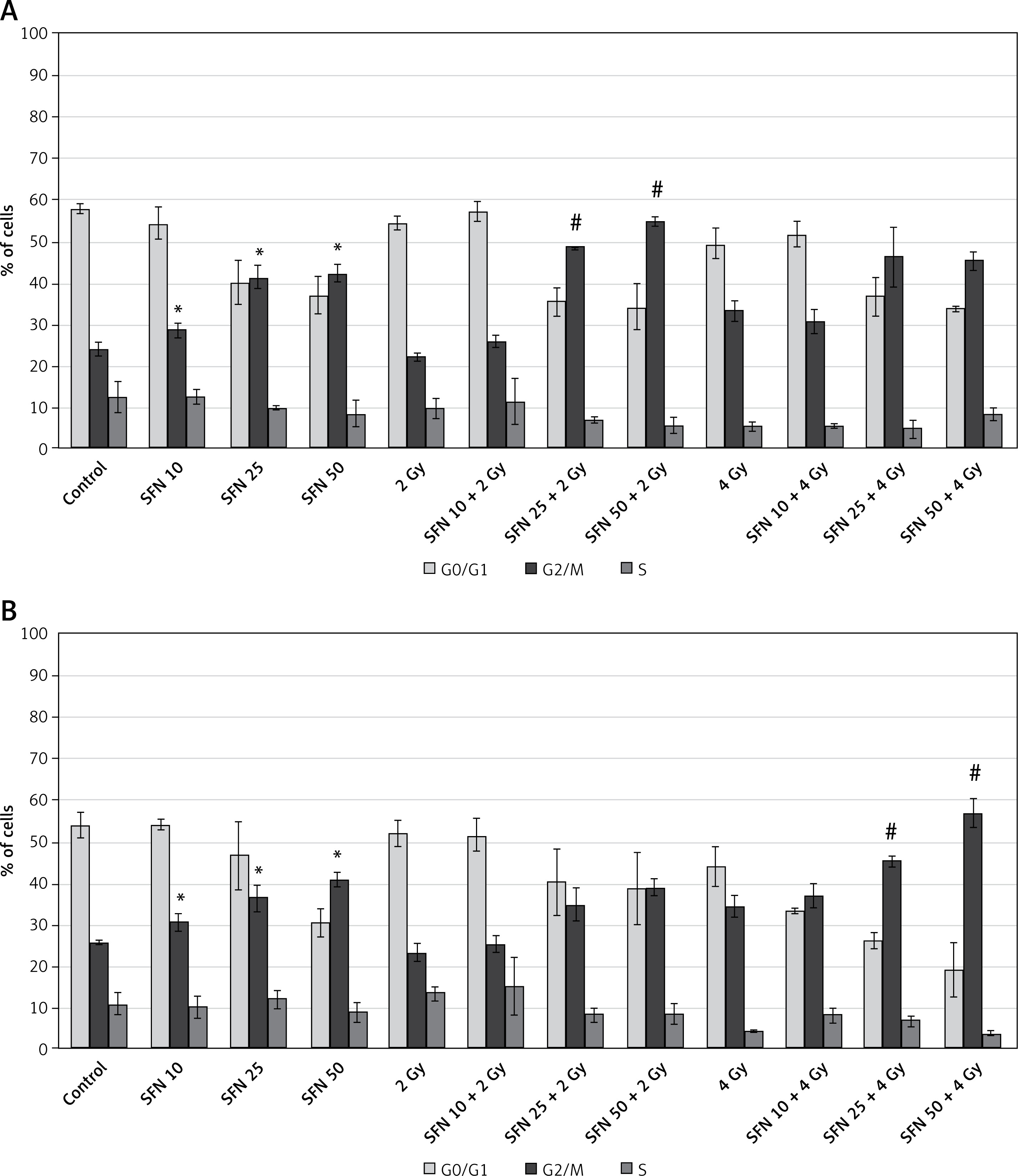
To investigate whether breast cancer cell growth inhibition by SFN and IR was caused by changes in cell cycle progression, we performed flow cytometric cell cycle analysis. When treating both cell lines with SFN alone, we observed an increase in the number of cells in the G2/M phase. These increases were much more pronounced in the MCF-7 cell line when the 2 Gy radiation was combined with 25 μM and 50 μM concentrations of SFN. However, 2 Gy radiotherapy treatment alone did not cause an increase in the number of cells in the G2/M phase. In the MDA-MB-231 cell line, the accumulation of cells in the G2/M phase was observed after 4 Gy radiation with SFN concentrations of 25 μM and 50 μM. As compared to SFN-only or IR-only therapies, no other treatment combinations showed statistically significant difference. G2/M accumulation occurs mostly at the cost of cells in the G1 phase; therefore, a reduction of cells in this phase was detected. No significant differences were observed in the S phase (Figure 4).
Changes in expression of apoptosis-related genes after exposure to SFN and IR
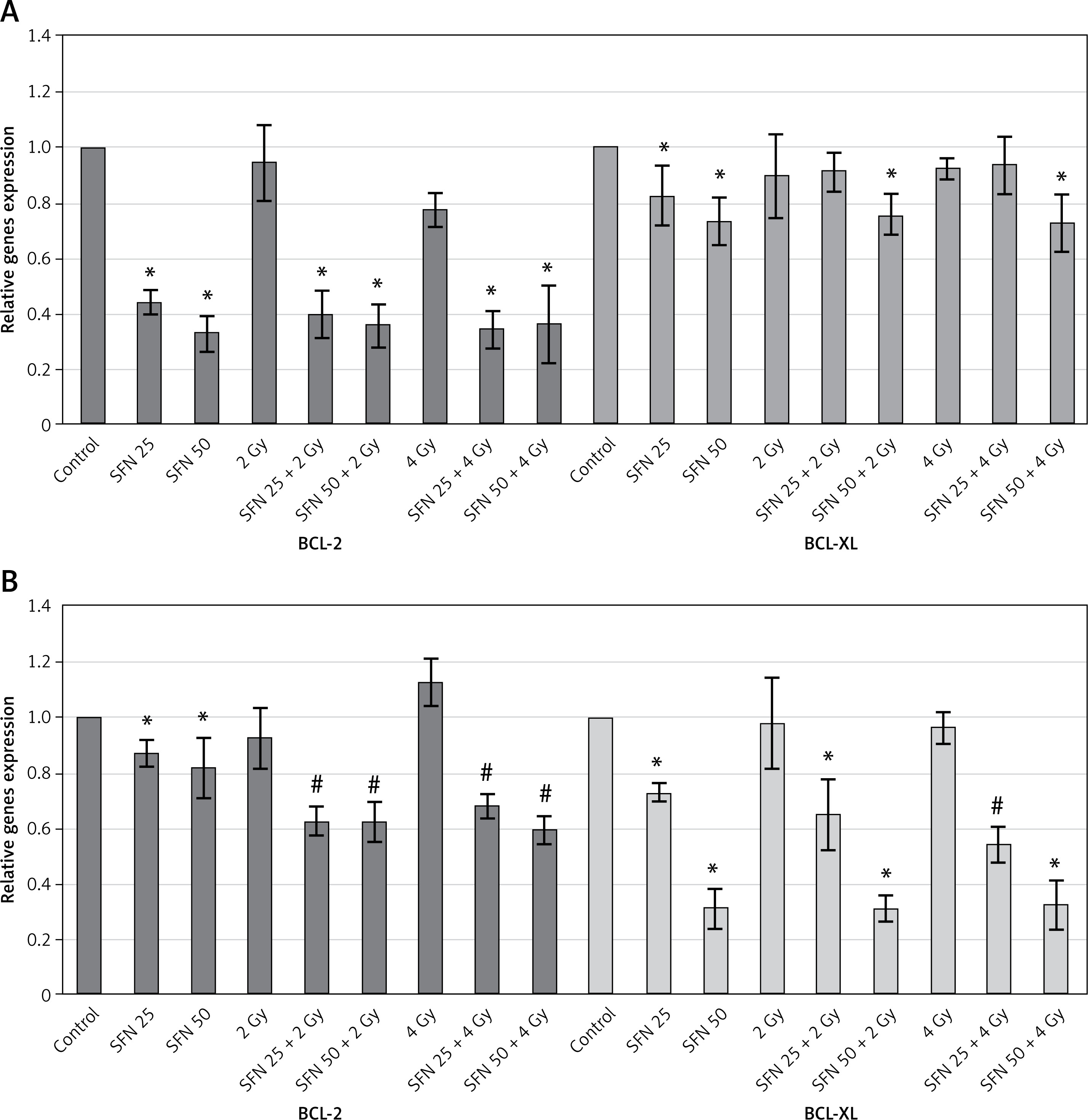
The impact of SFN and IR on BCL-2 and BCL-XL genes linked to apoptosis was then investigated in the research that followed. In MCF-7 cells, RT-qPCR analysis showed that SFN reduced expression of all examined genes. The treatment of SFN + IR combinations did not cause greater decreases in expression of either gene and was not more effective than SFN-only or IR-only exposure. In the study with the MDA-MB-231 cell line, the results showed that the combination SFN + IR reduced BCL-2 gene expression more than only-SFN treatment. Similar outcomes were noted following the use of 4 Gy exposure. Also, SFN reduced the expression of the BCL-XL gene in both concentrations. Following combination exposure, only the SFN 25 μM + 4 Gy treatment statistically significantly decreased BCL-XL gene expression. The other combinations of treatments were similar to those that used only SFN (Figure 5).
Levels of BCL-2 and BCL-XL proteins following SFN and IR treatment
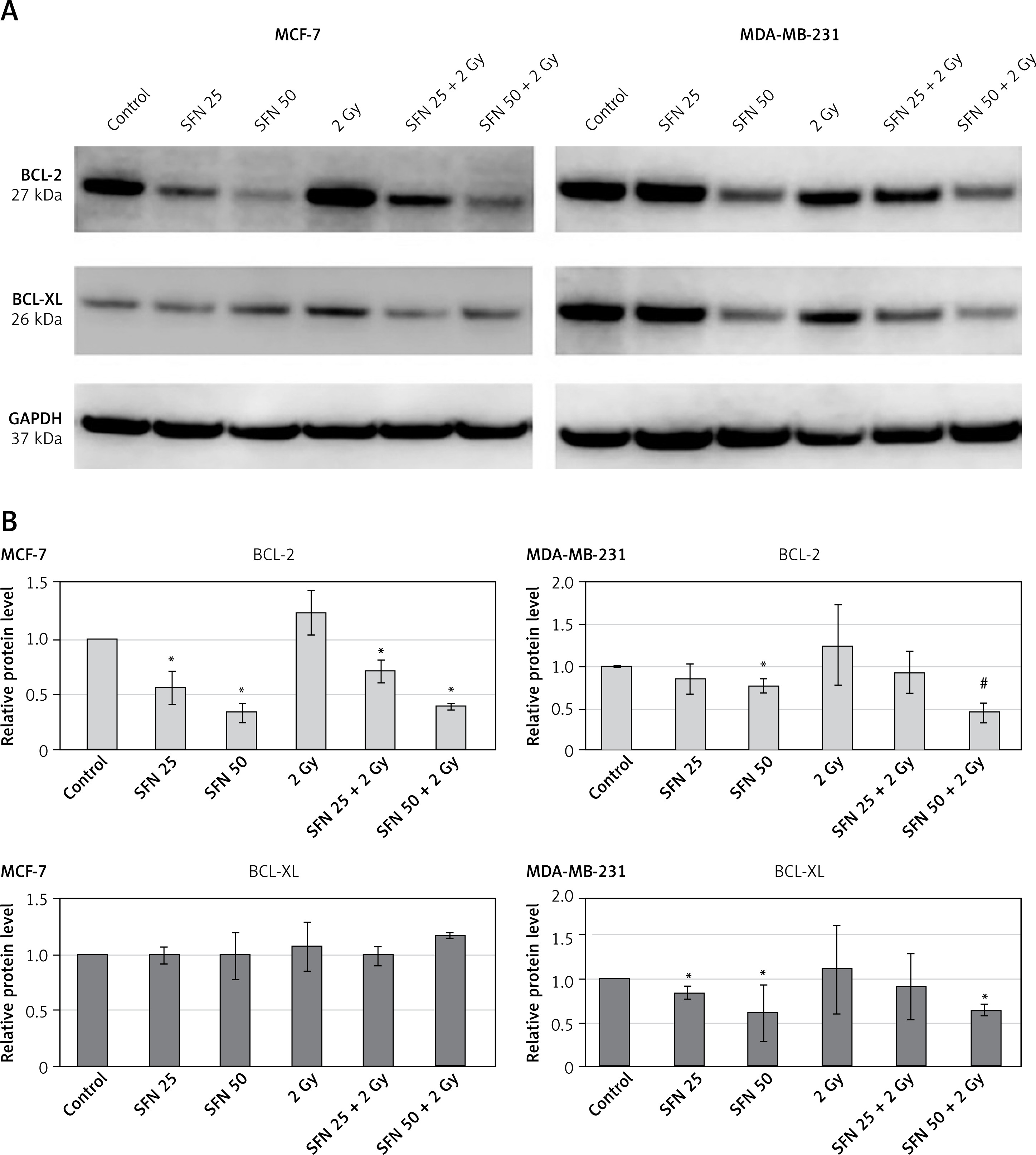
Finally, in both cell lines, we determined the levels of BCL-2 and BCL-XL proteins. Western blot analysis confirmed the decrease in BCL-2 level in MCF-7 cells treated with SFN. The effects of SFN and IR combinations were similar to those of SFN alone exposure. No reduction of the BCL-XL protein was noted after SFN-only or combination therapy in MCF-7 cells. In MDA-MB-231 cells, the combination of SFN 50 μM + 2 Gy reduced BCL-2 gene expression more than only-SFN treatment. Combination treatment had a similar effect on BCL-XL protein level as SFN alone (Figure 6).
Discussion
Recently, sulforaphane has received attention from scientists for its cancer-preventive attributes [19], anticancer effects [20], and as a radiosensitizer in radiotherapy [14, 21]. Sulforaphane is one of the most frequently researched isothiocyanates. Isothiocyanates, which are produced after the hydrolysis of glucosinolates, are currently believed to be responsible for some of the anticancer effects of cruciferous vegetables, such as broccoli, kale, and cauliflower [22]. In this study, we investigated sulforaphane as a radiosensitizer in breast cancer cell lines, with a focus on the apoptosis molecular mechanism.
For this investigation, two breast cancer cell lines, MCF-7 and MDA-MB-231, were selected due to their many phenotypic and genotypic differences. MCF-7 cells are widely used as a good model for ER-positive breast tumors because this cell subtype is hormone-dependent and estrogen and progesterone receptor-positive (ER+ and PR+). In addition, MCF-7 has mutations typically reported in human luminal breast tumors, such as PIK3CA E545K and a GATA3 frameshift mutation, and is TP53 wild type [23]. MDA-MB-231 cells are triple negative, meaning they do not have hormone epidermal growth factor 2 (HER2), progesterone, or estrogen receptors [24]. It also contains a mutant p53 gene and mutations in KRAS, BRAF, and NF1 genes. These mutations often contribute to their aggressive phenotype and have a worse prognosis [25]. It is believed that different subtypes of breast cancer may respond differently to radiation. Therefore, it is essential to understand the underlying variety in radiation sensitivity in breast cancer subtypes to maximize the benefits of radiotherapy [26].
Using the above-mentioned cell models, we assessed the impact of sulforaphane on cell viability. The MTT assay showed that SFN inhibited the growth of both cell lines in a dose- and time-dependent manner, but there was no significant difference in variation amongst the cell lines. These findings corroborate those previously reported by other researchers. Zhou et al. noted that MDA-MB-231 cells exhibited the same SFN inhibitory effect as MCF-7 cells [13]. Pledgie-Tracy et al. examined the effects of sulforaphane on the growth of four breast cancer cell lines, which are representative of a wide range of breast cancer phenotypes. The findings demonstrated no significant differences in SFN inhibitory potency amongst the cell lines [22]. Licznerska et al. obtained the opposite outcome, demonstrating that MCF-7 cells were more sensitive to SFN than MDA-MB-231 cells [27]. A potential explanation for this disparity is that in this study, the L-SFN isomer was used, while in our study a synthetic analog of the naturally occurring D,L-SFN was applied.
Interestingly, in control of non-tumorigenic MCF10A cells, the cytotoxic effect of SFN was very slight [28]. According to Pawlik et al., that cytotoxic effect of SFN is unique to cancer cells, and it is confirmed by other reports [29–33]. In the Licznerska et al. study, MCF10A cells had an IC50 value greater than 80 μmol/l and were less susceptible to the cytotoxic action of SFN after 72 h than breast cancer cell lines [27]. We did not investigate the impact of SFN on non-cancerous cells because we are interested in processes occurring only in cancer cells.
Subsequently, it was demonstrated that irradiation plus SFN improved the anticancer efficacy of breast cancer cells. Our results showed that SFN acts as a radiosensitizer in MCF-7 and MDA-MB-231 cells. Although only-SFN and only-IR demonstrated similar efficacy in both cell lines, the combination therapy induced a greater inhibitory effect in MDA-MB-231 cells. Other investigations have reported the ability of SFN to act as a radiosensitizer in various types of malignant tumor cells. According to the findings of Kotowski et al.’s research, sulforaphane could radiosensitize and inhibit the proliferation of head and neck cancer cells [14]. Wang et al. established that SFN could exert irradiation sensitization in cervical cancer cells via activating LATS2 and blocking Rad51 and MDC1 nucleus recruitment and DNA damage repair [15]. Ren et al.’s study indicated that SFN increases the radiosensitivity of hepatocellular carcinoma by blocking the NF-κB pathway [16]. In this study, we found that the combinations of SFN and radiation treatment enhanced apoptosis and increased the cell population in the G2/M phase. Our results were consistent with most of the research reviewed showing that SFN has apoptosis-inducing properties and can cause cell cycle arrest in the G2/M phase. Cells are typically the most radiosensitive in this phase [34]. Thus, agents (in this study, SFN) that cause G2/M phase cell cycle arrest can demonstrate strong radiosensitivity [35]. However, only combinations with a dose of 2 Gy showed an increase in the number of apoptotic cells. It is believed that other cell death processes, such as mitotic catastrophe, may have occurred after the application of a dose of 4 Gy.
Overexpression of anti-apoptotic proteins, which inhibit apoptosis, is one of the primary mechanisms behind acquired resistance and/or insensitivity to cancer [36]. It has been demonstrated that using key regulators of the BCL-2 protein family as a radiosensitization technique can overcome apoptosis resistance in a variety of cancer types [37]. Based on this, two anti-apoptotic genes, BCL-2 and BCL-XL, were selected for this study. Our results demonstrated that SFN could reduce the expression of these BCL-2 family members at gene and protein levels in MDA-MB-231 and MCF-7 cells. Other investigations have shown that after SFN treatment, BCL-2 expression levels were diminished in various cancer cell lines, including breast cancer cells [1, 38, 39], pancreatic cancer cells [40], prostate cancer cells [40–42], glioblastoma cells [43], human cervical carcinoma cells, and hepatocarcinoma cells [37, 43], etc. However, Yasunaga et al. reported that BCL2 expression was not altered after SFN therapy in breast cancer cells [44]. These contradictory results may be due to the low SFN concentration (2 μM) that they used for the experiment. Furthermore, they used another breast cancer cell line, MDA-MB-468. Our data were consistent with the results of Kim et al.’, who also found that SFN could downregulate the BCL-2 level in MDA-MB-231 cells [38]. We have performed too few studies to confirm the exact molecular mechanism of action of SFN-induced apoptosis. Our findings, however, imply that apoptosis was most likely induced by the intrinsic apoptosis pathway in both MCF-7 and MDA-MB-231 cell lines, given that the BCL-2 protein family is known to control activation of the intrinsic apoptotic pathway [45].
We also investigated the potential relationship between these anti-apoptotic molecules and the radiosensitivity of cells with a combination of SFN and radiotherapy. Following combination treatment, there was no significant effect on the expression of the studied genes in MCF-7 cells at either the gene or protein level (compared to only-SFN and only-IR). In MDA-MB-231 cells, the combined therapy resulted in greater downregulation of only the BCL-2 gene at both gene and protein levels. It is possible that the radiosensitivity of MDA-MB-231 cells can be enhanced by reducing BCL-2 expression, thereby initiating apoptosis and inhibiting cancer cell proliferation. The combination treatment of MCF-7 cells also initiates apoptosis and causes cell death, but probably through other molecules related to apoptosis, rather than those we studied. This difference may be due to varying gene expression patterns that are characteristic of different subtypes of breast cancer. Despite these differences, combinations of SFN and IR increased cellular radiosensitivity and enhanced radiotherapy efficacy in MCF-7 and MDA-MB-231 cells.
In conclusion, SFN and radiotherapy together may be a useful method for making breast cancer cells more sensitive to radiation for improved effectiveness and outcomes of treatment. Additionally, we highlight the potential of BCL-2 family proteins for triple-negative breast cancer subtype treatment.