Current issue
Archive
Manuscripts accepted
About the Journal
Editorial office
Editorial board
Section Editors
Abstracting and indexing
Subscription
Contact
Ethical standards and procedures
Most read articles
Instructions for authors
Article Processing Charge (APC)
Regulations of paying article processing charge (APC)


ACUTE CORONARY SYNDROME / RESEARCH PAPER
Association of AT1R-Mediated TGF-β1/Smad Axis Downregulation with Antifibrotic and Functional Benefits of Extended Exercise After Myocardial Infarction
1
Guangdong Provincial People’s Hospital (Guangdong Academy of Medical Sciences), Southern Medical University, Guangdong Cardiovascular Institute, China
Submission date: 2026-04-14
Final revision date: 2026-05-27
Acceptance date: 2026-06-18
Online publication date: 2026-09-05
Corresponding author
Moussa Ide Nasser
Guangdong Provincial People’s Hospital (Guangdong Academy of Medical Sciences), Southern Medical University, Guangdong Cardiovascular Institute, China
Guangdong Provincial People’s Hospital (Guangdong Academy of Medical Sciences), Southern Medical University, Guangdong Cardiovascular Institute, China
KEYWORDS
Exercise trainingMyocardial infarctionCardiac fibrosisTGF-β1Smad signalingAngiotensin II type 1 receptorRat modelCardiac pathologyEchocardiographySystolic function
TOPICS
ABSTRACT
Introduction:
Exercise training has cardioprotective effects after myocardial infarction (MI), but the associated molecular pathways remain incompletely defined. The novel aspect of this work is the comparison of short-term and extended post-MI ET while examining the angiotensin II type 1 receptor (AT1R)/TGF-β1/Smad axis as an associated anti-fibrotic pathway.
Material and methods:
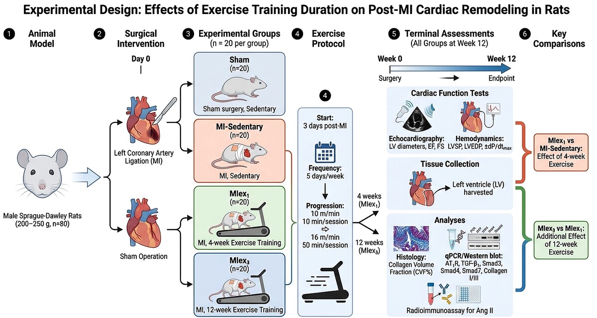
Sprague-Dawley rats were assigned to four groups: sham control, MI-sedentary, MI with 4-week Exercise training (MIex1), and MI with 12-week of exercise training (MIex3). ET began three days after coronary artery ligation. After 12 weeks, echocardiography and hemodynamic measurements were performed, and left ventricular (LV) tissue was collected for histological and molecular analyses.
Results:
Compared with MI-sedentary rats, MIex1 rats showed increased LV systolic pressure, ±dP/dtmax, ejection fraction, and fractional shortening, along with decreased LV end-diastolic pressure and LV diameters. Twelve-week Exercise training further enhanced functional outcomes. Both ET groups reduced collagen volume fraction and collagen I/III expression. ET was also associated with lower cardiac Ang II, AT1R, TGF-β1, and phosphorylated Smad2/3, with stronger effects after 12 weeks of Exercise training.
Conclusions:
Exercise reduces myocardial fibrosis following MI and preserves cardiac function, an effect that may be amplified by extended exercise training. Downregulation of AT1R-mediated TGF-β1/Smad signaling may be associated with these cardioprotective effects.
Exercise training has cardioprotective effects after myocardial infarction (MI), but the associated molecular pathways remain incompletely defined. The novel aspect of this work is the comparison of short-term and extended post-MI ET while examining the angiotensin II type 1 receptor (AT1R)/TGF-β1/Smad axis as an associated anti-fibrotic pathway.
Material and methods:
Sprague-Dawley rats were assigned to four groups: sham control, MI-sedentary, MI with 4-week Exercise training (MIex1), and MI with 12-week of exercise training (MIex3). ET began three days after coronary artery ligation. After 12 weeks, echocardiography and hemodynamic measurements were performed, and left ventricular (LV) tissue was collected for histological and molecular analyses.
Results:
Compared with MI-sedentary rats, MIex1 rats showed increased LV systolic pressure, ±dP/dtmax, ejection fraction, and fractional shortening, along with decreased LV end-diastolic pressure and LV diameters. Twelve-week Exercise training further enhanced functional outcomes. Both ET groups reduced collagen volume fraction and collagen I/III expression. ET was also associated with lower cardiac Ang II, AT1R, TGF-β1, and phosphorylated Smad2/3, with stronger effects after 12 weeks of Exercise training.
Conclusions:
Exercise reduces myocardial fibrosis following MI and preserves cardiac function, an effect that may be amplified by extended exercise training. Downregulation of AT1R-mediated TGF-β1/Smad signaling may be associated with these cardioprotective effects.
REFERENCES (45)
1.
Gerber Y, Weston SA, Enriquez-Sarano M, et al. Mortality Associated With Heart Failure After Myocardial Infarction. Circ Hear Fail. 2016;9(1):1-21. doi:10.1161/circheartfailure.115.002460.
2.
Bulluck H, Dharmakumar R, Arai AE, Berry C, Hausenloy DJ. Cardiovascular magnetic resonance in acute st-segment-elevation myocardial infarction: Recent advances, controversies, and future directions. Circulation. 2018;137(18):1949-1964. doi:10.1161/CIRCULATIONAHA.117.030693.
3.
Prabhu SD, Frangogiannis NG, Service M, Einstein A. The Biological Basis for Cardiac Repair After Myocardial Infarction: From Inflammation to Fibrosis. Circu. 2017;119(1):91-112. doi:10.1161/CIRCRESAHA.116.303577.
4.
Talman V, Ruskoaho H. Cardiac fibrosis in myocardial infarction—from repair and remodeling to regeneration. Cell Tissue Res. 2016;365(3):563-581. doi:10.1007/s00441-016-2431-9.
5.
Frangogiannis NG. Pathophysiology of myocardial infarction. Compr Physiol. 2015;5(4):1841-1875. doi:10.1002/cphy.c150006.
6.
Aras O, Messina SA, Shirani J, Eckelman WC, Dilsizian V. The role and regulation of cardiac angiotensin-converting enzyme for noninvasive molecular imaging in heart failure. Curr Cardiol Rep. 2007;9(2):150-158. doi:10.1007/BF02938342.
7.
Rosenkranz S. TGF-β1 and angiotensin networking in cardiac remodeling. Cardiovasc Res. 2004;63(3):423-432. doi:10.1016/j.cardiores.2004.04.030.
8.
Meng XM, Tang PMK, Li J, Lan HY. TGF-ß/Smad signaling in renal fibrosis. Front Physiol. 2015;6(MAR):1-8. doi:10.3389/fphys.2015.00082.
9.
Miyazawa K, Miyazono K. Regulation of TGF-β family signaling by inhibitory smads. Cold Spring Harb Perspect Biol. 2017;9(3):1-25. doi:10.1101/cshperspect.a022095.
10.
He X, Zhang K, Gao X, et al. Rapid atrial pacing induces myocardial fibrosis by down-regulating Smad7 via microRNA-21 in rabbit. Heart Vessels. 2016;31(10):1696-1708. doi:10.1007/s00380-016-0808-z.
11.
He X, Gao X, Peng L, et al. Atrial fibrillation induces myocardial fibrosis through angiotensin II type 1 receptor-specific Arkadia-mediated downregulation of Smad7. Circ Res. 2011;108(2):164-175. doi:10.1161/CIRCRESAHA.110.234369.
12.
Zhang K, He X, Zhou Y, et al. Atorvastatin Ameliorates Radiation-Induced Cardiac Fibrosis in Rats. Radiat Res. 2015;184(6):611-620. doi:10.1667/rr14075.1.
13.
Giallauria F, Acampa W, Ricci F, et al. Effects of exercise training started within 2 weeks after acute myocardial infarction on myocardial perfusion and left ventricular function : a gated SPECT imaging study. 2011. doi:10.1177/1741826711425427.
14.
Tucker WJ, Beaudry RI, Liang Y, et al. Meta-analysis of Exercise Training on Left Ventricular Ejection Fraction in Heart Failure with Reduced Ejection Fraction: A 10-year Update. Prog Cardiovasc Dis. 2019;62(2):163-171. doi:10.1016/j.pcad.2018.08.006.
15.
Lawler PR, Filion KB, Eisenberg MJ. Efficacy of exercise-based cardiac rehabilitation post-myocardial infarction: A systematic review and meta-analysis of randomized controlled trials. Am Heart J. 2011;162(4):571-584.e2. doi:10.1016/j.ahj.2011.07.017.
16.
Zucker IH, Schultz HD, Patel KP, Wang H. Modulation of angiotensin II signaling following exercise training in heart failure. Am J Physiol - Hear Circ Physiol. 2015;308(8):H781-H791. doi:10.1152/ajpheart.00026.2015.
17.
Wan W, Powers AS, Li J, Ji L, Erikson JM, Zhang JQ. Effect of post-myocardial infarction exercise training on the renin-angiotensin-aldosterone system and cardiac function. Am J Med Sci. 2007;334(4):265-273. doi:10.1097/MAJ.0b013e318068b5ed.
18.
Puhl SL, Müller A, Wagner M, et al. Exercise attenuates inflammation and limits scar thinning after myocardial infarction in mice. Am J Physiol - Hear Circ Physiol. 2015;309(2):H345-H359. doi:10.1152/ajpheart.00683.2014.
19.
Jia D, Hou L, Lv Y, Xi L, Tian Z. Postinfarction exercise training alleviates cardiac dysfunction and adverse remodeling via mitochondrial biogenesis and SIRT1/PGC-1α/PI3K/Akt signaling. J Cell Physiol. 2019;234(12):23705-23718. doi:10.1002/jcp.28939.
20.
Kraljevic J, Marinovic J, Pravdic D, et al. Aerobic interval training attenuates remodelling and mitochondrial dysfunction in the post-infarction failing rat heart. Cardiovasc Res. 2013;99(1):55-64. doi:10.1093/cvr/cvt080.
21.
Kröpfl JM, Spengler CM, Frobert A, Ajalbert G, Giraud MN. Myocardial infarction does not affect circulating haematopoietic stem and progenitor cell self-renewal ability in a rat model. Exp Physiol. 2018;103(1):1-8. doi:10.1113/EP086643.
22.
Xu X, Wan W, Ji L, et al. Exercise training combined with angiotensin II receptor blockade limits post-infarct ventricular remodelling in rats. Cardiovasc Res. 2008;78(3):523-532. doi:10.1093/cvr/cvn028.
23.
Sahn DJ, DeMaria A, Kisslo J, Weyman A. Recommendations regarding quantitation in M-mode echocardiography: Results of a survey of echocardiographic measurements. Circulation. 1978;58(6):1072-1083. doi:10.1161/01.CIR.58.6.1072.
24.
Rennison JH, McElfresh TA, Okere IC, et al. High-fat diet postinfarction enhances mitochondrial function and does not exacerbate left ventricular dysfunction. Am J Physiol - Hear Circ Physiol. 2007;292(3):1498-1506. doi:10.1152/ajpheart.01021.2006.
25.
Luo J, Gao X, Peng L, Sun H, Dai G. Effects of hydrochlorothiazide on cardiac remodeling in a rat model of myocardial infarction-induced congestive heart failure. Eur J Pharmacol. 2011;667(1-3):314-321. doi:10.1016/j.ejphar.2011.06.012.
26.
Livak KJ, Schmittgen TD. Analysis of relative gene expression data using real-time quantitative PCR and the 2-ΔΔCT method. Methods. 2001;25(4):402-408. doi:10.1006/meth.2001.1262.
27.
Anderson L, Oldridge N, Thompson DR, et al. Exercise-Based Cardiac Rehabilitation for Coronary Heart Disease Cochrane Systematic Review and Meta-Analysis. J Am Coll Cardiol. 2016;67(1):1-12. doi:10.1016/j.jacc.2015.10.044.
28.
Belardinelli R, Georgiou D, Cianci G, Purcaro A. 10-year exercise training in chronic heart failure: A randomized controlled trial. J Am Coll Cardiol. 2012;60(16):1521-1528. doi:10.1016/j.jacc.2012.06.036.
29.
Wang Y, Tian M, Mi C, et al. Exercise protects the heart against myocardial infarction through upregulation of miR-1192. Biochem Biophys Res Commun. 2019;(xxxx). doi:10.1016/j.bbrc.2019.11.019.
30.
Guizoni DM, Oliveira-Junior SA, Noor SLR, et al. Effects of late exercise on cardiac remodeling and myocardial calcium handling proteins in rats with moderate and large size myocardial infarction. Int J Cardiol. 2016;221:406-412. doi:10.1016/j.ijcard.2016.07.072.
31.
Marijianowski MMH, Teeling P, Becker AE. Remodeling after myocardial infarction in humans is not associated with interstitial fibrosis of noninfarcted myocardium. J Am Coll Cardiol. 1997;30(1):76-82. doi:10.1016/S0735-1097(97)00100-9.
32.
Van Aelst LNL, Voss S, Carai P, et al. Osteoglycin prevents cardiac dilatation and dysfunction after myocardial infarction through infarct collagen strengthening. Circ Res. 2015;116(3):425-436. doi:10.1161/CIRCRESAHA.116.304599.
33.
Yengo CM1, Zimmerman SD, McCormick RJ TD. Exercise training post-MI favorably modifies heart extracellular matrix in the rat. Med Sci Sport. 2012;44(June):1005-1012. doi:10.1249/MSS.ObO.
34.
Solomon SD, Skali H, Anavekar NS, et al. Changes in ventricular size and function in patients treated with valsartan, captopril, or both after myocardial infarction. Circulation. 2005;111(25):3411-3419. doi:10.1161/CIRCULATIONAHA.104.508093.
35.
Granger CB, McMurray JJ V, Yusuf S, et al. CHARM-Alternative trial (ARB CHF). Lancet. 2003;362(9386):772-776. doi:10.1016/S0140-6736(03)14284-5.
36.
Lim B-K, Park J, Park S-J, et al. Fimasartan for Remodeling after Myocardial Infarction. J Clin Med. 2019;8(3):366. doi:10.3390/jcm8030366.
37.
Huang BS, Chen A, Ahmad M, Wang HW, Leenen FHH. Mineralocorticoid and AT1 receptors in the paraventricular nucleus contribute to sympathetic hyperactivity and cardiac dysfunction in rats post myocardial infarct. J Physiol. 2014;592(15):3273-3286. doi:10.1113/jphysiol.2014.276584.
38.
Haack KKV, Engler CW, Papoutsi E, Pipinos II, Patel KP, Zucker IH. Parallel changes in neuronal at1r and grk5 expression following exercise training in heart failure. Hypertension. 2012;60(2):354-361. doi:10.1161/HYPERTENSIONAHA.112.195693.
39.
Lee Y, Kwak HB, Hord J, Kim JH, Lawler JM. Exercise training attenuates age-dependent elevation of angiotensin II type 1 receptor and Nox2 signaling in the rat heart. Exp Gerontol. 2015;70:163-173. doi:10.1016/j.exger.2015.07.016.
40.
Liang ZG, Yao H, Xie RS, Gong CL, Tian Y. MicroRNA-20b-5p promotes ventricular remodeling by targeting the TGF-β/Smad signaling pathway in a rat model of ischemia-reperfusion injury. Int J Mol Med. 2018;42(2):975-987. doi:10.3892/ijmm.2018.3695.
41.
Matsumoto J, Takada S, Kinugawa S, et al. Brain derived neurotrophic factor attenuates cardiac fibrosis through suppression of TGF β1/Smad signaling. J Mol Cell Cardiol. 2022;162:1 10. doi:10.1016/j.yjmcc.2021.09.001.
42.
Okada M, Oikawa M, Yamashita T, et al. BDNF inhibits cardiac fibroblast activation and collagen synthesis via TrkB ERK signaling. Cardiovasc Res. 2023;119(4):987 998. doi:10.1093/cvr/cvac178.
43.
Green DJ, Hopman MTE, Padilla J, et al. Exercise training and arterial function in humans: a meta analysis. J Physiol. 2024;602(3):451 470. doi:10.1113/JP284567.
44.
Chen K, Pittman RN, Popel AS. Nitric oxide in the vasculature: where does it come from and where does it go? Am J Physiol Heart Circ Physiol. 2023;324(5):H589 H606. doi:10.1152/ajpheart.00622.2022.
45.
Zhou Y, Wang X, Sun L, et al. Nitric oxide upregulates Smad7 expression to limit TGF β1 induced cardiac fibrosis. J Biol Chem. 2025;301(2):107654. doi:10.1016/j.jbc.2024.107654.
Share
RELATED ARTICLE
| eISSN: | 1896-9151 |
| ISSN: | 1734-1922 |

We process personal data collected when visiting the website. The function of obtaining information about users and their behavior is carried out by voluntarily entered information in forms and saving cookies in end devices. Data, including cookies, are used to provide services, improve the user experience and to analyze the traffic in accordance with the Privacy policy. Data are also collected and processed by Google Analytics tool (more).
You can change cookies settings in your browser. Restricted use of cookies in the browser configuration may affect some functionalities of the website.
You can change cookies settings in your browser. Restricted use of cookies in the browser configuration may affect some functionalities of the website.
