

Introduction
Hepatitis E virus (HEV) was discovered in 1983 by the Soviet virologist Mikhail Balayan [1, 2]. Initially, it seemed that the virus occurred locally, in regions with poorly developed sanitary infrastructure – in Central Asia, Central America and Africa. However, at the end of the last century, a shift in understanding of HEV epidemiology occurred due to the identification of a new genotype present in developed countries worldwide [3].
HEV is currently considered one of the most common causes of acute hepatitis in humans globally [4]. The epidemiological and clinical significance of HEV infections in Europe is growing: in just 5 years, a 10-fold increase in the number of infections reported to epidemiological surveillance has been observed, which reflects two factors – increased access to diagnostics, but also a higher incidence of infections, which was confirmed in countries with greater availability of HEV tests [5].
In the current paper we review literature on HEV epidemiology in Poland, in both the environment and human population, including patients and blood donors. We analyzed manuscripts indexed in PubMed using the search terms HEV (hepatis E virus) and Poland. Data for Poland were compared to other regions of the world and discussed in the context of current knowledge on clinical significance, transmission routes, and biology of the virus crucial for understanding the effectiveness of infection prevention and the natural course of infection, and in view of optimal diagnostic and therapeutic procedures.
Taxonomic classification and biology
HEV was initially classified into the Hepeviridae family, genus Orthohepevirus, within which four species – Orthohepevirus A-D – were distinguished. According to the latest taxonomic division, viruses infecting humans have been classified into the genus Paslahepevirus [6].
Within the species Orthohepevirus A, seven genotypes (1–7) were distinguished [7]. It is known that genotypes 1–4 infect humans, but recent observations indicate that other genotypes within Orthohepevirus A and species of Orthohepeviruses also have a significant zoonotic potential – crossing the interspecies barrier and causing hepatitic manifestations. There are documented symptomatic human cases of infection with genotype 7, previously detected exclusively in camels [8], in Africa, and in Spain and in Hong Kong infections with Orthohepevirus C, previously identified only in rats [9, 10]. Genotypes 1 and 2 are human-specific pathogens, responsible for the spread of large epidemics in developing countries – mainly in Asia, Africa, and Central America. The spread of infections is facilitated by poor sanitary conditions, contaminated water and, to a lesser extent, contaminated food. Infections with genotypes 3 and 4 are recorded mainly in developed countries in North and South America and in Europe and are most often a consequence of meat consumption from infected animals – pork and game – and in some infected persons result in hepatic or extrahepatic symptoms [11].
The biological properties of HEV, similar to hepatitis A virus (HAV), determine the course of infection and also explain resistance to external factors and infectivity through blood and transfusion and frequent widespread distribution of infections in humans, in some part of the world. HEV is a relatively small (27–34 nm in diameter), spherical virus, whose genetic material is in the form of short RNA (approx. 6.4–7.2 kb). The virus genome includes three partially overlapping open reading frames: ORF1 encodes non-structural genes – helicase, protease and RNA-dependent RNA polymerase; ORF2 encodes the capsid protein; and ORF3 encodes a small phosphoprotein that binds to the cytoskeleton [3, 10].
It has been shown that the virus isolated from plasma, in contrast to virions from feces, is not neutralized by immune serum containing antibodies against ORF2 protein, and has higher density and diameter. These differences are related to the presence of enveloped virions in the blood and non-enveloped virions in feces. Some authors describe the structure of HEV virions as quasi-enveloped and suggest different routes of entry of the mentioned morphological forms of the virus into the cell [12, 13].
It is believed that the structure of virions determines the high resistance of HEV to (extreme) factors/physicochemical conditions. Disinfectants containing high concentrations of isopropanol or ethanol have been shown to be ineffective. Only the addition of phosphoric acid to alcohol improves the effectiveness of this type of disinfection [14]. The relationship between temperature and virus infectivity seems to be more limited than in the case of other viruses, especially enveloped ones: infectivity can be maintained at refrigerator temperature (4°C) for many weeks (at least 56 days) and at room temperature (approx. 22°C) for up to 4 weeks; only exposure to a temperature of at least 80°C almost immediately destroys the replicative potential of the virus, preventing its transmission [15].
Recent work by a research team from Catalonia indicates an even more complex biology of HEV. It was found that in blood of infected donors, in addition to the enveloped form of virions, the non-enveloped form also occurs in a minority. This indicates that the presence of this morphological form of the virus is not limited to the bile ducts and feces, as previously thought. Importantly, the proportion of enveloped virions fluctuates during the course of infection, increasing in seropositive individuals and those with elevated liver enzyme activity [16]. This observation seems to explain the high infectivity of HEV by transfusion, even in inactivated components.
Transmission routes
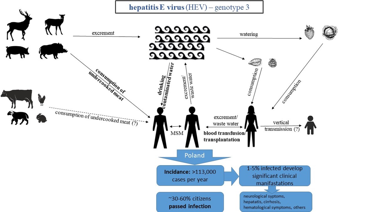
It is well documented that HEV infection is transmitted to humans via the fecal-oral route, through the consumption of contaminated food, as well as through blood transfusion and transplantation. Although vertical transmission was reported for genotype 1, it remains uncertain for genotype 3, as there is a shortage of data clearly confirming or excluding this route of transmission for autochthonous polymorphic forms present in developed countries. Sexual transmission is suspected based on a population study of serological markers in a group of men who have sex with men (MSM), but the role of this route of transmission in spread of infection still requires further studies [17, 18] (Figure 1). HEV transmission through drinking water contaminated with feces of infected people mainly concerns genotypes 1 and 2 of HEV and is dominant in developing countries [11]. Consumption of fruits (e.g. strawberries), vegetables watered with contaminated water, as well as seafood (e.g. oysters, mussels) is also a risk factor for HEV infection [19, 20]. HEV transmission through the consumption of infected meat concerns genotypes from animals (3 and 4), the reservoir of which are mainly domestic pigs and wild boars, as well as deer, rabbits, hares, cows, and goats [11, 19, 21]. Consumption of raw or undercooked meat, such as pork or game, is particularly risky. Contact with sick animals may also be a source of HEV infection. Cases of HEV infection have been described in France as a result of frequent contact with domestic pigs, and higher seroprevalence of HEV has also been observed in Swedish, Dutch, and French veterinarians and hunters in Germany and France [20]. Infection with genotype 7 HEV has also been reported in the Middle East as a result of consumption of camel meat and milk [7]. HEV transmission may occur vertically from mother to child, with the risk of death due to the development of severe liver failure being particularly high, up to 20%, for genotypes 1 and 2, and not elevated for genotypes 3 and 4 [19, 22, 23].
Figure 1
Routes of HEV transmission (based on [11, 17, 19, 20, 21, 23, 24, 27, 28, 29])
MSM – men who have sex with men, MID – minimal infection doze, IU – international units.
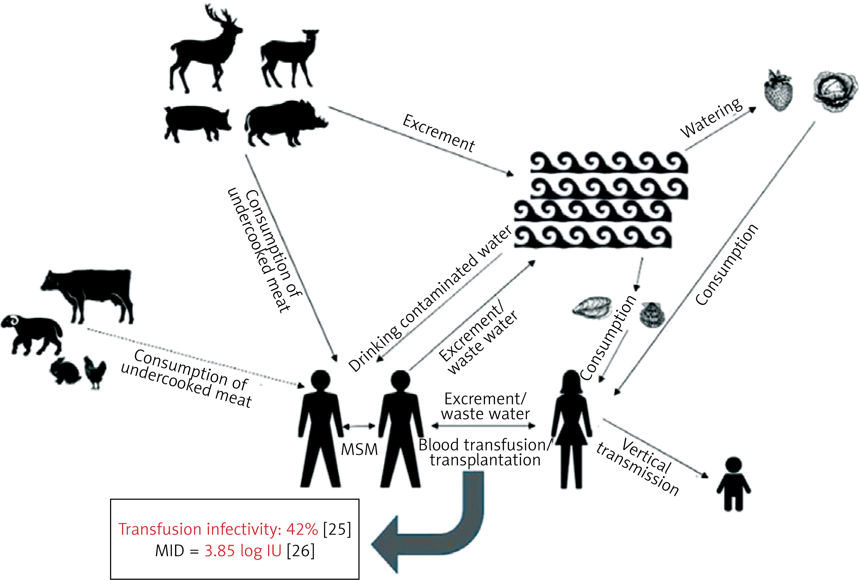
Transmission of HEV infection by transfusion of blood and its components has been repeatedly documented [21, 24]. Based on the analysis of transfusion cases of blood and its components infected with HEV, the average risk of transmission was determined at about 42% (the highest for plasma, lower for platelet concentrates and the lowest for red blood cell concentrates) [25], and the minimum infectious dose (MID) was estimated at 3.85 log IU, although it is emphasized that transmission is more likely to occur when the dose exceeds 4 log IU [26].
HEV transmission through transplantation has also been described, including liver [27] and kidney [28]. Transmission of infection through sexual contact seems likely, especially between men (MSM), as indicated by the higher frequency of anti-HEV IgG in the MSM population in Italy compared to a control group [29], but this route of HEV spread has not been confirmed so far [19].
HEV infection course and diagnosis
The infection course, taking into account appearance of infection markers, is schematically presented in Figure 2. The incubation period of HEV infection is usually 2–6 weeks. After about 3 weeks from infection, HEV RNA is detected in blood and feces, in which RNAemia persists for about 3–6 and 4–6 weeks, respectively. HEV RNA can be detected shortly before the onset of symptoms. The virus level doubles on average within 2.4 days, and the half-life is 1.6 days. Usually about 3 weeks after the onset of clinical symptoms, HEV RNA becomes undetectable in the blood, although it continues to be excreted in the feces for another 1–2 weeks. In studies of blood donors, HEV RNA was detected for up to 6 months [30–32].
Figure 2
HEV – typical infection profile (antibodies appearance in immunocompetent person) (based on [23, 41])
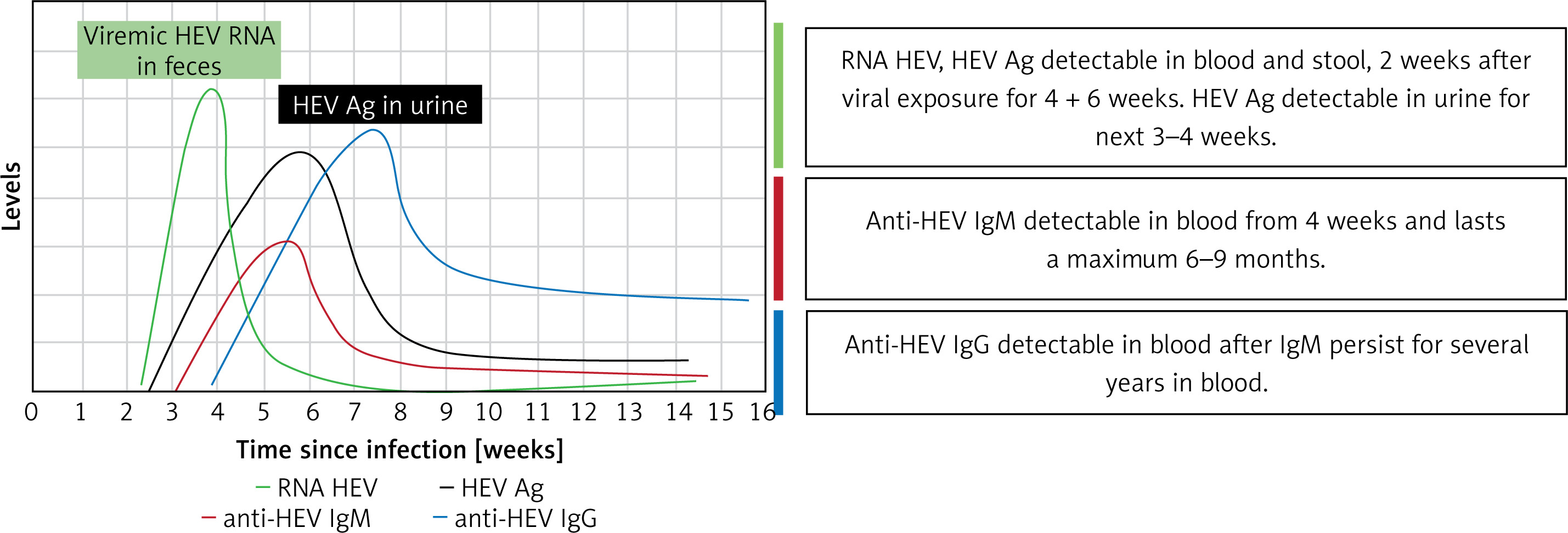
In immunocompetent persons, infection usually follows a similar course. Clinical symptoms may be accompanied by an increase in the activity of biochemical markers, and then antibodies – IgM and IgG – also start to appear at the same time [23]. The level of anti-HEV IgM starts to increase on average around day 33 and reaches its maximum concentration on day 36 from the detection of RNA. In most immunocompetent patients, this antibody isotype was no longer detectable after 6 months. The concentration of anti-HEV IgG starts to increase on average around day 32 and reaches a maximum concentration around day 53 from the appearance of RNA in blood. This antibody isotype was still detectable in most patients after 1 year [31, 33].
Another marker of infection, the diagnostic use of which is discussed in the literature, is the hepatitis E virus antigen (HEV-Ag) – a viral capsid protein detectable in blood during the serological window period and the acute phase of infection, persisting for 3 to 4 weeks after the symptoms of infection have subsided [34]. Recently, it was discovered that HEV Ag is specifically taken up from blood by renal cells and eliminated in urine, which is why the Ag concentration is > 10 times higher in urine than in blood and results in higher diagnostic sensitivity. Detectable Ag in urine was observed 6 days earlier than in serum and persisted longer than RNAemia and antigenemia in blood. In studies on rabbit models, detectability of Ag in blood showed good agreement with detectability of RNA in feces [35, 36].
In immunocompromised patients who develop chronic infection, HEV RNA persists in blood, feces, and body fluids for more than 3 months. It is believed that in this group there is a low or undetectable level of anti-HEV antibodies [37].
The phenomenon of HEV reinfection is not well understood. Based on previous observations, it seems to occur, although its scale remains unknown. Cases of HEV reinfection have been described in solid organ transplant recipients. Importantly, in this group, HEV reinfection may lead to chronic infection [20, 38, 39]. The results of an 8-year follow-up of blood donors in the German population showed that, among 495 HEV RNA(+) donors, as many as 78.4% did not have IgM or IgG antibodies against HEV, while 8.5% had only IgG antibodies. Interestingly, only 26.6% of HEV RNA(+) donors showed an increase in ALT activity. These data may indicate ongoing recurrent reinfections in the studied population [40].
Available diagnostic tests and their clinical application for differentiation of phases of infection and limitations, especially in patients with immunodeficiency, are presented in Table I. Diagnosis of HEV infection is based on the examination of a molecular (HEV RNA) and serological markers (IgM and IgG antibodies and HEV antigen). The detected markers allow differentiation between acute, chronic, and past infection phase; however, their usefulness differs between immunocompetent and immunodeficient patients (Table I) [30, 41, 42].
Table I
Diagnostic tests and their application [based on 30, 42]
| Test (method) | Phase of infection | Application in patients | Material for testing | |
|---|---|---|---|---|
| immunocompetent | with immunodeficiency | |||
| IgM anti-HEV (ELISA or rapid test) | Acute | Diagnostics# | Support in phase determination; however, application is limited (results should be interpreted with caution)* | Serum |
| IgG anti-HEV (ELISA or rapid test) | Acute, persistent, past | Diagnostics | Support in phase determination; however, application is limited (results should be interpreted with caution)* | Serum |
| HEV RNA | Acute, persistent | Diagnostics | Diagnostics | Plasma, serum, feces |
| Monitoring of response for treatment | Monitoring of response for treatment | Plasma, serum, feces | ||
| HEV-Ag (ELISA) | Acute, persistent | Differential diagnosis – Acute (lower level) – Persistent (higher level) | Diagnostics**, if HEV RNA not available | Serum, feces, urine^ |
^ current studies on effectiveness of urine tests mainly concern immunocompetent individuals [42],
The gold standard for diagnosis of acute and chronic infection in both immunocompetent and immunodeficient individuals is the HEV RNA test. HEV RNA in the acute phase can be detected in both blood and feces from the third to the sixth week of infection. HEV RNA present for more than 3 months indicates chronic infection [43, 44]. The HEV RNA test also allows us to monitor the reduction of infection and the effectiveness of antiviral treatment, as well as reinfections [45, 46]. It should be noted that molecular tests (both qualitative and quantitative) may differ in terms of analytical and clinical sensitivity declared by manufacturer. Analytical sensitivity of tests (the lowest RNAemia detected by the test) can be reliably determined and compared by analyzing the results of dilution tests of international WHO standards and secondary standards [47–49], and clinical sensitivity (the efficiency of detecting various polymorphic forms) can be compared by analyzing the results of reference panel tests (IRP) [50]. The most sensitive molecular tests detect even several IU of HEV RNA/ml [20].
Anti-HEV antibody tests remain an important element of HEV diagnostics in immunocompetent individuals. Anti-HEV IgM and IgG antibodies can be detected in up to 98% of this group of patients in the acute phase, provided that tests with high sensitivity and specificity are used [51].
High sensitivity of some tests, e.g. Wantai, results from the use of the µ chain coating strategy, which is used by a few manufacturers of anti-HEV tests [52–54]. The results of some studies indicate that the sensitivity of the so-called rapid anti-HEV IgM tests, e.g. All Diag, can be comparable to ELISA tests [51, 55]. Otherwise, in people with acute hepatitis E and reduced immunity, anti-HEV IgM and IgG antibodies were not detected in as many as 20% and 85% of infected people, respectively; therefore, in this group of patients, antibody testing should not be the basis for HEV diagnosis [56, 57]. Moreover, when analyzing the results of IgM isotype tests, it should be taken into account that cases of their persistence for more than 1 year were observed, and in people infected with e.g. EBV, CMV, HCV, there is a risk of a false reactive anti-HEV result; therefore, diagnosis and differentiation of the phase of HEV infection, even in immunocompetent people, should not be based solely on the anti-HEV IgM test [58–60]. The presence of only anti-HEV IgG antibodies indicates a past infection or vaccination. In people with past infection, the sensitivity of anti-IgG tests is estimated at 57.5–75% [51].
HEV ORF2 antigen is detected in blood and urine in both acute and chronic phases of infection, which may be helpful in diagnostics. HEV-Ag tests performed in serum allow for detection of infection with RNAemia at the level of 102–105 copies/ml, and are characterized by high sensitivity (80–94%) and specificity (86–100%) [61–63]. The results of studies using the new HEV-Ag test in urine indicate that its effectiveness in diagnosing HEV may be high compared to HEV RNA and anti-IgM tests, because the HEV-Ag level in urine is over 10 times higher than in blood [36]. However, it should be noted that in immunocompetent individuals a positive HEV-Ag result does not always correlate with presence of HEV RNA. For example, HEV-Ag was detected in patients treated with ribavirin even several dozen months after the elimination of HEV RNA [30]. Studies on HEV cell cultures suggest that the reason for the lack of correlation between the results of HEV antigen and RNA is the detection of glycosylated forms of ORF2 secreted in infected patients at high concentrations by the HEV-Ag test, in addition to the less abundant non-glycosylated form of ORF2 present in infectious virions [64]. The use of HEV antigen testing in diagnostics is still not well established, as it has been noted that this marker may be undetectable during the period of antibody development, and, moreover, little is known about its detection in immunocompromised individuals [65].
Epidemiological situation in Poland
Presence of the virus in the ecosystem
Since HEV infections are mainly spread by the oral route, data on the presence of the virus in food are particularly important. Our knowledge in this area is limited and largely indirect. Studies conducted on liver samples (n = 100) and blood (n = 146) of pigs obtained from slaughterhouses and retail outlets in central and eastern Poland showed the presence of HEV RNA in 1 and 5 samples, respectively [66]. In addition, viral RNA was detected in the blood of wild boars (25.8%) [67]. The presence of HEV genetic material on the surface of fruits and vegetables was also demonstrated – it was identified on 1 to 2% of leafy vegetables and strawberries, respectively [68, 69].
Detection of HEV infection markers in humans
HEV RNA and antigen
The results of tests of markers of ongoing (acute) infection (viral RNA and antigen) in humans and animals in Poland are presented in Table II [66, 67, 70–74]. The data on incidence based on HEV RNA testing to date come only from blood donor testing. The nationwide analysis conducted in 2015 covered nearly 13 thousand donations from first-time donors, which were tested individually. The genetic material of the virus (confirmed reactive result of the screening test) was found in 6 donors (1/2,109 donors/donations) [70]. From mid-2018 to the end of 2019, the Regional Blood Transfusion Center (RBTC) in Poznan tested donors whose plasma was intended for clinical use in England, where HEV RNA is a mandatory qualification test in blood donors. The tests were conducted in mini-pools, initially from 16 donations using the transcription mediated amplification (TMA) method, and then from 24 donations using the real-time PCR method. During screening of almost 40 thousand donations, a total of 10 infected donors were identified (1/approx. 3,900 donations) (data from the Institute of Hematology and Transfusion Medicine in Warsaw [IHTM] and RBTC Poznan, A. Bukowska, PhD). In Poland, according to the current recommendations [71], HEV RNA testing is not obligatory, but is recommended as it increases the safety of transfusions. In April 2024, the RBTC in Warsaw started testing for HEV RNA in IDT – 35 infections were detected in 51,782 donations (confirmed repeatably reactive [RR] results; frequency 1/1,479 donations) (data from IHTM and RBTC Warsaw, J. Gdowska, MSc).
Table II
Detection of markers of active HEV infection (RNA or Ag) in Poland
| Period of research | Area Tested group | Available demographic data | References | Method (format) | Number (frequency) of donations | |
|---|---|---|---|---|---|---|
| tested | reactive/confirmed | |||||
| 2012–2014 | North-west Poland Wild boars (blood) | Not applicable | Dorn-In et al. Foodborne Pathog Dis 2017 [67] | RNA, nested RT-PCR (IDT) | 163 | 42 (25.8%) |
| 2018–2019 | Central and eastern Poland Pigs | Not applicable | Bigoraj et al., Food Environ Virol 2021 [66] | RNA, rt-PCR (MP) | 146 | 5 (3.4%) |
| 2015 | Poland Blood donors | Random donors, ~60% > 40 years old, ~70% males | Grabarczyk et al. Transfusion 2018 [70] | RNA, TMA (IDT) | 12,664 | 6 (0.047%; 1 : 2,109) |
| 2018–2019 | Greater Poland Blood donors | As above | Bukowska A. (RCKiK Poznań) | RNA, TMA (MP16), rt-PCR (MP24) | 38,716 | 10* (0.026%; 1 : 3,872) |
| V–VIII 2024 | Mazovia Blood donors | As above | Gdowska J. (RCKiK Warszawa) | RNA, TMA (IDT) | 51,782 | 35 (0.1%, 1 : 1,438) |
| 2013–2016 | Podlasie With liver cirrhosis After transplantation With HIV | Age: mean 50.35; range: 21–80 years; 63.56% males | Parfieniuk-Kowerda et al. Arch Med Sci 2021 [72] | ELISA Ag | 180 180 90 | 3 (1.7%) 5 (2.8%) 0 (0%) |
| 2015–2018 | Greater Poland Hemodialyzed | Age: median 65.6; 57.1% males | Bura et al. J Med Virol 2020 [73] | ELISA Wantai HEV-Ag ELISA Plus | 189 | 0% |
| 2019–2023 | Mazovia Hematological After allo-HSCT | Age: median 62, mean 59, range 8–89 years; 56.8% males Age: median 49, mean 49, range 18–71 years; 52.34% males | Kalińska et al. Acta Haematologica Polonica 2024 [74] | RNA, rt-PCR ELISA Ag RNA, rt-PCR ELISA Ag | 148 166 | 0 (0%) 0 (0%) 0 (0%) 0 (0%) |
In several other studies conducted locally, the presence of the virus was also analyzed using direct methods (RNA or antigen detection). Infection markers (Ag by ELISA without verification) were detected in single patients from Podlasie with liver cirrhosis (1.7%) and after transplantation (2.8%) [72], but they were not present in hemodialysis patients from Wielkopolska [73] or in hematological patients, including after allo-HSCT [74].
The number of studies in which the polymorphic form of the virus occurring in Poland was determined is still limited. As expected, HEV donors were carriers of genotype 3 infections – subtypes 3i and 3c [70]. Subtype 3i was identified in wild boars, while subtypes 3f and 3e were identified in the liver and blood of pigs [66, 75]. Moreover, subtypes 3i and 3c and mixed infections with different/two subtypes were detected in pig feces [76].
IgM anti-HEV isotype
Knowledge about the incidence can be supplemented by the results of tests for anti-HEV IgM antibodies appearing in the early phase of infection and persisting for the first few months. Data for populations of blood donors (A), other symptomless person (B), and patients (C) are summarized in Table III [70, 72–74, 77–82]. In the only nationwide study conducted so far, the serological marker of acute infection was detected in 0.8% of donations collected in 2015 [70].
Table III
Detection of serological markers of acute HEV infection (anti-HEV IgM) in Poland
| Period of research | Area Tested group | Available demographic data | References | Method of | Number (%) of samples | ||
|---|---|---|---|---|---|---|---|
| Screening | Confirmatory testing | Tested | Positive | ||||
| A. | |||||||
| 2015 | Poland Blood donors | Age: 50 donors from each group 18–27, 28–37, 38–47, 48–57, > 57 years; ~70% males | Grabarczyk et al. Transfusion 2018 [70] | Wantai HEV-IgM ELISA | recomLine HEV IgM/IgG immunoblot test, Mikrogen | 3,079 | 39 (0.8%) |
| Before 2018 | Greater Poland Blood donors | Age: mean ± SD 44.1 ±6,5, range 29–58, median 43.5 | Bura et al. Adv Clin Exp Med 2018 [82] | Anti-Hepatitis E Virus ELISA [IgM], Euroimmun | No | 90 | 0 (0%) |
| 2020 | Mazovia Blood donors | IHTM data | Rapid IgM HEV Test (Colloidal Gold Device), Wantai | Wantai HEV-IgM ELISA | 650 | 3 (0.5%) | |
| B. | |||||||
| Before 2008 | Białystok Indian students | Age: mean 24.4 ±0.56, range 18–48; n = 45 males | Jaroszewicz et al. Przegląd Epidemiologiczny 2008 [81] | DI.PRO, Diagnostics Bioprobes Sri. | No | 45 | 7 (15.6%) |
| 2010–2012 | Poland Hunters | No data | Sadkowska-Todys Przegląd Epidmiologiczny 2015 [77] | recomWell HEV IgM, Mikrogen | recomLine HEV IgM/IgG immunoblot test, Mikrogen | 1,027 | 3 (0.3%) |
| 2010–2012 | Poland Hunters | Age: 38–312 person per each age group (< 30, 31–40, 41–50, 51–60, 61–70, > 71); 97.7% males | Baumann-Popczyk et al. Med Microbiol Immunol. 2017 [78] | recomWell HEV IgM, Mikrogen | recomLine HEV IgM/IgG immunoblot test, Mikrogen | 1,021 | 5 (0.5%) |
| 2016 | Poland Soldiers | Age: range 26–57 years; 93.7% males | Korzeniewski et al. Int Marit Health. 2018 [79] | Anti-Hepatitis E Virus ELISA [IgM], Euroimmun | recomLine HEV IgM/IgG immunoblot test, Mikrogen | 253 | 2 (0.8%) |
| Before 2018 | Greater Poland foresters | Age: mean 45 ±9.6; median 44.5 years; 70.8% males | Bura et al. Adv Clin Exp Med 2018 [82] | Anti-Hepatitis E Virus ELISA [IgM] Euroimmun | MP Diagnostocs ASSURE HEV IgM Rapid Test | 48 | 1 (2.1%) |
| C. | |||||||
| 2013–2016 | Podlasie With liver cirrhosis After transplantation With HIV | Age: mean 50.35, range 21–80 years; 63.56% males | Parfieniuk-Kowerda et al. Arch Med Sci 2021 [72] | Wantai HEV-IgM ELISA | No | 180 180 90 | 9 (5%) 2 (1.1%) 1 (1.1%) |
| 2015–2018 | Greater Poland Hemodialyzed | Age: median 65.6 years; 57.1% males | Bura et al. J Med Virol 2020 [73] | Wantai HEV-IgM ELISA | No | 189 | 0% |
| 2015–2019 | Mazovia With AIH | Age: median 34, range 18–83 years; 68% females | Janik et al. Pol Arch Intern Med 2024 [80] | Anti-Hepatitis E Virus ELISA [IgM] Euroimmun | No | 379 | 5 (1.3%) |
| 2021–2023 | Mazovia Hematological | Age: median 62, mean 59, range 8–89 years; 56.8% males | Kalińska et al. Acta Haematologica Polonica 2024 [74] | Wantai HEV-IgM ELISA | No | 148 | 0 (0%) |
In other studies on healthy individuals from different regions of Poland, a comparable percentage of positive results was obtained: in 0.3% to 0.5% of hunters (samples collected slightly earlier (2010–2012) [77, 78] than in the case of the above-mentioned donors) [70] and in 0.8% of soldiers (2016) [79].
More varied results were observed in patients. Anti-HEV IgM ranged from 0% in hemodialysis [73] and hematological patients [74], through about 1% in immunocompromised patients (with HIV and after transplantation) [72] and with autoimmune hepatitis [80], up to 5% in patients with liver cirrhosis [72]. Differences in results between blood donors and patients should be interpreted with caution, because in the second group, unlike donors, the specificity of reactive results in ELISA was not verified in WB. Usually, the percentage of confirmed positive results is lower among unverified samples than among samples verified by WB. Moreover, a large proportion of patients had, to a greater or lesser extent, impaired immunity and the ability to produce antibodies in response to infection. For that reason, we cannot exclude the possibility that a larger proportion than indicated by the test results had been infected in the months preceding the sample collection.
The frequency of anti-HEV IgM (15.6%) identified in the group of Hindus studying in Bialystok clearly differs from the range of the results quoted. During this first population study of specific anti-HEV antibodies in Poland, an appropriate confirmatory test was not yet available. Additionally, the authors note that seropositivity was most likely a consequence of infections that occurred during stays in Asia [81].
IgG anti-HEV isotype
When analyzing the results of anti-HEV IgG antibody studies, several associations from previous observations, including Polish research, should be taken into account. The frequency of antibodies increases with age and, as shown by the results of some studies, is higher in men [70]. Therefore, the demographic structure of the studied group may have a significant impact on the obtained results. Moreover, as shown in the section on diagnostics, the percentage of seropositive results depends on the test used. It was noted that some tests may be characterized by higher sensitivity and therefore give a higher percentage of reactive results in population studies than tests with lower sensitivity.
A summary of screening results of anti-HEV IgG antibodies, indicating a past infection in Poland in animal and human populations (both healthy individuals and patients), is presented in Table IV [67, 70, 72–74, 77–80, 82–89]. The results of tests in humans are discussed in the following section. Especially, we focused on the results obtained using the most specific and sensitive Wantai test, which has been used in numerous studies worldwide for assessment of donor populations.
Table IV
Detection of serological markers of past HEV infection (anti-HEV IgG) in Poland
| Period of research | Area Tested group | Available demographic data | References | Method of | Number of (%) samples | ||
|---|---|---|---|---|---|---|---|
| Screening | Confirmatory testing | Tested | Positive | ||||
| Animal populations | |||||||
| 2012–2013 | Poland Wild boars Deer Roe deer Bison Chamois Bear | Not applicable | Larska et al. Zoonoses Public Health 2015 [83] | ID Screen Hepatitis E multi species indirect ELISA (ID.vet, France) | No | 261 118 38 68 4 1 | 116 (44.4%) 0% 0% 0% 0% 0% |
| 2012–2014 | North-west Poland Wild boars (blood) | Not applicable | Dorn-In et al. Foodborne Pathog Dis 2017 [67] | 163 | 28 (17.2%) | ||
| 2014–2015 | Poland Wild boards Pigs | Not applicable | Weiner et al. Vet Res 2016 [84] | PrioCHECK HEV Ab porcine (Prionics, Switzerland) | No | 290 143 | 90 (31%) 63 (44.1%) |
| Healthy human populations | |||||||
| 2015 | Poland Blood donors | Age: 50 donors from each group: 18–27, 29–37, 38–47, 48–57, > 57; ~70% males | Grabarczyk et al. Transfusion 2018 [70] | Wantai HEV-IgG ELISA | No | 3,079 | 1340 (43.5%) |
| 2015 | Greater Poland Blood donors | Age: mean 37.7 ±7.7, range 18–55 years; 84.4% males | Bura et al. Adv Clin Exp Med 2017 [85] | Anti-Hepatitis E Virus ELISA [Ig] Euroimmun | No | 105 | 4 (3.8%) |
| 2015 | Greater Poland Blood donors | Age: mean 38.4 ±7.7, median 40 years; 68.3% males | Bura et al. Pol J Microbiol 2018 [86] | Wantai HEV-IgG ELISA | No | 110 | 67 (60.9%) |
| 2015–2016 | Greater Poland Blood donors | No data on age, 65.5% males | Bura et al. Int J Infect Dis 2017 [87] | Wantai HEV-IgG ELISA | No | 246 | 122 (49.6%) |
| Before 2018 | Greater Poland Blood donors | Age range: 18–55 years | Bura et al. J Med Virol 2018 [88] | Anti-Hepatitis E Virus ELISA [Ig] Euroimmun Cut of ≥ 2.2 IU/ml Cut of ≥ 0.8 IU/ml Wantai HEV-IgG ELISA | No | 153 | 7 (4.6%) 35 (22.9%) 86 (56.2%) |
| 2010–2012 | Poland Hunters | No data | Sadkowska-Todys et al. Przegląd Epidemiologiczny 2015 [77] | recomWell HEV IgG (Mikrogen) | recomLine HEV IgM/IgG immunoblot test (Mikrogen)** | 1,027 | 206 (25%) |
| 2010–2012 | Poland Hunters | Age: 38–312 person per each age group (> 30, 31–40, 41–50, 51–60, 61–70, > 71); 97.7% males | Baumann-Popczyk et al. Med Microbiol Immunol. 2017 [78] | recomWell HEV IgG (Mikrogen) | No | 1,021 | 227 (22.2%) |
| 2014 | Greater Poland Foresters | Age: mean 45 ±9.6, range 29–65 years, median 4.5 | Bura et al. Adv Clin Exp Med 2018 [82] | Anti-Hepatitis E Virus ELISA [Ig] Euroimmun | No | 48 | 0 (0%) |
| 2016 | Poland Soldiers | Age range 26–57; 93.7% males | Korzeniewski et al. Int Marit Health 2018 [79] | Anti-Hepatitis E Virus ELISA [Ig] Euroimmun | recomLine HEV IgM/IgG immunoblot test (Mikrogen)** | 253 | 16 (6.3%) |
| Patient populations | |||||||
| 2013 | Greater Poland From the Infectious Diseases Clinic | Age: mean 47.2 ±14.2, range 19–85 years, 55.5% males | Bura et al. Postepy Hig Med Dosw 2015 [89] | EIA-gen HEV IgG kit (Adaltis) | No | 178 | 29 (16.3%) |
| 2013–2016 | Podlasie With liver cirrhosis After transplantation With HIV | Age: mean 50.35; range 21–80; 63.56% males | Parfieniuk-Kowerda et al. Arch Med Sci 2021 [72] | Wantai HEV-IgG ELISA | No | 180 180 90 | 87 (48.3%) 73 (40.6%) 34 (37.7%) |
| 2015 | Greater Poland With HIV | Age: mean 37.7 ±7.7; range 18–55 years; 84.8% males | Bura et al. Adv Clin Exp Med 2017 [85] | Anti-Hepatitis E Virus ELISA [Ig] Euroimmun | No | 105 | 1 (0.95%) |
| 2015–2016 | Greater Poland With HIV | Age: range 18–55; 84% males | Bura et al. Int J Infect Dis 2017 [87] | Wantai HEV-IgG ELISA | No | 244 | 124 (50.8%) |
| 2015–2018 | Greater Poland Hemodialyzed | Age: median (Q1–Q3) 65.6 (55.6–74.2); 57.1% males | Bura et al. J Med Virol 2020 [73] | Wantai HEV-IgG ELISA | No | 189 | 94 (49.7%) |
| 2015–2019 | Mazovia With AIH^ | Age: median (IQR): 34 (18–83) | Janik et al. Pol Arch Intern Med 2024 [80] | Anti-Hepatitis E Virus ELISA [IgM] Euroimmun | No | 374# | 55 (14.7%) |
| 2021–2023 | Mazovia Hematological | Age: median 62, mean 59; 56.8% males | Kalińska et al. Acta Haematol Pol 2024 [74] | Wantai HEV-IgM ELISA | No | 148 | 66 (44.6%) |
So far, the results of two nationwide studies on detection of the anti-HEV IgG isotype in animals have been published. They indicate the commonness of infections in wild boars and pigs/swine. Anti-HEV IgG was detected using tests from different manufacturers in 31% to 44.4% of wild boars and in 44.1% of pigs; however, it was not detected in other wild animals (deer, roe deer, bison, etc.) [83].
Population studies of IgG anti-HEV in humans
The only nationwide analysis of humans showed anti-HEV IgG seropositivity in an average of 43.5% of donors – from 30% in Podlasie to 60% in Greater Poland [70]. Other studies performed locally in donors in Greater Poland confirmed a high prevalence of antibodies to HEV (from 49.6% to 60.9%) [85–88].
A high percentage of antibodies indicating a past infection was also identified among patients treated in Poznań. Specific IgG were detected in 37.7-50.8% of HIV patients [72, 87], 40.6% of transplant patients [72], 48.3% of patients with liver cirrhosis [72], and 49.7% of hemodialysis patients [73]. In Mazovia, despite treatment and the underlying disease significantly reducing immunity, anti-HEV IgG was detected in 44.6% of samples collected from hematological patients in 2021–2023 [74].
Other nationwide studies were conducted using tests from other manufacturers (Mikrogen, Euroimmun) than in the case of the abovementioned studies of donors and patients (Wantai). These analyses showed HEV IgG seropositivity in 22.2–25% of hunters [77, 78] and 6.3% of soldiers [79].
Epidemiological situation in Poland compared other European regions and globally
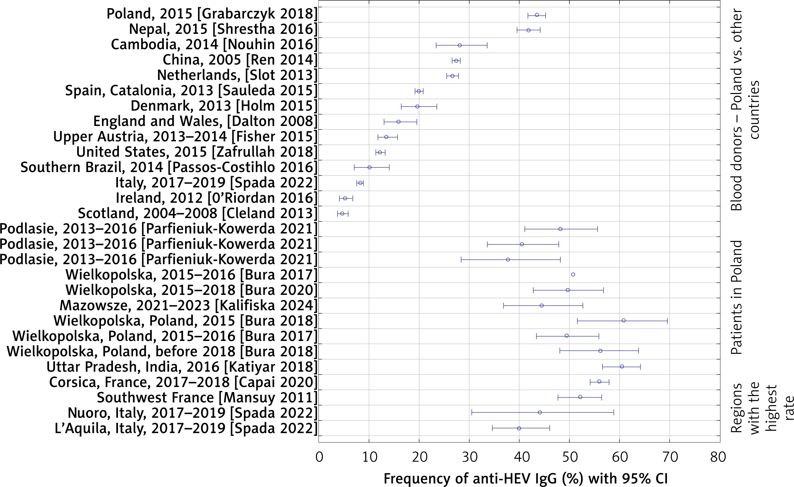
The prevalence of anti-HEV IgG (Wantai test) in patients and in blood donors in Poland, compared with other countries and regions worldwide, is presented in Figure 3. Based on the highest seroprevalence rates reported to date in blood donors using the Wantai assay, which is considered one of the most sensitive available tests, Poland appears to have the highest seroprevalence in Europe. Over 40% of blood donors nationwide have experienced prior infection, which represents the highest seroprevalence among European countries and one of the highest worldwide [90–93]. The Polish results are comparable only to Nepal [94], where testing was performed shortly after the catastrophic earthquakes, which probably influenced the epidemiological situation. Moreover, it can be assumed that the dominant genotype transmitted in Nepal was genotype 1, while in Poland only genotype 3 infections have been documented so far. Also, the frequencies of anti-HEV IgG antibodies observed locally, in the Greater Poland province (approx. 60%) are among the highest. The seroprevalence in this area exceeds the extreme frequencies observed in southern France (39.1%) [95] and two provinces in Italy (40–44%) [96] and are comparable only with Corsica (56.1%) [97] and northern India (60.5%, expected genotype 1) [98] (Figure 3).
Figure 3
Frequency of anti-HEV IgG (Wantai test) in patients and blood donors in Poland and in selected countries and regions of the world (country/region, year of study, references)
The epidemiological data presented here allow for a preliminary estimate of HEV incidence in Poland. For this purpose, seroprevalence of specific IgM is helpful, as it is a marker of acute infection and remains detectable in human blood for approximately 6 months. Extrapolating results of nationwide studies of donors (0.8%) to the whole population of Poland (38 million), one of the pioneers of research on genotype 3 HEV in Europe, Dr. John Dalton, estimated the number of new/acute HEV infections per year at approximately 400 thousand [99]. More conservative estimates can be obtained using the lower percentage of IgM positive results obtained in other locally conducted studies (0.3–0.5% – Table III) and assuming that this isotype of specific antibodies may persist for up to 12 months rather than an average of 6 months. Under such assumptions, the number of predicted infections would range from about 113 to 189 thousand cases per year (1 year × 37.72 million inhabitants [100] × 0.3–0.5% [percentage of IgM positive infections per year] = 113,160–188,600). Therefore, based on more or less conservative estimates, we can expect from about 113 to 400 thousand infections per year in Poland, respectively.
It is worth noting that in Poland, unlike in most European countries [101], HEV is not subject to mandatory epidemiological surveillance. Therefore, in our country we do not have reliable data on the epidemiology of infections, especially symptomatic ones. It is known that acute infections identified in blood donations and at least some infections diagnosed in infectious disease clinics (RNA-positive) are reported to epidemiological surveillance and then presented in the category of “other and unspecified hepatitis” (item 107) in the NIZP-PZH report published every 2 weeks [102]. Even if we assume that all cases reported in this way concern HEV (and in 2023 there were 59 reports classified as such) and cautiously estimate that no more than 1% of infections have a significant clinical course requiring treatment (1% out of 113,160–400,000 estimated infections per year in the whole country = 1,130–4,000 symptomatic cases), it appears that the number of diagnosed infections is significantly underestimated (from 19 to 68 times).
Currently, no more than several dozen cases of hepatitis E or other symptomatic HEV infections are diagnosed in Poland each year, while in Western European countries with significantly lower seroprevalence, many times more symptomatic cases are reported. Over the period 2005–2015, a total of 21,018 confirmed hepatitis E cases were reported from 22 countries. The largest numbers of confirmed cases, accounting for 80% of all cases reported, were from 3 countries – Germany, France, and the United Kingdom [5].
Clinical significance
HEV genotype 3 can lead to asymptomatic infections in the majority of patients. Less than 5% may develop acute self-limiting hepatitis, sometimes with jaundice. After an incubation period of 3–8 weeks (on average about 4 weeks), prodromal general symptoms may occur. These include a subfebrile state or fever, increasing fatigue, weakness, loss of appetite, nausea, vomiting, and muscle and joint pain, which are accompanied by pain in the right hypochondrium. In some patients, jaundice with pruritus, hepatomegaly, dark urine, and discolored stools may appear. Laboratory tests performed during this period show a constant increase in transaminase activity. The peak of HEV viremia usually precedes the peak of transaminase activity by about 6 weeks. The course of the disease is significantly influenced by the status of immune response and pre-existing liver diseases. In immunocompetent patients without concomitant liver diseases, acute HEV infection is self-limiting. Symptoms usually subside after 1–2 weeks along with transaminase activity, which usually normalizes after several weeks. Single cases of HEV viremia lasting more than 2 years have been observed in immunocompetent patients, but this was not associated with the progression of liver fibrosis. Symptoms that may accompany HEV infection were grouped in Table V into the category of different clinical manifestations: hepatic, hematologic, neurological, nephrological, and other [21, 23, 103].
Table V
Clinical symptoms of HEV infection [based on: 21, 23, 103]
A significant clinical problem is the overlap of acute HEV infection with underlying liver disease, especially in the phase of advanced fibrosis or hepatic cirrhosis. This often leads to decompensation of liver function, which sometimes may progress to acute, life-threatening organ failure [22, 23].
In acute infection with HEV genotype 1 during pregnancy, a particularly severe hepatitis progressing to fulminant liver failure and death was noted in up to 20–25% of infected women [41].
Immunosuppression of various etiology, particularly after solid organ transplantation, has a significant impact on the immunological control of HEV replication. In 50–60% of immunocompromised patients, infection with HEV genotype 3 or 4 becomes chronic, which is defined as prolonged detectability of HEV RNA in blood for over 6 months. However, subsequent observational studies have shown that in some solid organ recipients, spontaneous elimination of HEV RNA occurred up to 3 months after infection. The likelihood of spontaneous elimination of HEV RNA in the period from 3 to 6 months after infection was very low [104]. Hence, the current recommendations of experts from the European Association for the Study of the Liver (EASL) indicate that patients in whom HEV replication lasts > 12 weeks should be considered as chronically infected with HEV [23]. Symptoms of chronic HEV infection are non-specific. Most frequently (in 1/3 of patients), progressive, prolonged fatigue was observed. In the majority of patients, a chronic moderate increase in transaminase activity was observed. In some, transaminase activity remained within the normal range, serological markers of HEV infection were undetectable, and the only test confirming the infection was the detection of HEV RNA in blood [105]. It is estimated that approximately 10% of patients with chronic hepatitis E will develop liver cirrhosis within 1–2 years, which may result in liver failure and death of the patient or the need for liver transplantation [23].
Hepatological symptoms of acute or chronic hepatitis E may be accompanied by extrahepatic manifestations, which reflect the extrahepatic HEV replication and immunological damage to tissues and organs. Neurological symptoms such as bilateral amyotrophic neuralgia (pain, paresthesias, and muscle weakness in the shoulder girdle and arm) or Guillain-Barré syndrome more frequently affect immunocompetent individuals infected with genotype 3 HEV. However, these complications have also been observed in immunosuppressed patients with chronic hepatitis E. In patients with immunodeficiencies, extrahepatic nephrological complications (glomerulonephritis, IgA nephropathy) and hematological complications (monoclonal gammopathy in 25% of individuals with acute HEV infection, thrombocytopenia, cryoglobulinemia, aplastic anemia, or hemolytic anemia) are more common. Additionally, cases of pancreatitis, arthritis, myocarditis, or thyroiditis have been reported for genotype 1 HEV infections [21, 23] (Table V).
It is estimated that 3–13% of patients with suspicion of drug-induced liver injury (DILI) are infected with HEV. Therefore, testing for HEV is highly recommended in this group [103]. In our own material (unpublished data), 25% of patients diagnosed with viral hepatitis E were admitted to hospital with an initial diagnosis of acute drug-induced liver injury.
The disease caused by HEV is self-limiting, and treatment is primarily symptomatic, based on maintaining the water and electrolyte balance, vitamin supplementation, ursodeoxycholic acid (UDCA) in patients with cholestatic hepatitis and pruritus, and avoiding paracetamol and other potentially hepatotoxic drugs. Specific therapeutic interventions, such as ribavirin, may be considered only in patients with acute liver failure or chronic hepatitis E in the immunocompromised population. Single reports indicate that in severe acute hepatitis E in patients with underlying liver diseases, ribavirin usage shortened the period of HEV viremia and accelerated convalescence. Patients with hepatitis E and fulminant liver failure may require liver transplantation [22, 23]. Clinical reports indicate the efficacy of pegylated interferon α in inhibiting HEV replication; however, due to its immunomodulatory effect, most patients with chronic, complicated hepatitis E have contraindications to this therapy. Single reports indicate a potential beneficial effect of sofosbuvir in inhibiting HEV replication; however, emerging resistant variants are of specific concern [22, 23]. Several compounds that inhibit HEV replication by targeting either host or viral factors are currently in the pipeline.
Infection prevention
Infection prevention is particularly important in the case of groups at risk of more significant complications in the course of HEV infection: in patients with liver disease (including cirrhosis, hepatitis B and C, etc.) and patients with reduced immunity due to the underlying disease (cancer), immunosuppressive treatment (especially in transplant patients) and HIV infection. The significance of HEV infection in pregnant women has been discussed. It is known that genotype 1 infections are particularly dangerous, but the significance of genotype 3 identified in developed countries, including Poland, in this group is not fully known [22, 41].
The primary prevention of diseases transmitted through the fecal-oral route involves improving sanitary conditions, access to clean drinking water, proper sewage disposal, and educational measures that prevent virus transmission within the population, such as the habit of washing hands with clean water before meals.
Prevention of zoonotic infections (HEV genotypes 3 and 4) includes maintaining good hygiene, washing hands after contact with animals or their feces, and proper cooking of meat. Particular caution is recommended regarding consumption of pork and wild game that has not been adequately fried or baked. For these meat products, careful and as long as possible heat treatment is recommended [22, 41].
Another potentially significant source of infection is transfusions of blood, its components, and blood products. Thus, blood services in many countries, mainly European ones, have introduced HEV RNA screening for donations intended either for all recipients or only those belonging to risk groups. In Poland, there is no such requirement so far; however, some RBTCs (e.g. in Warsaw) have introduced multiplex tests for donor screening, which, in addition to detecting HBV DNA, HCV RNA and HIV RNA, identify HEV RNA. An additional measure to reduce the risk of HEV transmission through transfusion may be the use of inactivation methods. A significant limitation is the lack of such a method for whole blood and red blood cell concentrates and limited effectiveness ensuring a smaller reduction in infectivity/replication capacity (2–3 log) than in the case of enveloped viruses such as HBV, HCV, or HIV [21, 23, 25, 92, 103].
Intensive work on an effective vaccine against hepatitis E is underway. So far, four recombinant vaccines have been developed, all of which use ORF-2 antigens administered in three doses over 6 months for immunization. Three vaccines were constructed using the genotype 1 antigen, and one using genotype 4. The most advanced work is on the Chinese Hecoline vaccine using the genotype 4 antigen. The results of four phases of clinical trials indicate high effectiveness against viruses belonging to genotype 4; it has a weaker effect against genotypes 1 and 2; and the effectiveness of the developed vaccines against genotype 3 remains unknown. Vaccination of risk groups and people in areas of HEV epidemics is being considered. An important observation is that vaccinations do not lead to the development of long-lasting immunity, but prevent progression of HEV infection to symptomatic hepatitis. Furthermore, it has been shown to be safe for pregnant women [106].
Discussion
The presented data indicate a high incidence of HEV among people in Poland, with significant underestimation of epidemiological surveillance data. Therefore, it is necessary to expand knowledge of the epidemiology and clinical significance among medical personnel, increase the availability of diagnostics, and implement mandatory separate reporting of hepatitis E. In terms of prevention, primary efforts should be directed toward protecting high-risk groups of patients at high risk of clinical complications from HEV infection – transfusion and transplant recipients (by implementing HEV RNA screening in donors of blood and blood components, as well as organs and tissues). To explain the phenomenon of extremely high seroprevalence in Poland and develop strategies to limit the infection epidemic, further research on the sources of HEV infection in Poland – food, water, etc. – is desirable. More accurate forecasting of the infection epidemic in Poland and other countries will be possible once more detailed data become available to confirm or rule out reinfection and characterize its features. The Polish experience appears to be broadly applicable and may be useful for other countries, especially developed ones.

