Introduction
Common bile duct (CBD) disease presents a momentous risk to the patient, since it may lead to biliary colic, pancreatitis or cholangitis and obstructive jaundice. Traditionally, surgeons used T-tube drainage for removal of CBD lesion or stones. Several researchers suggest that a T-tube drainage system allows the edema or spasm of the sphincter with the trauma of exploration. Despite the advantage of the T-tube, several disadvantages are associated with it, including bile leakage and CBD obstruction seen after CBD surgery. Common bile duct surgery by T-tube requires the patient to be under constant observation with restriction of food habit. There is always a high chance of infection after CBD treatment, especially when the T-tube is removed, a reason for more risk to the patient. For avoidance of T-tube complications, the surgeons and researchers now focus on an alternative to treat CBD.
Day by day, biomaterial technology is gaining more popularity due to the lack of adverse reaction in and on the tissue. Biodegradable implant material has the advantage that it can easily be absorbed, gradually dissolved, and there is no need for surgical removal after surgery. Traditional metallic materials such as cobalt-chromium, stainless steel and nitinol alloys have been commonly used in biomaterial technology [1]. Biomaterial such as magnesium (Mg) and zinc (Zn) both have good biocompatibility and consequently make good potential material for medical implants [2]. Biodegradable Mg alloy has more potential due to its excellent mechanical properties and good biocompatibility with the tissues [3]. Nowadays magnesium based biomaterial has been most commonly used as a biodegradable medical implant device due to excellent mechanical properties [4, 5].
The flow rate plays an important role during biodegradation of Mg and its alloys. The various investigations reported that reduced flow rate compositions may result in excellent corrosion resistance in a bare mate, involving the application in other non-vascular fluid conditions [6–8]. On the basis of the above facts, the researchers focused their research mainly on Mg and its alloy (non-vascular endoluminal stents). Pure Mg has a corrosion rate of 0.01594 ml/cm2/h for 10 days mainly in the esophageal stent. Another report suggests that Mg has a relaxing factor in airway smooth muscles of rabbits. One study on a stent (Mg-3Y) implanted into the trachea of a canine rodent model showed no adverse effect found in the structure of adjacent cartilage after 8 weeks [4, 5, 7, 9]. JDBM alloy was implanted for 1–2 years as a tracheal stent in rodent (rabbits) and induced tracheomalacia via coating the multiple layers. Also, Mg-based alloys used as an intestinal stent have exhibited a significantly higher apoptosis rate when implement as intestinal epithelial cells. In vitro investigation of AZ31 stent used in an artificial urine system has shown good antibacterial activity and it is used as a ureteral stent [4, 9].
It is necessary to keep supporting material in the CBD to protect it from post-operative CBD stenosis. This has led to adverse immunological and inflammatory responses [10, 11]. These reactions commonly trigger apoptosis necessary to remove the inflamed and damaged tissue and maintain the regeneration and healing mechanisms [11, 12]. Necrosis is generally induced by cell poison effects and apoptosis showed the effect on the cell biological and morphological features. Apoptosis plays a defensive role in immune reaction or cell damage via noxious or disease agents [10–12]. Dissimilar to apoptosis, the proteolytic enzyme (cytoplasmic content) of secretion of necrotic cells enters into the surrounding tissue and induces the inflammation. Necrosis frequently arises in pathological changes, whereas apoptosis is a normal physiological event [13–15].
Materials such as titanium, polymer and silicone rubber are most commonly employed as CBD supporting systems. Silicon rubber stent (T-tube) can cause complications such as dehydration, biliary fistula, and incapability to clean the drain tube in CBD [16, 17]. Another stent such as polymeric and titanium alloy can induce the accumulation of bacteria, epithelial hyperplasia, and bio-film sludge that need to be eliminated. Due to the limitation of the above-discussed stent, different types of biodegradable CBD stent have been intended to replace the non-degradable stents [18, 19]. The biodegradable stent has the advantage in cleaning, reduction in deposition of biofilm with size alteration. Several types of research suggest that magnesium alloy is the best biodegradable implanting material for its biocompatibility and excellent mechanical property. Magnesium alloy stents are mostly used in surgical clips, vascular stents, bone implants, surgical sutures, and staples.
There are limitations for the animal experimental studies, as they are often used in big animals such as dogs or pigs (usually they use 4–6 animals). A big animal has two disadvantages: first, they are too expensive and limit animal use in the study, and second they have a limited number of antibodies against the big animals, which adds a more difficult molecular experiment in CBD. In the current investigation, we tried to explore the effect of AZ31 alloy on necrosis or apoptosis via in vitro and in vivo experimental studies. Primary mouse extrahepatic bile epithelial cells (MEBECs) were able to show a concentration base effect of AZ31 alloy stent to investigate the apoptotic gene expression such as tumor necrosis factor-α (TNF-α), B-cell lymphoma 2 (Bcl-2), Bax, NF-κB and caspase-3. Further, we investigated the long term effect of AZ31 alloy stent on different biochemical, hematological and non-hepatic parameters.
Material and methods
Material and extract preparation
Biodegradable stent AZ31 tubes were prepared as extruded AZ31 alloy, with purity of Zn 99.999% and Mg 99.99%. The composition of AZ31 alloy is presented in Table I.
Table I
Chemical composition of AZ31 alloys bile duct stent and composition of AZ31 stent
| Material | Chemical compositions (wt%) | |||||
|---|---|---|---|---|---|---|
| Al | Zn | Mn | Si | Fe | Ca | |
| AZ31 | 3.1 | 0.73 | 0.25 | 0.02 | 0.005 | 0.0014 |
A high purity crucible with high temperature (700–750°C) was used for the preparation of AZ31 alloy in the muffle furnace. After 30 min, temperature 700°C, metal was melted and transferred into the steel mold at a high temperature. Cover gas such as argon (99.99% purity) was employed throughout the casting and melting processes. The solid solution of AZ31 alloy treated at 350°C for 120 min followed by water treatment. Finally, the hot treated alloy sample was hot extruded at 250°C at an extrusion ratio (8 : 2). Finally, the AZ31 was placed into tube-shaped samples for experimental study. Tube shaped samples of AZ31, with dimensions of 1.0 mm (luminal diameter), 0.1 mm (thickness) and 5 mm (length), were used throughout the experiment. In a prior experimental study, all the tubes were polished with hydrochloric acid (2 ml), glycerol (20 ml), acetic acid (5 ml) and nitric acid (3 ml) and washed with distilled water and ethanol. Subsequently, the tubes were re-sterilized and weighed [20, 21].
In vitro model
Briefly, modified Dulbecco’s Eagle’s medium (DMEM) suspended into glycerine and fetal bovine serum was used as extraction medium. The extraction medium (1.25 cm2/ml) and the immersed sample (AZ31) were kept in a humidified area (CO2 5%) at 37°C for 1 day. After that, the extraction medium was collected and the supernatant was successfully removed, and centrifuged and stored at 4°C in a refrigerator.
For observing the concentration-dependently effect, the extract was serially diluted (100–20) with DMEM. We used inductively coupled plasma-atomic emission spectroscopy (ICP-AES) for the determination of the concentration of released Zn and Mg ions in the AZ31 alloy extract [22].
Surgery
New Zealand rabbits (3.0 ±0.5 kg) were used in our animal study with approval from the institutional ethical committee. The rabbits were kept in a single cage and received the standard diet and water ad libitum.
Animals were fasted overnight (10 h) before surgery. Rabbits were anesthetized by iv injection of sodium pentobarbital (30 mg/kg, b.w.). The epigastrium tissue was cut (layer by layer), the gastrointestinal tract (GI tract) was pushed and the duodenum was picked up to locate the common bile duct, with the expanded connection of the duodenum and common bile duct visible. A bile duct stent (AZ31) was placed in the common bile duct through an angle incision (2 mm) at the anterior wall of the bile duct. The incision and epigastrium were sealed by suture followed by 5 days of intramuscular injection of penicillin. After the surgery, the rabbits were sacrificed at a regular time interval for analysis of different biochemical and hematological parameters [23–25].
Cell apoptosis
Cell apoptosis was detected by kit as per the manufacturer’s instruction. Briefly, AZ31 alloy with different concentrations (40–100%) was treated with MEBDEC’s cultured cells along with the control group. Trypsin (0.25%) was added for recovery cells and recovered cells were re-suspended in buffer (Annexin-binding Buffer). After 15 min Annexin-binding Buffer was added, samples were kept in an ice-cold water bath and estimation was made through fluorescence emission at 530 nm [26, 27].
mRNA isolation and quantification
AZ31 alloy with concentration 40–100% treated with MEBDEC’s cultured cells along with a control group and Trizol reagent was used to determine the quantity and concentration of ribonucleic acid (RNA). Briefly, primers presented in Table II were designed and synthesized and real-time polymerase chain reaction (PCR) was used for estimation of mRNA quantity [28, 29].
Table II
Primer for real time PCR
Hematological profile
Hematological parameters white blood cells (WBC), hemoglobin (Hb) and platelets were estimated via using the standard kits as per the manufacturer’s instructions (Sigma Aldrich, USA).
Hepatic parameters
Hepatic parameters such as alanine aminotransferase (ALT) and aspartate aminotransferase (AST) were estimated through standard kits as per the manufacturer’s instructions (Sigma Aldrich, USA).
Non-hepatic parameters
Non-hepatic parameters such as creatinine, urea nitrogen and total bilirubin estimated via using the standard kits as per manufacturer instructions (Sigma Aldrich, USA).
Statistical analysis
Statistical analysis was performed with the GraphPad Prism software package (USA). The experimental values were analyzed using paired sample tests and presented as the mean ± standard deviation. One-way analysis of variance was performed to evaluate differences between groups. The Dennett test was used when equal variances were not assumed. P < 0.05 was considered to indicate a statistically significant difference.
Result
Effect on apoptosis
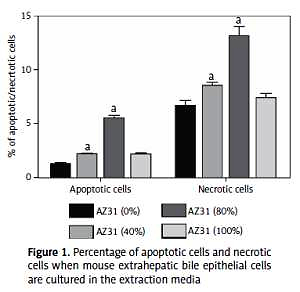
Figure 1 exhibits the fluorescence intensity of AZ31 extracts. The cells are divided into 3 distinct quadrants. The first quadrant (left) demonstrated the normal visible cells and the 2nd quadrant (lower) illustrated the apoptotic cells and the 3rd quadrant exhibited the necrotic and apoptotic/necrotic cells (data not shown). Dose-dependent treatment of AZ31 extract exhibited an effect on the apoptotic and necrotic cells. On day 1, AZ31(80%) extract concentration showed a larger apoptotic effect and the same results were observed in the necrotic cells treated with AZ31 (80%) (Figure 1). After 1 day, AZ31 (100%) showed significantly (p < 0.05) less Bax mRNA expression than the other groups. On day 3, the mRNA expression of Bax was significantly (p < 0.05) increased in AZ31 (40%) extract compared to other groups. On the other hand, no difference was observed in the Bcl-2 mRNA expression in the AZ31 (40, 80 and 100%) and control group.
Figure 1
Percentage of apoptotic cells and necrotic cells when mouse extrahepatic bile epithelial cells are cultured in the extraction media
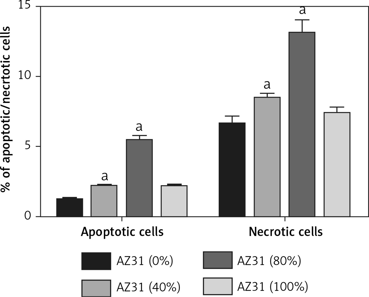
Figure 2 demonstrates the ratio of Bax to Bcl-2 with more accuracy and reflects the cell sensitivity to apoptotic stimuli than the individual expression of these two genes. AZ31 (40%) extract treated with MEBECs for 72 h – this group has a higher level of Bax/Bcl-2 ratio as compared to other groups.
Figure 2
Effect on relative mRNA expression. A – Bax, B – Bcl-2 and C – Bax/Bcl-2 when mouse extrahepatic bile epithelial cells are cultured in media for a period of 1 and 3 days
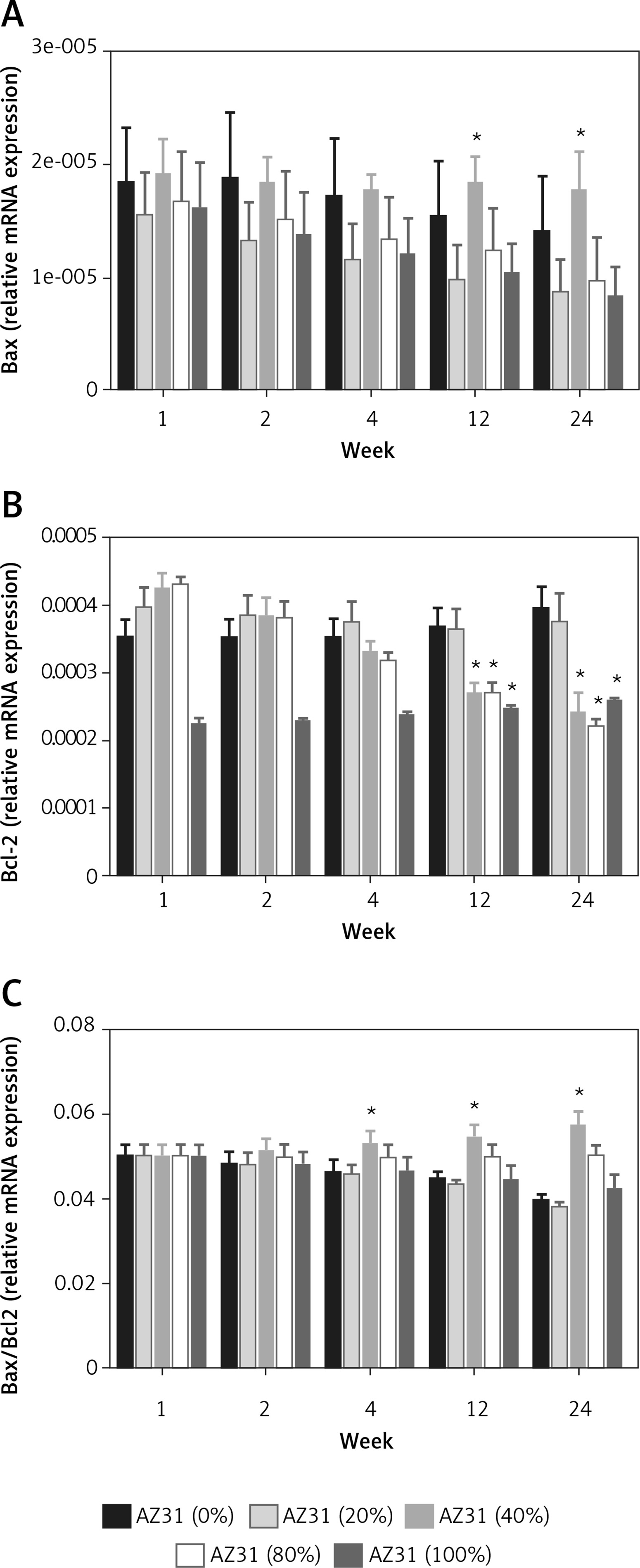
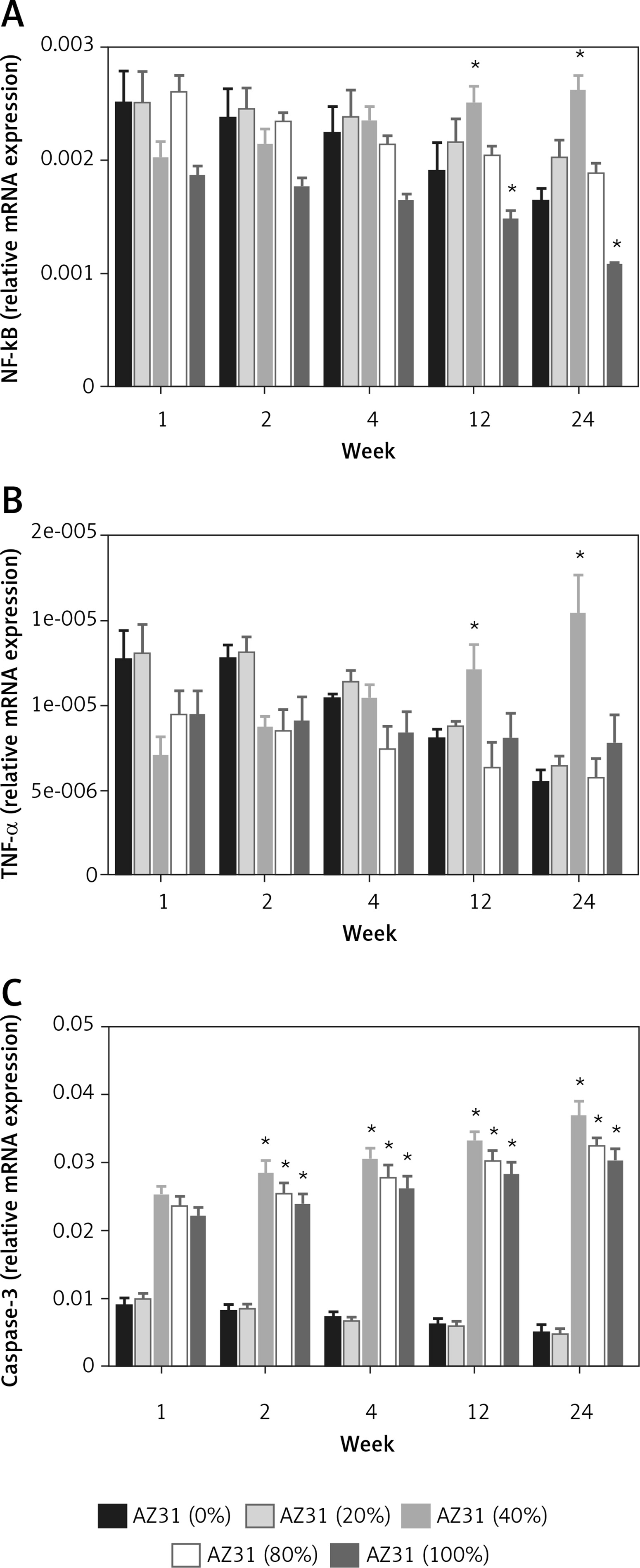
Figure 3 displays the relative mRNA expression of TNF-α, caspase-3 and NF-κB of cultured MEBECs. There was no significant difference between extract groups and control group on day 1 in expression of NF-κB. After day 3, the 40% extract group showed a higher expression level of NF-κB in comparison to the control group. In the case of TNF-α mRNA expression, no significant differences were observed between extract groups and the control group on both day 1 and day 3. The expression of caspase-3 on day 1 was higher in 80 and 100% extracts than in the control group, and after day 3 all the experiment groups exhibited increased activity in caspase-3 expression as compared to the control group.
Effect on apoptotic and pro-apoptotic gene expression
Figure 2 exhibits the effect of AZ31 extract on apoptotic gene expression at different time intervals. Figure 3 A shows the effect of AZ31 extract on Bax gene expression at a different time intervals. Week 1 showed almost the same level of Bax in all concentrations of AZ31 (0%, 20%, 40%, 80% and 100%). As time increases, the relative expression of Bax changes in all concentration groups, AZ31 (40%) showed the increased relative gene expression of Bax at the end of the experimental study. A similar pattern was observed in the Bcl-2 relative gene expression. AZ31 (40%) showed the enhanced expression of Bcl-2 as compared to other groups (Figure 2 B). Figure 3 C illustrated the relative expression of Bax/Bcl-2 in AZ31 extract treatment. AZ31 (40%) extract showed increased expression.
Figure 3 A shows the effect of different concentrations of AZ31 extracts on the NF-κB expression. AZ31 (40%) showed down-regulation of NF-κB expression. A similar trend was observed in TNF-α and caspase-3; the expression of TNF-α and caspase-3 decreased in the AZ31 (40%) treated group.
Effect of AZ31 on hematological parameters
Figure 4 showed the effect of AZ31 stent on the hematological parameters. Hemoglobin level was significantly (p < 0.05) increased during the first week and was reduced the next week. After that, the hemoglobin level significantly (p < 0.05) increased in the next week and continued to increase until the last week of the experimental study (Figure 4 A). Figure 4 B demonstrates the WBC effect; initially, WBC content increased and in the next week the level was boosted and after that, the level of WBC was normal at the end of the experimental study. Figure 4 C demonstrates that the platelet count increased from the first week and remained enhanced until the end of the experimental study. Figure 4 D illustrated the serum magnesium concentration and data showed that the level of Mg2+ significantly (p < 0.05) decreased in first 2 weeks and after that, it remained at a normal level as compared to the initial level.
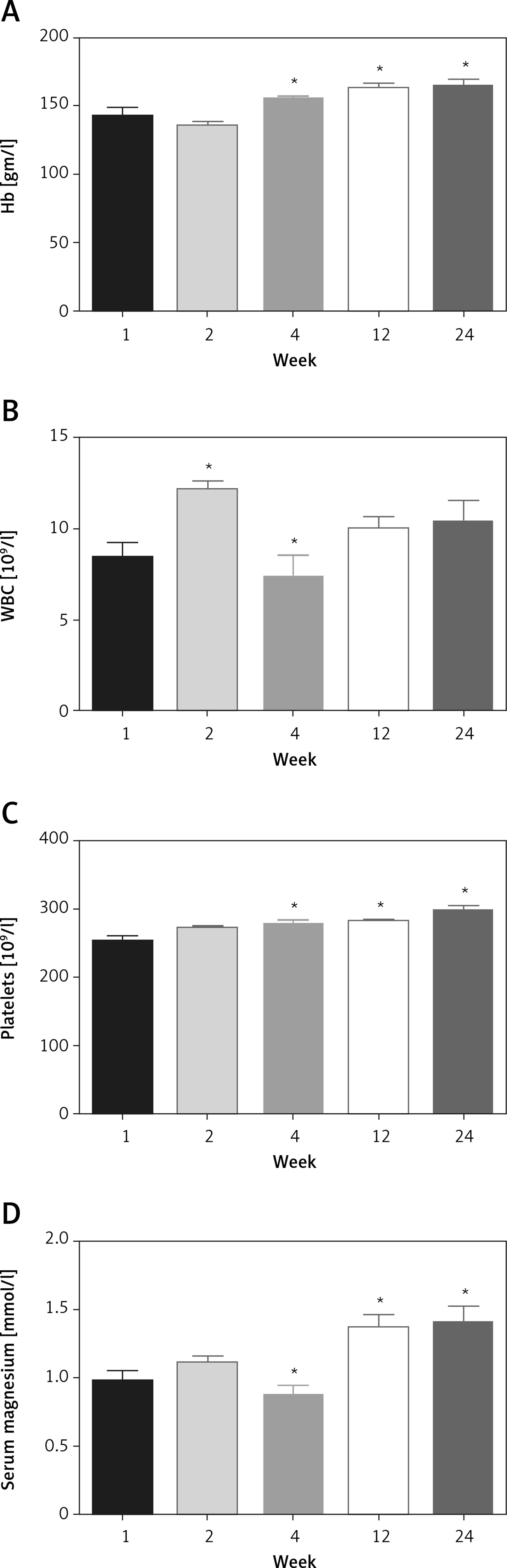
Effect of AZ31 on hepatic parameters
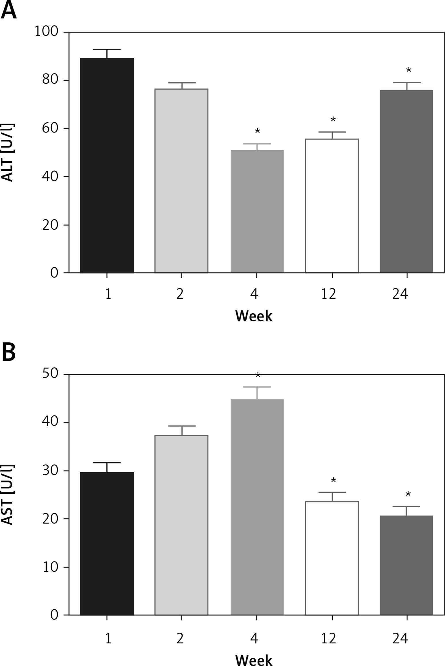
Figure 5 A shows the ALT level its level slightly reduced in the first 12 weeks but after that, the level of ALT increased (week 24). An opposite trend was observed in the AST, the AST level was increased in the 1 to 4 week and after that the level of AST going to reduced and reached to the normal level.
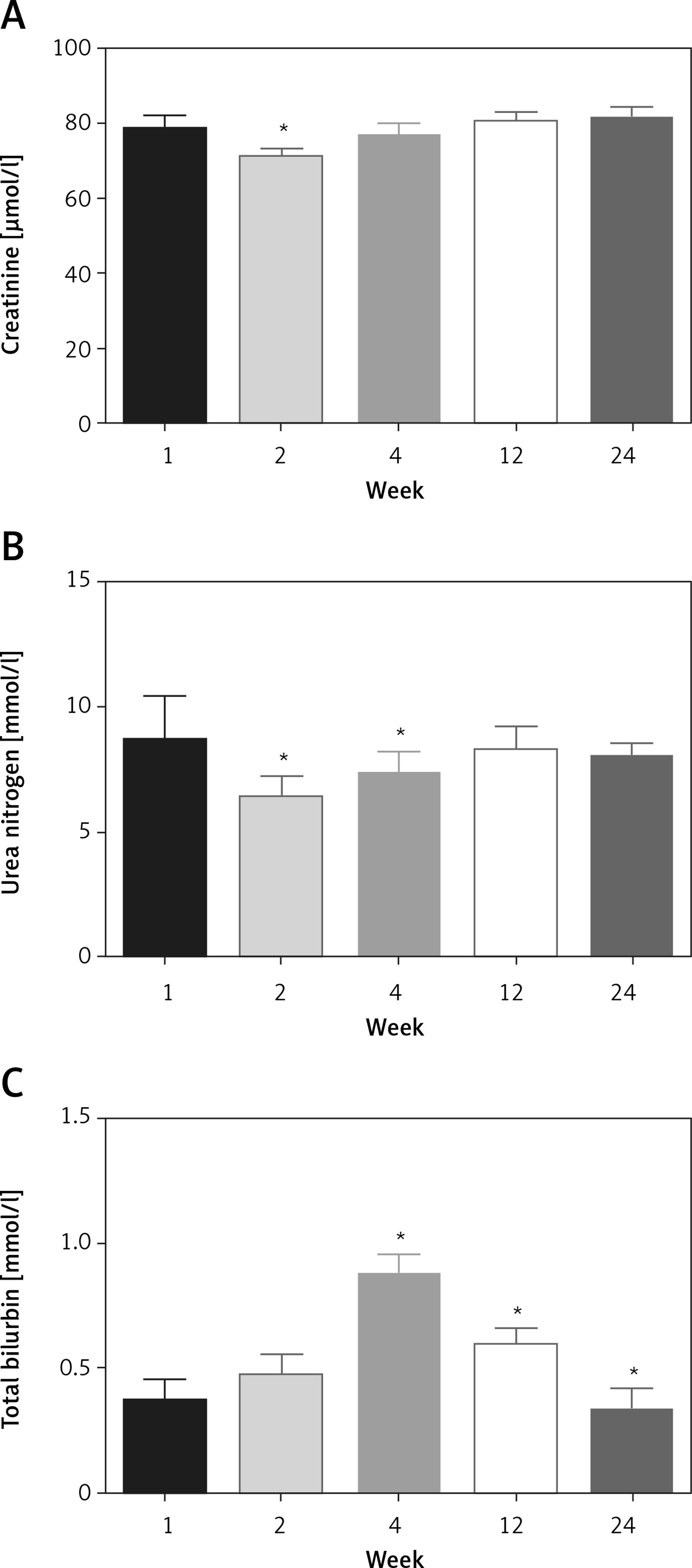
Effect of AZ31 on non-hepatic parameters
Figure 6 exhibited the effect AZ31 on non-hepatic parameters. According to Figure 6 at the creatinine level was unaltered by the AZ31 stent until the end of the experimental study. Figure 6 B exhibits the content of urea nitrogen; urea nitrogen level was significantly (p < 0.05) decreased in the first week and in the next week the level of urea nitrogen increased and reached almost the initial level at end of the experimental study. Figure 6 C demonstrates the total bilirubin; the level of bilirubin significantly (p < 0.05) increased at the start of the week (week 4) and after that the content of total bilirubin significantly (p < 0.05) decreased at the end of the experimental study.
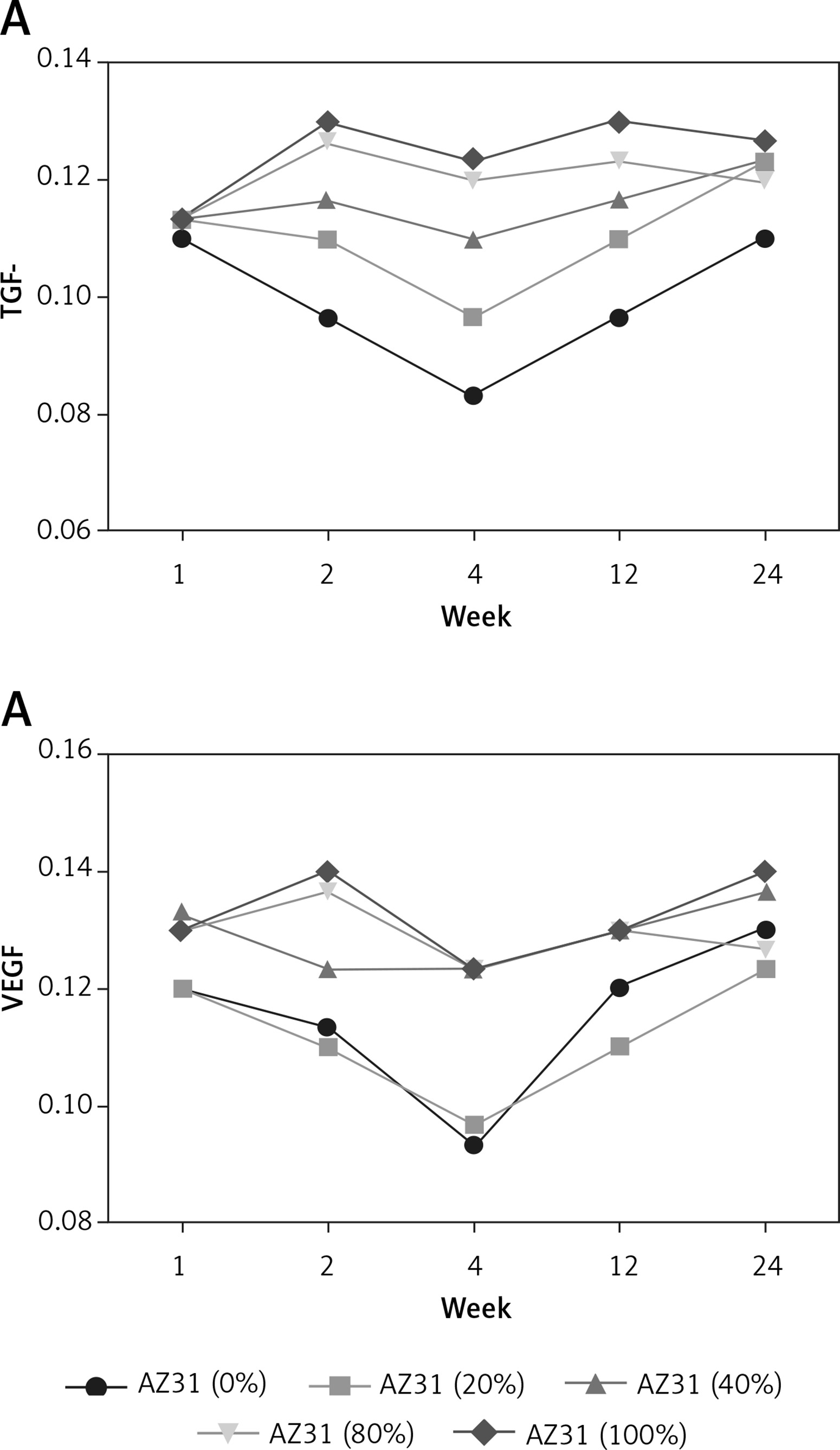
Effect of AZ31 on TGF-β and VEGF
Figure 7 exhibits the effect of AZ31 on inflammatory mediators such as vascular endothelial growth factor (VEGF) and transforming growth factor β (TGF-β). The level of TGF-β decreased in the first week to the fourth week and recovered in the last 24 weeks. A similar result was observed in the case of VEGF level.
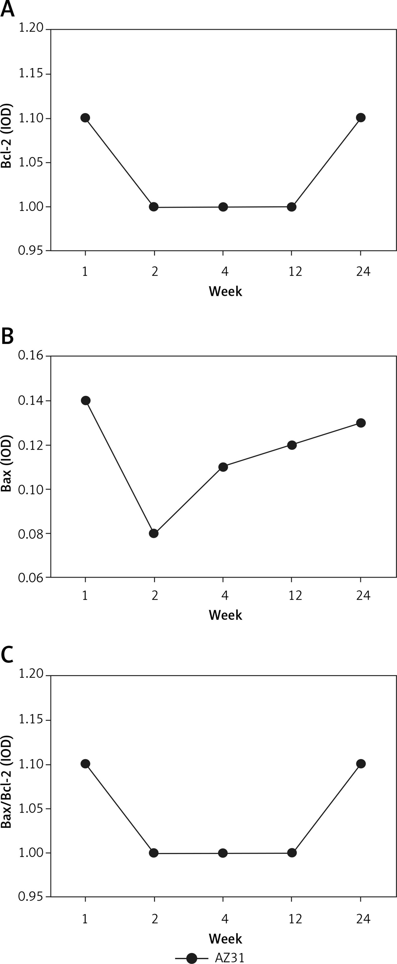
Effect of AZ31 on apoptosis
Figure 8 represents the experiment on BAX, Bcl-2 and BAX/Bcl-2 levels. In the first 2 weeks, the levels of BAX, Bcl-2 and BAX/Bcl-2 were decreased and after that, the levels of BAX, Bcl-2 and BAX/Bcl-2 increased until 24 weeks.
Discussion
Following CBD examination, the T-tube was inserted in the CBD to provide support and maintain the open CBD. Conversely, the treatment cost is higher due to a prolonged stay in the hospital. Another fact is the risk of T-tube placement, such as increased mortality or morbidity and increased biliary infection, migration of the T-tube inducing the bile duct obstacle, peritonitis, bile leakage following the removal of the T-tube [30]. Now the researchers and surgeons focus on the new therapy for the CBD and the choice of the researchers and surgeons is a biodegradable stent. The advantage of a biodegradable stent is that it is easy to implant, prolong the duration of action, and there is no need for surgery to remove it from the body. Biomaterial technology is an emerging topic due to clinical use with less or no adverse effect. Magnesium and aluminum are both metals having excellent biocompatibility with tissues or organs and due to this property they are widely used for medical implants. Due to the high demand of bimetallic magnesium and aluminum, they are widely used in various clinical therapies [31]. Various previously published literature suggest that the biomaterial might persuade the monocytes, inflammation reaction, phagocytosis and macrophages migration [16, 32]. Consequently, it should be taken into consideration that excessive deposition of degradation products due to excessive loading of burst release or organism regulation would in turn depreciate the local physical condition [33–35]. In the current investigation, we scrutinized the long term effect of biomaterial stent (AZ31) in the common bile duct. Before implanting the stent into the tissue, the medical professional mainly focuses on the safety issue. Before we start the experimental study, we take the safe combination of bimetal which is presented in Table I. We used the AZ31 alloy stents with 1.0 mm diameter, length 5 mm and thickness 0.1 mm which were implanted into the rabbits and were screened at regular time intervals using CT scanning. On scanning, the stents degraded in the first week (~67%), second week (~38%), fourth week (~24%), twelfth week (~16%) and finally completely degraded at twenty weeks after the surgery. In the current study, we observed the effect of AZ31 stent on hematological, hepatic, non-hepatic parameters and apoptosis parameters.
The term apoptosis was firstly used by Kerr Wyllie (non-classic paper) and Currie to define the morphological as discrete from the cell death [36, 37]. Another apoptosis is defined as having a preventive effect viz., exclusion of unwanted cells and upholds the host stability [36, 37]. The mechanism of apoptosis involved in the process of transpired the mammalian cells from cell death occurred during the expansion of nematode. In vitro investigations suggest that AZ31 alloy stent extract boosts MEBECs apoptosis via using the low concentration and on the country AZ31 alloy stent extract significantly enhanced the necrosis/apoptotic necrosis in a concentration-dependent manner [38, 39]. 80% concentration of AZ31 alloy stent extract exhibited considerably higher apoptosis of MEBECs cultured as compared to other treatment. When the concentration reached 100% the apoptotic and necrosis/apoptotic necrosis level enhanced and supported the apoptosis. Conversely, the result exhibited that no difference was observed in between the apoptotic necrosis and necrotic cells. In long culture treatment, apoptotic cells will undergo secondary necrosis, which is called late apoptosis or post-apoptotic necrosis [40]. In the current experimental study, after the cultures, the cells were found as necrotic and further converted into the secondary necrosis. As a result, our experimental result showed that the necrosis may be affected via late apoptosis or post-apoptotic necrosis.
Bax is a pro-apoptotic protein [41] and Bcl-2 is an anti-apoptotic protein [42] and these are discovered to be mammalian cell death regulators [43, 44]. The researchers suggest that the Bcl-2 protein is involved in the negative and positive regulation of apoptotic cell death [43, 44]. Bcl-2 (anti-apoptotic members) regulates the negative cell death and protects the cells from undergoing apoptosis induced via numerous stimuli of various cells [45]. On the other hand, pro-apoptotic proteins such as Bax accelerate or boost cell death [46, 47]. In vitro studies demonstrated that after 1 week the Bax and Bax.Bcl-2 mRNA expression was significantly higher in 40% of extracts and remained higher until 24 weeks. On the basis of the results, we can say that 40% of AZ31 alloy stent extract induced the apoptosis in MEBEGs in an in vitro atmosphere [47, 48].
In the in vivo experimental study, AZ31 alloy extract did not exhibit any significant difference in the apoptosis relative genes. Therefore at 1-week post-operation, the AZ31 alloy group exhibited higher Bax expression as compared to the control group and no significant difference was observed in the Bax/Bcl-2 ratio in the AZ31 alloy and control group. The apoptosis mechanism is very complex. Several apoptosis mediators such as caspase-3, TNF-α and NF-κB play an important role in the apoptosis mechanism [49, 50]. The apoptosis mediators are activated via transactivating gene expression of anti-apoptosis [51–54]. Several researchers suggest that the NF-κB pathway is a crucial factor in the cell reaction to apoptotic stimuli [55–57]. During the NF-κB pathway, TNF-α (downstream protein) can start the apoptosis reaction via cross-link protein or ligand binding. Our experimental study suggests the alteration of the NF-κB concentration-dependent manner of AZ31. AZ31 alloy extract (40%) showed significantly increased expression of NF-κB at a regular time interval. A similar result was observed in the TNF-α concentration; AZ31 alloy extract (40%) demonstrated increased expression [52–54, 58]. Another apoptosis parameter, caspase-3, is considered as a significant marker of inflammation and plays an important role in the apoptosis pathway [57]. In our experimental study, caspase-3 expression was more sensitive to changes in the AZ31 alloy concentration. The data revealed that the expression of caspase-3 was significantly increased in AZ31 alloy extract (40%, 80% and 100%) treatment. In short, we have found that AZ31 alloy extract (40%) boosted the apoptosis relative genes such as Bax, Bax/Bcl-2 ratio and critical apoptotic protein viz., caspase-3 and NF-κB. In the current experimental investigation, AZ31 alloy extract (40%) could induce the alteration in the apoptosis genes in both intestinal epithelial cells and intestinal tract tissues. On the basis of the result, we can say that aluminum plays an important role in the activation of NF-κB expression [59–61].
In conclusion, collectively, we can say that the advancement of the stent reduces the risk of infection. In the current experimental study, in vitro and the in vivo study was performed to scrutinize the biocompatibility of AZ31 with CBD. Our study suggests that the AZ31 stent gradually degraded in the CBD in the rabbits. AZ31 stent does not influence the biochemical parameters. The reduced level of VEGF expression may be related to lowering the stimulation resulting from the degradation of AZ31 alloy. Thus, it can be concluded that the AZ31 alloy stent is a safe biocompatible material for CBD.