

Introduction
Malignant ascites (MA) is a finding of peritoneal carcinomatosis in which malignant cells are present in the peritoneal cavity. Peritoneal carcinomatosis often develops secondary to peritoneal surface malignancies. They usually arise from intra-abdominal cancers such as ovarian, colorectal, pancreatic, gastric, and uterus or extra-abdominal lymphoma, lung and breast tumors [1]. MA constitutes approximately 10% of all ascites cases [2]. It is a common complication associated with numerous cancers, including pancreatic, gastric, colorectal, hepatic, and ovarian cancers. MA, which is a sign of advanced cancer and poor prognosis, also significantly reduces the quality of life [3].
Although tuberculosis (TB) is not common in developed countries, it is still endemic in developing countries. An increase has been reported in developed countries due to socio-economic reasons (such as communal living, homelessness, substance abuse), migration from endemic regions, increase in secondary immunosuppression, and diagnostic delays due to the COVID-19 pandemic [4]. TB most commonly infects the lungs, but can spread to any organ in the body. The abdomen (gastrointestinal tract, peritoneum, internal organs, and lymph nodes) is the most commonly involved extrapulmonary site [5]. TB can spread to the peritoneum via the bloodstream, from the gastrointestinal tract via the mesenteric lymph node, or directly from the fallopian tubes [6]. The most common causes of exudative ascites with serum-ascites albumin gradient (SAAG) < 1.1 include MA followed by TB peritonitis-associated ascites (TBpA). Difficulty may be experienced in the differential diagnosis of peritoneal carcinomatosis, especially in older individuals [7]. Due to difficulties in microbiological diagnosis in people suspected of having TBpA, laparoscopic peritoneal biopsy is recommended for early diagnosis [5, 6, 8]. Delays in diagnosis may negatively affect morbidity and mortality by causing a delay in effective treatment for both diseases, and invasive diagnostic procedures may have some serious complications [6, 9].
The neutrophil-to-lymphocyte ratio (NLR) is a simple, easy-to-access biomarker of inflammation status that represents the ratio of neutrophils to lymphocytes calculated from a complete blood count [10]. NLR has been shown to be an effective biomarker in various diseases including cardiac and inflammatory conditions, especially infectious diseases [11–13]. There are many studies showing the relationship of NLR with advanced stage disease and poor prognosis in colorectal, gastric, pancreatic, and ovarian cancers, which are the leading causes of MA [11, 14–17].
This study investigates the discrimination of NLR between MA and TBpA, leading causes of exudative ascites.
Material and methods
Patients
This study was conducted in an 800-bed medical faculty hospital located in the southeast of Turkey. A total of 100 patients over 16 years old, 50 with TBpA and 50 with MA, who were consecutively diagnosed between September 2015 and December 2020, were included in this retrospective cohort study.
The diagnosis of TBpA was confirmed by a complete clinical and laboratory response after antituberculous therapy. All patients had a benign cytological examination of ascites. All patients were receiving isoniazid (300 mg/day; for 9 months), rifampicin (10 mg/kg/day; for 9 months), ethambutol (15–20 mg/kg/day; for 2 months), and pyrazinamide (20–25 mg/kg/day; for 2 months).
The patients’ primary cancer diagnoses were confirmed through biopsies obtained from lesions on endoscopic examination or via computed tomography or ultrasound. The diagnosis of MA was confirmed in all patients by demonstrating the presence of malignant epithelial cells in the cytological examination of ascitic fluid.
Exclusion criteria
Those with two primary cancers, those with undetected primary cancer, those diagnosed with peritoneal mesothelioma, those receiving chemotherapy, those with end-stage renal disease, those with liver cirrhosis, those with heart failure, and those who were HIV positive were excluded from the study.
SAAG calculation
To obtain SAAG, serum albumin and ascites fluid albumin concentrations were measured simultaneously, followed by subtraction of ascites fluid albumin from serum albumin.
NLR calculation
Leukocyte count, formulation, and C-reactive protein (CRP) values were taken before cytology was performed. Hematological parameters were studied using an automatic analyzer system (Abbott Cell-Dyn 3700; Abbott laboratory, Abbott Park, Illinois). Absolute neutrophil count was divided by absolute lymphocyte count to obtain the NLR. White blood cell count (WBC) differential count, NLR, and CRP level were compared between patients with MA and patients with TBpA.
Statistical analysis
Scale variables were presented as mean ± standard deviation (SD), and categorical variables were presented as absolute and relative abundance (%). Comparisons between groups were made using Student’s t-test when numerical data showed a normal distribution, and the Mann-Whitney U test when numerical data did not show a normal distribution. The distribution of the ratios of the levels of categorical variables was evaluated with the χ2 test. ROC curve analysis was performed to identify optimal cut-off levels for NLR, WBC count, neutrophil count, and lymphocyte count to identify the cutoff values for optimal sensitivity and specificity for distinguishing MA from TBpA. The ability of NLR, WBC count, neutrophil count, and lymphocyte count to distinguish MA from TBpA was compared using the area under the curve (AUC). A bidirectional p-value of ≤ 0.05 was considered a statistically significant. All statistical analyses were performed using SPSS Statistics 21.0 for Windows (IBM Corp., USA).
Results
Demographic and clinical characteristics, and laboratory and tumor marker results of each patient group are summarized in Tables I–III.
Table I
Patients’ demographic and clinical characteristics
Table II
Laboratory characteristics of the study population
| Variable (normal range) | MA group (N = 50) | TBpA group (N = 50) | P-value |
|---|---|---|---|
| Hemoglobin (12.9–14.2 g/dl) | 12.12 ±1.76 | 12.35 ±1.89 | 0.544† |
| RBC (4.06–4.69 × 10⁶/µl) | 4.48 ±0.81 | 4.63 ±0.68 | 0.294† |
| RDW (11.5–14.5%) | 15.50 ±2.65 | 15.52 ±2.90 | 0.986§ |
| WBC (3.7–10.1 × 10³/µl) | 9.311 ±3.060 | 6.281 ±1.878 | < 0.001§ |
| Neutrophil (1.63–6.96 × 10³/µl) | 6.534 ±2.751 | 4.161 ±1.648 | < 0.001§ |
| Lymphocyte (1.09–2.99 × 10³/µl) | 1.784 ±911 | 1.445 ±517 | 0.149§ |
| NLR (0.78–3.53)* | 4.65 ±3.17 | 3.17 ±1.74 | 0.007§ |
| Albumin (35–52 g/l) | 2.51 ±0.74 | 3.86 ±0.65 | < 0.001† |
| Globulin (20–30 g/l) | 3.53 ±0.86 | 3.77 ±0.74 | 0.114§ |
| Serum cholesterol (112–200 mg/dl) | 170.2 ±52.8 | 178.3 ±40.8 | 0.391† |
| Serum ferritin (22–322 µg/l) | 157.7 ±381.9 | 102.7 ±104.6 | 0.363§ |
| LDH (0–250 U/l) | 429.52 ±426.71 | 193.91 ±52.54 | < 0.001§ |
| CRP (0–5 mg/l) | 7.49 ±7.63 | 5.60 ±7.22 | 0.247§ |
| ADA (5–35 IU/l) | 12.17 ±8.12 (n = 9) | 44.74 ±21.49 (n = 27) | < 0.001† |
* from Forget P, et al. What is the normal value of the neutrophil-to-lymphocyte ratio? BMC Res Notes 2017; 10: 12. MA – malign ascites, TBpA – Tuberculos peritonitis ascites, RBC – red blood cell, RDW – red blood cell distribution width, WBC – white blood cell, NLR – neutrophil-lymphocyte count ratio, CRP – C-reactive protein, LDH – lactate dehydrogenase, ADA – adenosine deaminase.
Table III
Mean laboratory values for tumor markers
| Parameter | MA group Mean ± SD (n) | TBpA group Mean ± SD (n) | P-value† |
|---|---|---|---|
| CEA (0–5 ng/ml) | 54.96 ±169.35 (50) | 1.01 ±0.61 (40) | < 0.001 |
| CA125 (< 35 U/ml) | 603.94 ±1.041.42 (50) | 281.15 ±359.49 (44) | 0.228 |
| CA19-9 (< 39 U/ml) | 63.11 ±196.37 (50) | 45.65 ±232.90 (42) | 0.003 |
The mean age of the MA group was 62.9 ±15.1 years (23-89) and 62% were female. All patients had SAAG < 1.1 and a malignant peritoneal fluid cytology. Sixty-eight percent of the MA group (n = 34) consisted of patients with gastrointestinal system cancer and 32% with ovarian cancer (n = 16). Thirty-two percent of gastrointestinal cancers originated from the stomach (n = 16), 26% from the colorectal region (n = 13) and 10% from the pancreas (n = 5).
The total leukocyte count was 9,311 ±3,060/mm3 (range: 3,170–19,220), the neutrophil count was 6,534 ±2,751/mm3 (range: 1,720–15,000), and the lymphocyte count was 1,784 ±911/mm3 (range: 470–4,590) in the MA group (Table II). The NLR value was found to be 4.65 ±3.17 (range: 1.08–17.37). CRP was 7.49 ±7.63 mg/l (range: 0.01–31.37).
The level of carcinoembryonic antigen (CEA) was 54.96 ±169.35 ng/ml, cancer antigen 125 (CA125) was 603.94 ±1.041.42 U/l, and cancer antigen 19-9 (CA 19-9) was 63.11 ±196.37 in the MA group (Table III).
The mean age of the TBpA was 32.4 ±12.3 years (17–74) and 60% were female. All patients had SAAG < 1.1 and peritoneal fluid cytology was benign. Peritoneal biopsy was performed in 14 (28%) patients, and granulomatous peritonitis was reported in all of them.
The total leukocyte count was 6.281 ±1.878/mm3 (range: 2.610–11.000), the neutrophil count was 4.161 ±1.648/mm3 (range: 1.540–8.790), and the lymphocyte count was 1.455 ±517/mm3 (range: 550–2.790) in the TBpA group (Table II). The NLR value was found to be 3.17 ±1.74 (range: 1.97–3.61). CRP was 5.60 ±7.22 mg/l (range: 0.33–38.94).
The level of CEA was 1.01 ±0.61 ng/ml, CA125 was 281.15 ±359.49 U/l, and CA 19-9 was 45.65 ±232.90 in the TBpA group (Table III).
The mean adenosine deaminase (ADA) value of 39 patients whose data were available was 51.9 ±25.14 U/l (range: 15.4–82.8).
There was no difference between the two groups in terms of gender and weight loss. Those with malignant ascites were older (p < 0.001) and those with tuberculous ascites were more likely to experience fever, night sweats, and abdominal pain (p < 0.001, p < 0.001 and p = 0.026, respectively) (Table I).
All tumor markers were found to be higher in the MA group than in the TBpA group, but statistically significant differences were found in only CEA and CA19.9 (p < 0.001 and p = 0.003, respectively).
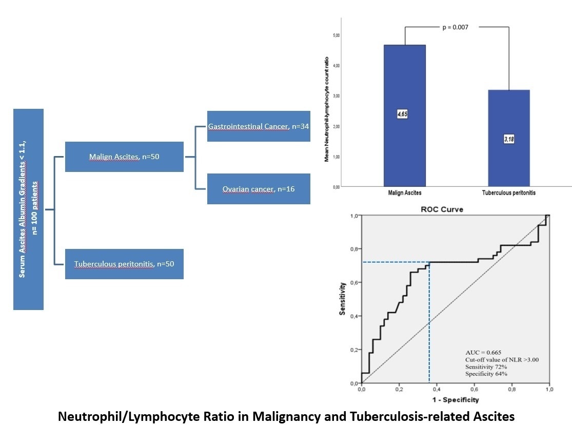
The mean NLR value of the MA group was found to be significantly higher compared to the TBpA group (4.65 ±3.17 and 3.17 ±1.74, respectively, p = 0.007).
Within the MA group, although NLR was higher in patients with ovarian cancer compared to patients with gastrointestinal cancer, the difference was no significant (5.14 ±3.42 & 4.24 ±3.08, respectively, p = 0.662). Considering the organ where the cancer originated, the highest NLR value was found in patients with pancreatic cancer (6.32 ±1.82), followed by ovarian (5.14 ±3.42), colorectal (4.34 ±4.05), and gastric (3.89 ±2.31) cancer patients, but no significant difference was found among them (p = 0.332).
In the correlation analysis between NLR and tumor markers, a statistically significant but weak-to-moderate correlation was detected between CA19.9 (r = 0.271; p = 0.009) and CA125 (r = 0.219; p = 0.034) and NLR.
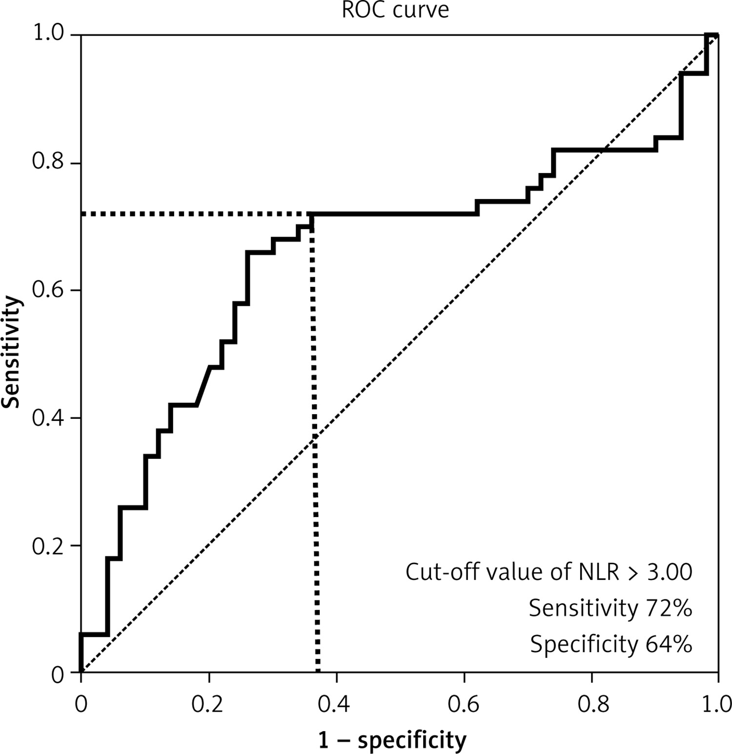
ROC analysis was used to determine the diagnostic NLR threshold for predicting MA. The area under the curve value of NLR was 0.655 (95% confidence interval = 0.544–0.767, p = 0.007) (Figure 1). The optimal threshold value for NLR in MA prediction is > 3.00, and the sensitivity and specificity at this threshold level were 72% and 64%, respectively.
Figure 1
ROC curves of neutrophil-lymphocyte ratio (NLR) for discriminating malignant ascites from tuberculous peritonitis secondary ascites. The area under the curve (AUC) for NLR was 0.665 with an 95% confidence interval [CI] of 0.544–0.767 (p = 0.007)
The effectiveness of the NLR > 3.00 threshold value in predicting MA in different age groups was evaluated (Table IV). Having an NLR > 3.00 over the age of 40 had the highest specificity (88.0%), positive predictive (85.4%), negative predictive (74.6%), and accuracy (79%) values in predicting MA, with similar sensitivity (70%).
Table IV
Specificity, sensitivity, positive predictive value (PPV), negative predictive value (NPV) and accuracy rates of NLR alone and with different age groups in predicting malignant ascites
Discussion
Despite regional differences in the etiology of ascites in those with SAAG < 1.1, peritoneal carcinomatosis and TBpA (approximately 12% and 2% of all ascites, respectively) are responsible for the majority of cases [18, 19]. MA is a common complication associated with many cancers, including pancreatic, gastric, colorectal, hepatic, and ovarian cancers, and are a sign of advanced cancer and poor prognosis [3]. Tuberculosis has a moderate burden in Turkey, with a prevalence of 23/100,000 people and an incidence rate of 22/100,000 people [20]. In a study conducted in our country, the proportion of TBpA among all ascites cases (7.8%) was found to be approximately four times that of western countries [18, 21]. In these two diseases, whose treatments are completely different, early diagnosis can provide timely appropriate treatment for those with MA, which has a short life expectancy, and can provide a positive impact on morbidity and mortality in those with TBpA [9].
A timely diagnosis of the cause of ascites is a challenge. Cytology and peritoneal biopsy are used in differential diagnosis. These methods have disadvantages such as being invasive, causing serious complications, high cost, and limited diagnostic effectiveness [6, 22]. There is an ongoing search for simple tests that can contribute to overcoming these difficulties. In various studies on the differentiation of MA, it has been reported that markers such as vascular endothelial growth factor, telomerase activity, endostatin, insulin like growth factor-1, and cytokines have high diagnostic value [23–25]. However, the fact that these markers are not available worldwide prevents their widespread clinical use. Therefore, there is a need for simple, cheap, and rapid tests that can be used worldwide.
The increase in total leukocytes and neutrophils reflects an inflammatory reaction [26]. The relationship between inflammation and cancer has been known for a long time. The role of neutrophils in this inflammation in solid tumors is very important [27–29]. Depending on the cytokine pool in the cancer, neutrophils can be manipulated by the tumor to promote or inhibit tumor growth [28]. A high neutrophil count in the blood has been found to be associated with poor prognosis in cancer patients [30]. In addition, it has been reported that in various tumor types, there is a more significant decrease in the number of lymphocytes in patients with advanced disease compared to those with localized disease [31]. The association between the increase in circulating neutrophils and the increase in NLR, seen in combination with inflammation in solid tumors, has been shown to be associated with poor prognosis and low recurrence-free survival in various cancer types [14–17, 28, 32, 33].
In this study, NLR was found to be a useful biomarker to distinguish MA from TBpA. In this study we found that serum: a) NLR levels were significantly higher in patients with MA than in patients with TBpA, b) that the optimal NLR cut-off value to distinguish MA from TBpA was > 3, c) and that the optimal cut-off value was > 3 in patients over the age of 40.
In our study, the gender distributions of the TBpA group and the MA group were similar, but the mean age was significantly lower in the TBpA group. The frequency of clinical findings including fever, night sweats, and abdominal pain was significantly higher in the TBpA group compared to the MA group. These demographic findings and clinical symptoms are very similar to the results of previous studies that included similar patient groups [7, 34]. In addition, in our study, the mean CEA and CA19.9 levels were significantly higher in the MA group compared to the TBpA group, and these results are also consistent with the results of previous studies [7, 34]. In older individuals with ascites but without fever and night sweats, the presence of elevated serum CEA and CA19.9 and elevated NLR may further support the possibility of MA.
An increase in NLR has been found to be associated with poor prognosis and low relapse-free survival in cancers [14–17, 28, 32, 33]. Among these, subgroup analyses regarding peritoneal metastasis are included in gastric and ovarian cancer studies [17, 34, 35]. In the study of Magdy et al., which included 61 gastric cancer patients, 19.6% of whom had peritoneal carcinomatosis, high NLR (> 2.4) was found to be strongly associated with poor survival [35]. A significant correlation was found only between the presence of ascites and high NLR [35]. In the study by Feng et al., in which they evaluated 875 ovarian cancer patients, high preoperative NLR level (≥ 3.24) was correlated with diffuse ascites, and was found to be an independent prognostic factor of progression-free survival [17]. In this study, a high NLR was found to increase in direct proportion to the presence and amount of ascites [17]. In another study where high NLR was found to be strongly associated with advanced stage, higher CA125 level, and ascites in surgery, the median (IQR) NLR value was found to be significantly higher in ovarian cancer patients with ascites compared to those without ascites [36].
In a study including patients with exudative pleural effusion, both blood and pleural fluid NLR were found to be significantly higher in patients with pleurisy due to malignancy compared to those with pleurisy due to tuberculosis [37]. In another study evaluating patients with pneumonia, NLR was found to be significantly lower in patients with tuberculous pneumonia compared to patients with bacterial pneumonia [38]. Although these studies demonstrating the differentiation of NLR from TB include pleural effusion and pneumonia, the results obtained from our study support the view that it is a useful biomarker in the distinction between MA and TBpA. As a result of our study, we found that WBC count, neutrophil count, and NLR were statistically significantly lower in the TBpA group compared to the MA group (p < 0.001, p < 0.001, and p = 0.007, respectively). In our study, while the sensitivity of the NLR > 3.00 threshold value in distinguishing MA was 70% and the specificity was 64%, in people over 40 years of age it demonstrated higher specificity (80%) and accuracy (74.6%).
Low serum-ascites albumin gradient (< 1.1), high number of lymphocytes in the ascitic fluid, low glucose level, high LDH level, high protein level, and high ADA level are in favor of TBpA. However, all these tests do not definitively confirm the diagnosis of TBpA. At this point, it can be used as a diagnostic supporting biomarker in addition to initial examinations. Ultimately, when necessary, the diagnosis can be made using laparotomy, laparoscopy or histopathological examination of percutaneous peritoneal biopsy [8]. These diagnostic tools are invasive and have various serious complications, so NLR can accelerate the selection of patients for these invasive interventions in the early planning of the procedure.
This study has several important limitations. First, it is a single-center, retrospective study with a relatively small number of patients. Secondly, there is no analysis regarding the quality of life and life expectancy of the patients. Finally, the lack of ADA and tumor marker data in all patients weakens the power of analysis of these laboratory data. Therefore, more studies in multiple centers with sufficient sample sizes are needed to confirm the ascites NLR discriminatory capacity between MA and TBpA.
In conclusion, the current study demonstrated that NLR obtained from a simple blood test can be used as a useful biomarker to distinguish between MA and TBpA, leading causes of exudative ascites, at the initial diagnostic stage. For this reason, especially in places where the frequency of tuberculosis is not low, NLR can be included in the routine examination in patients with diagnostic uncertainty between malignancy and tuberculosis-related ascites.

