Introduction
Bladder cancer (BCa) is the ninth most common malignant tumor, accounting for 90–95% of all urothelial carcinomas, and the majority of BCa occurs in men [1, 2]. Global cancer statistics reveal that there were approximately 429,800 new cases of BCa and 165,100 deaths in 2012 worldwide [1]. Cancer statistics in China indicate that 80,500 cases were newly diagnosed and 32,900 deaths occurred in 2015 [3]. In fact, approximately 70% of BCa patients are diagnosed with a non-muscle invasive BCa (NMIBC), while 30% of BCa patients present with a muscle-invasive BCa (MIBC) or a metastatic disease [2]. Currently, transurethral resection of bladder tumor or radical cystectomy is recommended as first-line treatment for the primary therapy of NMIBC or MIBC respectively [4]. Generally, most patients with NMIBC have a good prognosis, but a small fraction of patients develop MIBC or a metastatic disease resulting in aggressive features and poor prognosis, with a 5-year survival rate less than 50% [5]. Therefore, it is extremely urgent to identify new prognostic biomarkers, which may be helpful for the treatment and management of BCa.
Long non-coding RNAs (lncRNAs) are originally transcribed from non-coding sequences, consisting of more than 90% of the human genome, and are longer than 200 nucleotides with lack of protein coding capability [6]. LncRNAs have been implicated in many biological processes, including carcinogenesis, and perform a dual role in tumor initiation, progression and metastasis by modulating oncogenic and tumor-suppressing genes [7–9]. Recently, lncRNAs have been reported as a class of biomarkers to predict the prognosis of patients with malignant neoplasms, such as gastric cancer [10], prostate cancer [11], esophageal squamous cell carcinoma [12] and hepatocellular carcinoma [13]. Several lncRNAs, including OIP5-AS1, DGCR5 and SNHG16, are identified as prognostic indicators and may be vital for the pathogenesis of BCa [14–16]. However, the roles of lncRNAs in BCa have not been completely clarified.
In the present study, we performed lncRNA microarray assays to explore lncRNA expression profiling in three pairs of BCa tissues and adjacent non-tumorous tissues. Microarray analysis revealed that lncRNA LINC00336 expression levels were significantly up-regulated in BCa tissues. Subsequently, we validated the expression of LINC00336 in ninety-eight pairs of BCa tissues and adjacent non-tumorous tissues using RT-qPCR analysis, and the association between LINC00336 and overall survival (OA) or disease-free survival (DFS) was investigated in BCa patients. Furthermore, in vitro experiments were performed to detect the roles of LINC00336 in BCa cell proliferation and apoptosis.
Material and methods
Patients and specimens
Sixty-two BCa patients were recruited from the Xiangya Hospital of Central South University (Changsha China), the Eighth People’s Hospital of Jinan City (Jinan China) and the Affiliated Hospital of Hebei University (Baoding China) from January 2010 to June 2013. Sixty-two pairs of BCa and adjacent non-tumorous tissues were obtained from BCa patients who had undergone surgery. All samples were immediately stored in an ultra-low temperature refrigerator. Signed informed consent forms were obtained from all BCa patients. The Ethics Committee of the Xiangya Hospital of Central South University (Changsha China), the Eighth People’s Hospital of Jinan City (Jinan China) and the Affiliated Hospital of Hebei University (Baoding China) approved our study.
Cell culture
Human normal bladder epithelial cell line (SV-HUC-1) and bladder cancer cell lines (5637, T24 and SW780) were purchased from the Cell Bank of Type Culture Collection of Chinese Academy of Sciences (Shanghai, China). Cells were cultured in Dulbecco’s modified Eagle’s medium (DMEM; Invitrogen, Carlsbad, CA, USA) with 5% fetal bovine serum (Thermo Scientific HyClone, Beijing, China), 5% CO2, 95% air atmosphere in a humidified incubator (Thermo, USA).
LncRNAs microarray
Differentially expressed lncRNAs in three pairs of BCa tissues and adjacent non-tumorous tissues were analyzed using the Agilent human lncRNA array V.2.0 platform (Agilent Technologies, Santa Clara, CA, USA).
Cell transfection
Short hairpin RNA (shRNA) was designed to specifically target LINC00336 using shRNA design tools (http://rnaidesigner.thermofisher.com/rnaiexpress/). Using BLAST (http://blast.ncbi.nlm.nih.gov/Blast.cgi), we verified that the designed shRNA targeted only the LINC00336. Sh-LINC00336 and sh-NC were synthesized by GenePharma (Shanghai, China).
CCK-8
5637, T24 and SW780 cell viability was measured using the CCK-8 Cell Proliferation/Viability Assay Kit (Dojindo, Japan) as described previously [17]. Absorbance was calculated at 450 nm using an Elx800 Reader (Bio-Tek Instruments Inc., Winooski, VT, USA).
Transwell assay
Invasion of bladder cancer cells was determined using a transwell insert (8 μm, Corning) as described previously [18]. Absorbance was determined at a wavelength of 570 nm using an Elx800 Reader (Bio-Tek Instruments Inc., Winooski, VT, USA).
Cell cycle analysis
5637, T24 and SW780 cell cycle distribution was measured using the CycleTESTTM PLUS DNA Reagent Kit (BD Biosciences, San Jose, CA, USA) as described previously [19].
Apoptosis
5637, T24 and SW780 cell apoptosis was analyzed using an Annexin V-FITC/PI double staining kit (Invitrogen, Carlsbad, Calif, USA) with flow cytometry (FACScan, BD Biosciences, San Jose, CA, USA) as described previously [20]. Apoptotic cell proportion was calculated using CELL Quest 3.0 software (BD Biosciences).
RT-qPCR
Total RNA was isolated using RNAiso (Takara, Dalian, China). The RT Kit (Toyobo Co. Ltd, Osaka, Japan) and KAPA SYBR Green FAST qPCR Kit (KAPA, Wilmington, MA, US) were used to measure the expression levels of LINC00336 using the Applied Biosystems 7300 Real-Time PCR System (Thermo Fisher Scientific, Inc.) and normalized to the internal control U6. LncRNAs expression levels were calculated using the 2–ΔΔCq method [21].
Ethics approval
Written informed consent was obtained from all of the participants prior to collection of samples. The Ethics Committee of the Xiangya Hospital of Central South University (Changsha China), the Eighth People’s Hospital of Jinan City (Jinan China) and the Affiliated Hospital of Hebei University (Baoding China) approved our study according to the Helsinki Declaration.
Statistical analysis
Data were presented as the mean ± standard error of the mean. Statistical analysis was performed using SPSS software version 19.0 (IBM Corp., Armonk, NY, USA). Student’s t-test or Kruskal-Wallis test was used to analyze two-group differences. Differences between multiple groups were analyzed by one-way analysis of variance, followed by a post-hoc Tukey test. Pearson’s χ2 test was used to evaluate differences between the clinical characteristics and LINC00336 expression levels in BCa patients. OS and DFS were calculated using the Kaplan-Meier method with the log-rank test applied for comparison. Univariate and multivariate regression analysis were performed to evaluate the correlation between clinical characteristics, LINC00336 expression and OS using a Cox proportional hazard model. P < 0.05 was considered to indicate a statistically significant difference.
Results
LncRNAs expression profiling in BCa tissues
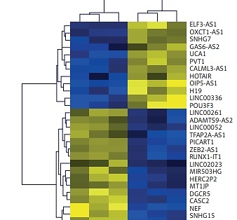
As shown in Figure 1 A, microarray probes were used to detect differentially expressed lncRNAs in three pairs of BCa tissues and adjacent non-tumor tissues. Based on the Log2|FC| ≥ 2.0, p < 0.001 and FDR < 0.001, a total of 29 lncRNAs were significantly differentially expressed in three BCa tissues compared with adjacent non-tumor tissues. Among them, 12 and 17 lncRNAs were up-regulated and down-regulated, respectively. To further validate the results of microarray, the top 6 differentially expressed lncRNAs, i.e. OIP5-AS1, H19, LINC00336, POU3F3, NEF and SNHG15, were chosen for RT-qPCR assays in sixty-two pairs of BCa tissues and adjacent non-tumor tissues. OIP5-AS1, H19, LINC00336 and POU3F3 were increased, while NEF and SNHG15 were decreased in sixty-two BCa tissues as compared to adjacent non-tumor tissues (Figure 1 B). These results were consistent with the microarray assay. The fold change (FC) of LINC00336 was 3.86 in BCa tissues compared with adjacent non-tumor tissues, which was higher than other lncRNAs. Therefore, we focused on LINC00336 in our study.
Figure 1
LncRNAs expression profiling in BCa tissues. A – Microarray and hierarchical cluster analysis were performed using the multiple experiment viewer 4.7.1 software, and 29 differentially expressed lncRNAs in three pairs of BCa tissues and adjacent non-tumorous tissues were selected out according to Log2|FC| ≥ 2.0, p < 0.001 and FDR < 0.001. B – Top 6 differentially expressed lncRNAs, i.e. OIP5-AS1, H19, LINC00336, POU3F3, NEF and SNHG15, were chosen for RT-qPCR assays in sixty-two pairs of BCa tissues and adjacent non-tumor tissues
*P < 0.05.
LINC00336 was an independent prognostic indicator in BCa patients
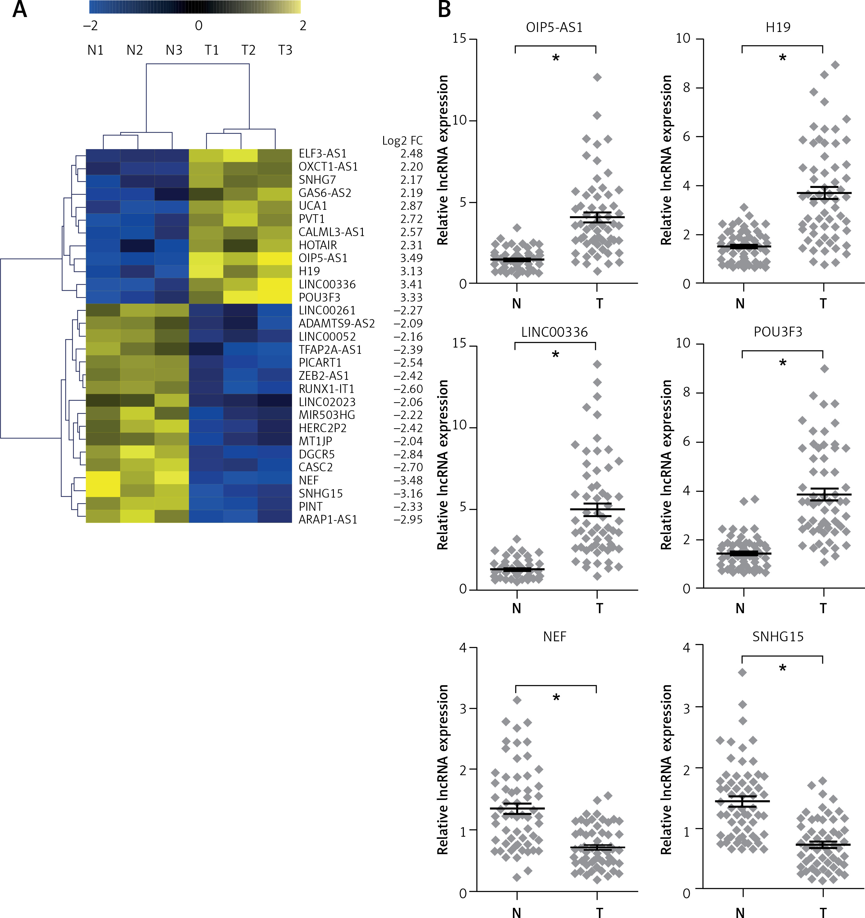
As shown in Table I, the up-regulation of LINC00336 expression level was significantly correlated with tumor stage T (p = 0.002), histological grade (p = 0.001) and vascular invasion (p = 0.043), while the expression level of LINC00336 were not associated with age, gender, tumor size or lymph node metastasis in BCa patients (Table I). We also found that age, gender, tumor size and lymph node metastasis had no obvious effect on OS in BCa patients (Figures 2 A–C, F). However, high tumor stage T (p= 0.014), histological grade (p = 0.004) and vascular invasion (p = 0.026) were significantly associated with poor prognosis of BCa patients (Figures 2 D, E, G). Kaplan-Meier analysis and log-rank tests revealed that high LINC00336 expression level showed worse OS and DFS than those of BCa patients with low LINC00336 expression (Figures 2 H, I). COX univariate and multivariate analyses revealed that LINC00336, vascular invasion and histological grade were independent risk factors for predicting the prognosis of BCa patients (Table II).
Table I
Correlation between LINC00336 expression and clinicopathological variables of BCa patients
Table II
Univariate and multivariate regression analysis of BCa patients for overall survival
Figure 2
LINC00336 was an independent prognostic indicator in BCa patients. Kaplan-Meier analysis and logrank test indicated that age (A), gender (B), tumor size (C) and lymph node metastasis (F) have no relevance for survival prognosis in BCa patients. Tumor stage T (D), Histological grade (E) and vascular invasion (G) are correlated with OS in BCa patients High LINC00336 expression was correlated with poor OS (H) and DFS (I) in BCa patients
Knockdown of LINC00336 repressed proliferation and invasion in BCa cells
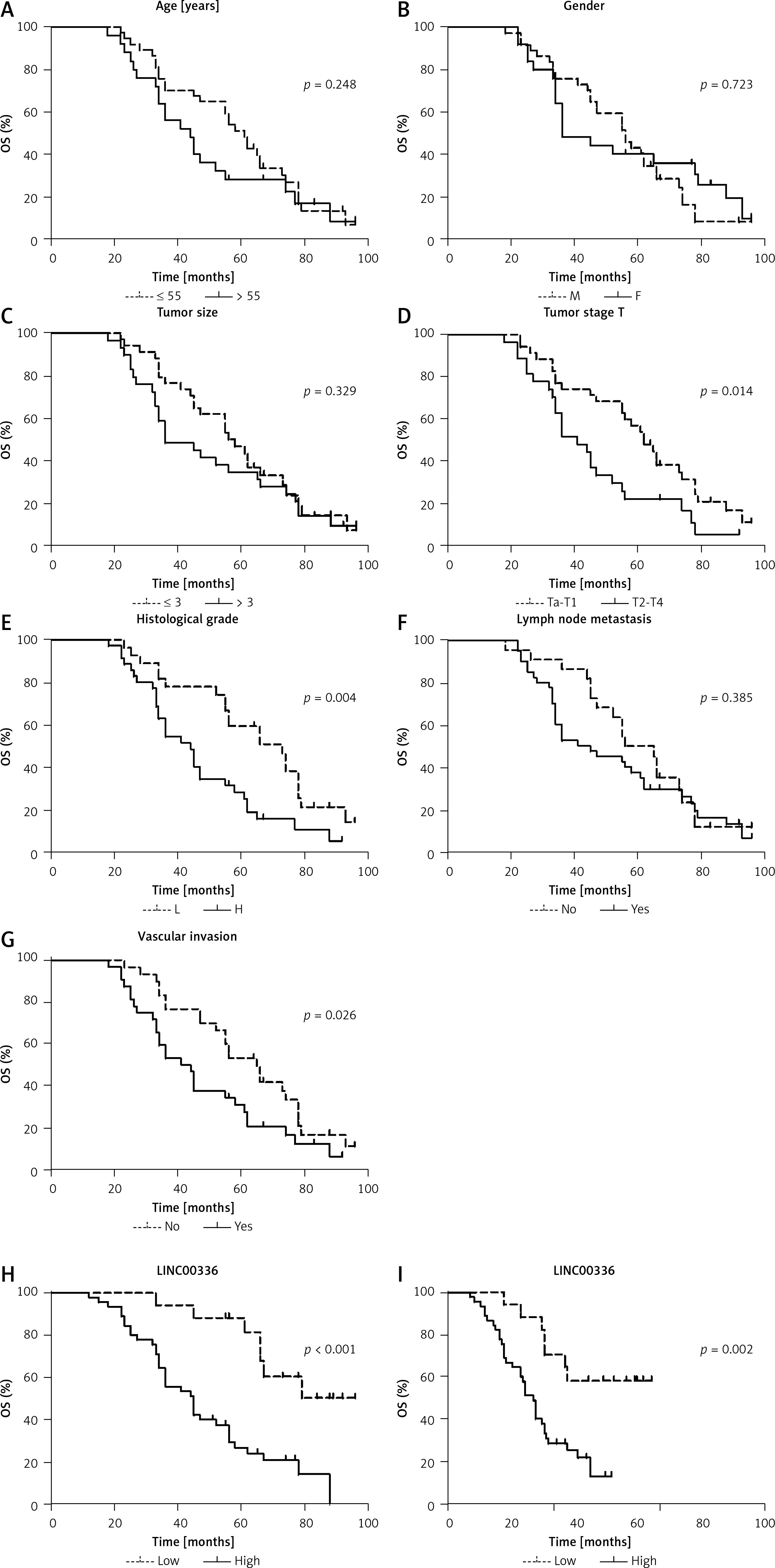
We further determined the functions of LINC00336 in BCa cells in vitro. First, we found significant up-regulation of LINC00336 expression level in BCa cells compared with human normal SV-HUC-1 cells (Figure 3 A). Next, the LINC00336 specific shRNA was designed to target inhibition of LINC00336 expression level in BCa cells, and the results demonstrated that the expression level of LINC00336 was significantly decreased in T24, 5637 and SW780 cells after transfection with sh-LINC00336 (Figure 3 B). After transfection with sh-LINC00336 into T24, 5637 and SW780 cells, the cell proliferation was markedly inhibited when the incubation time was more than 48 h (Figure 3 C). Transwell assay revealed that inhibition of cell invasion was observed in T24, 5637 and SW780 cells induced by silencing LINC00336 (Figure 3 D). These results indicated that LINC00336 had the ability to promote BCa cell proliferation and invasion.
Figure 3
Knockdown of LINC00336 repressed proliferation and invasion in BCa cells. A – Expression of LINC00336 in human normal bladder epithelial cell line (SV-HUC-1) and bladder cancer cell lines (5637, T24 and SW780) was measured using RT-qPCR. B – After transfection with sh-NC or sh-LINC00336 into 5637, T24 and SW780 cells, the expression of LINC00336 was measured using RT-qPCR. C – Cell proliferation changes of 5637, T24 and SW780 cells were determined using CCK-8 assay. D – Invasive abilities of bladder cancer cells were determined using transwell assay
*P < 0.05, **p < 0.01, ***p < 0.001 compared with control group.
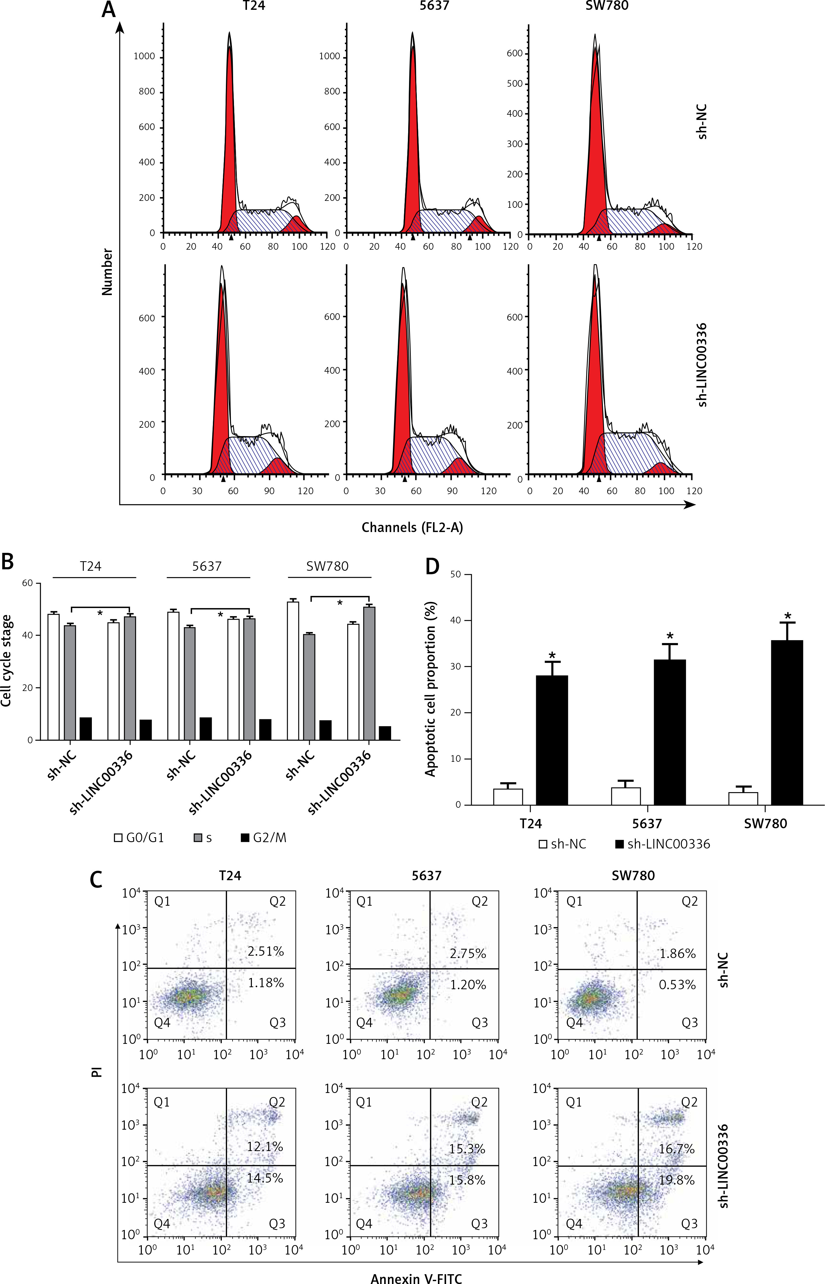
Knockdown of LINC00336 induced cell cycle arrest and apoptosis in BCa cells
Flow cytometry was used to analyze whether LINC00336 regulated cell cycle and apoptosis of BCa cells. The results showed that the percentage of S-phase cells was significantly increased in T24, 5637 and SW780 cells induced by silencing LINC00336 (Figures 4 A, B). We also found that the apoptotic cell proportion was significantly elevated in T24, 5637 and SW780 cells after transfected with sh-LINC00336 (Figures 4 C, D). These findings suggested that knockdown of LINC00336 induced S-phase cell cycle arrest and apoptosis in BCa cells.
Discussion
BCa usually exhibits an excellent prognosis, which critically depends on early diagnosis and treatment, after surgical resection combined with chemotherapy regimens [5]. However, the 5-year OS rate remains unsatisfactory for BCa patients with recurrence or metastasis [5]. Therefore, exploration of novel prognostic markers may be important for the treatment of BCa. At present, non-coding RNAs (ncRNAs), including microRNAs, lncRNAs and circular RNAs, as a novel class of molecular biomarkers, have been identified as prognostic biomarkers of several cancer types, including BCa [22, 23]. LncRNAs belong to the ncRNA family and play a crucial role in the progression of carcinogenesis [24]. With the rapid development of cancer genomics and high-throughput sequencing, thousands of lncRNAs are deregulated or mutated in various cancers [25]. In BCa, numerous lncRNAs, such as UCA1, HOXD-AS1, TUG1, MALAT1, MEG3 and H19, are associated with cell proliferation, migration, invasion, drug resistance and prognosis [25, 26]. Although lncRNAs are implicated in BCa, understanding the roles of tremendously large numbers of lncRNAs in the initiation and progression of BCa is a challenging task.
In our study, lncRNA microarray and RT-qPCR revealed that LINC00336 expression levels were significantly increased in BCa tissues and cell lines compared with the corresponding control group. Kaplan-Meier, COX univariate and multivariate analyses confirmed that LINC00336 could serve as an independent risk factor for predicting the prognosis of BCa patients. In vitro experimental measurements demonstrated that knockdown of LINC00336 showed the ability to inhibit cell proliferation and invasion and induce cell cycle arrest and apoptosis. These findings suggest than LINC00336 may act as an oncogene in the initiation and progression of BCa, and LINC00336 may be a valuable and promising therapeutic target for the treatment of BCa. Recently, He et al. reported that LINC00336 is up-regulated in chromophobe renal cell carcinoma tissues and was identified to be significantly correlated with OS [27]. Wang et al. also demonstrated that LINC00336 is upregulated and performs an important role in pulmonic tumorigenesis, and overexpression of LINC00336 promotes cell growth, colony formation and tumor formation [28]. Moreover, high LINC00336 expression level is associated with poor prognosis in non-small cell lung cancer [28]. These results confirm that LINC00336 functions as an oncogene to facilitate tumor cell growth.
Previous studies corroborate that lymph node metastases constitute a strong prognostic factor in BCa [29, 30]. Mechanistic investigations highlight that lymphovascular invasion may be an important predictor for lymph node metastasis in multiple cancer types [31–33]. The present study reveals a significant increase in LINC00336 expression level in BCa patients with vascular invasion. However, there is no obvious association between LINC00336 expression level and lymph node metastasis, which may be attributed to the small sample size in our study.
To determine possible mechanisms of LINC00336 loss-of-function induced BCa cell growth inhibition, we hypothesized that knockdown of LINC00336 might affect cell cycle and apoptosis of BCa cells. We silenced the expression of LINC00336 by transfection with specific shRNA into three BCa cell lines, i.e. T24, 5637 and SW780 cells, and the inhibition rate of LINC00336 expression level reached up to 50% in in vitro experiments. Subsequently, we observed that S-phase cell cycle arrest and an increase in apoptotic cell proportion occurred in T24, 5637 and SW780 cells after transfection with sh-LINC00336. Our results demonstrated that inhibition of cell growth rate might be associated with S-phase cell cycle arrest and elevation of apoptotic cell proportion in BCa cells after transfection with sh-LINC00336.
We need to make clear the limitations in our study. First, the roles of LINC00336 in cell growth have not been investigated in human BCa xenografts in nude mice. In addition, functions of LINC00336 with gene-gene interactions are not involved in the present study.
In conclusion, LINC00336 as an independent risk factor for predicting the prognosis of BCa was observed in our study, and knockdown of LINC00336 had the ability to suppress cell proliferation and invasion and induce cell cycle arrest and apoptosis in BCa cells. LINC00336 displayed aberrant expression of BCa tissues and showed strong potential for development of a novel therapeutic target for the treatment of BCa.