
Introduction
Myocardial infarction (MI) is a devastating condition in which the heart is deprived of blood supply, which results in necrosis of cardiomyocytes [1]. The burden of MI can be easily understood by the fact that approximately 290 million patients with cardiovascular disease were reported in China, among whom 2.5 million were myocardial infarction patients in 2014 [2]. However, the necrosis of myocardial tissues could be prevented by restoring the lost blood supply, but reperfusion of the tissues further worsens the situation. This leads to another phenomenon known as myocardial ischemic/reperfusion (MI/R) injury where tissues and organs deteriorate before re-establishment of the blood supply [3, 4]. In ischemic conditions, the cardiomyocytes and vascular endothelial cells undergo a series of numerous pathological events, such as production of reactive oxygen species (ROS). The high production of ROS causes generation of oxidative stress due to imbalance between the oxidative defense mechanism and induces lipid peroxidation of cellular membranes and release of various pro-inflammatory cytokines [4, 5]. The complex etiology of MI/R renders any single agent ineffective; thus, it is worthwhile to discover new agents which can provide protection against MI/R via multiple mechanisms.
Casticin (CST) is a polymethylflavone derived mainly from Vitex species of the family Verbenaceae. It shows a wide array of biological activities such as immunomodulatory, anti-hyperprolactinemia, anti-tumor, neuroprotective, anti-inflammatory and analgesic [6–8]. It has been found that CST causes blockade of nuclear factor κ-light-chain-enhancer of activated B cell (NF-κB) for anti-inflammatory action [9]. Nevertheless, no study has been performed to evaluate the effect of CST on MI/R injury. Therefore, in the present study we wish to assess the effect of CST in experimentally induced MI/R injury and to elucidate the possible mechanism underlying its effect.
Material and methods
Animals
Adult male Sprague-Dawley (SD) (240–270 g) rats were selected for the study and housed in strict hygienic conditions after receiving them from the institutional animal house. The experiment was duly approved by the animal ethical committee of the institute and performed according to the national guidelines of animal care and use of China.
Initiation of experimental myocardial ischemia/reperfusion injury
The MI/R injury in rats was induced through blocking of the left anterior descending coronary artery (LAD) for 30 min with subsequent reperfusion for 24 h. Briefly, after anesthesia, the rats underwent tracheal intubation, and the LAD was obstructed using silk ligature for a period of 30 min. After this, the ligature was removed and reperfusion was achieved for the next 24 h. The same surgical procedure was used in the control except blocking of LAD. After 24 h of reperfusion or sham operation, the rats were executed for further analysis.
Experimental design
Thirty SD rats were taken and randomly divided into five groups containing six animals in each group: Group 1 – normal control, Group 2 – ISO rats; Group 3 – CST (10 mg/kg) + ISO, Group 4 – CST (15 mg/kg) + ISO, Group 5 – CST (20 mg/kg) + ISO.
Casticin in the dosage as indicated above after being dissolved in normal saline (also 1% DMSO) was administered to rats 30 min before the induction of myocardial ischemia injury via intraperitoneal injection. At the end of the experiment, hemodynamic parameters were recorded as indicated below. Thereafter, the rats were killed under anesthetized conditions using urethane (1 g/kg, intra-peritoneally injected) to isolate blood. The heart tissues were rinsed in ice-cold 0.9% NaCl after harvesting.
Hemodynamic measurements
The hemodynamic parameters such as systolic, diastolic and mean arterial blood pressure (SAP, DAP and MAP, respectively) as well as HR were recorded using CARDIOSYSCO-101 (Experimentria, Hungary) with a pressure transducer connected to the right carotid artery.
Analysis of electrocardiogram and cardiac functional parameters
Electrocardiogram (ECG) recordings were obtained from the animals using a BL-420S biological function experimental system. For ECG recording, the standard limb lead II of the surface ECG was used for each rat by the PowerLab Data Acquisition System (AD Instruments, Australia) connected to a computer enabled with LabChart professional software version 8 (AD Instruments, Australia).
The ECG record exhibits the following parameters: heart rate in beats per minute, ST height, RR, and QRS complex.
Determination of serum lactate dehydrogenase and creatine kinase levels
The levels of serum lactate dehydrogenase (LDH) and serum creatine kinase (CK) myocardial band were recorded using commercial assay kits obtained from Nanjing Jiancheng Bioengineering Institute, China as per the given instructions.
Determination of tumor necrosis factor-α, cardiac troponin T and interleukin-6 levels in cardiac tissues
The serum levels of tumor necrosis factor-α (TNF-α) and interleukin (IL) 6 in were determined using commercial sandwich enzyme-linked immunosorbent assay (ELISA) kits obtained from Nanjing Jiancheng Bioengineering Institute, China. Moreover, a cardiac troponin T (Tn-T) ELISA kit (cat. No. ARB13662) was obtained from Rapidbio, West Hills, CA, USA [10–13].
Evaluation of lipid peroxidation and antioxidant enzyme levels
The supernatant of heart tissue homogenates was used to assay MPO (cat. no. A044), malondialdehyde (MDA) level (cat. no. A003-2), and superoxide dismutase (SOD) (cat. no. A001-3) and glutathione peroxidase (GPx) (cat. no. A005) activities as per the manufacturer’s instructions (Nanjing Jiancheng Bioengineering Institute, China) on a microplate reader (560 and 532 nm).
Western blot analysis
Using ice-cold RIPA lysis buffer, total protein was extracted. The dissolved proteins were then centrifuged at 12,000 rpm for 5 min at 4°C and the bicinchoninic acid protein assay (BCA) was used to determine the total protein concentration. The isolated proteins were loaded on SDS-PAGE and transferred onto the PVDF membrane and further surveyed with primary antibody. The expression of proteins was detected using an enhanced chemiluminescence reagent after incubating for 1 h at room temperature with HRP-conjugated goat anti-rabbit IgG. Quantification of protein bands was performed using ImageJ v1.42q software (National Institutes of Health, Bethesda, MA, USA).
Results
Effect of CST on hemodynamic parameters in MRI rats
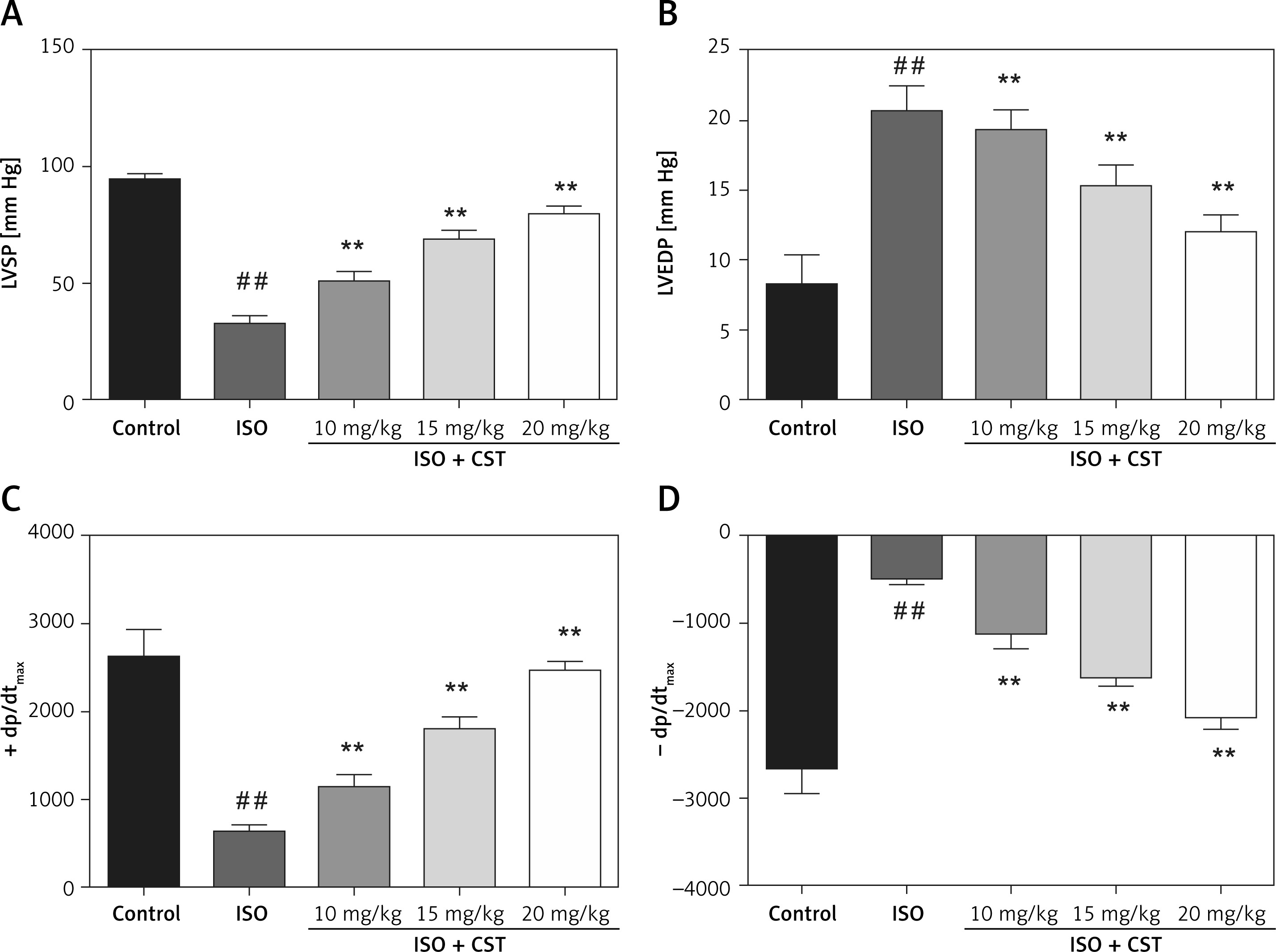
Initially, the effect of CST on left ventricular function in rats was determined. As shown in Figure 1, CST causes significant improvement in left ventricular systolic pressure (LVSP) and derivative of pressure over time of maximal rate of rise of (usually) left ventricular pressure (± dp/dtmax) with reduction of left ventricular end-diastolic pressure (LVEDP) in MRI rats.
Figure 1
Effect of CST on hemodynamic parameters of MRI rats, (A) LVSP (B) LVEDP, (C and D) ± dp/dtmax. Values represent the mean ± SD and are representative of three independent experiments
##p < 0.05 vs. control; *p < 0.05, **p < 0.01 vs. model.
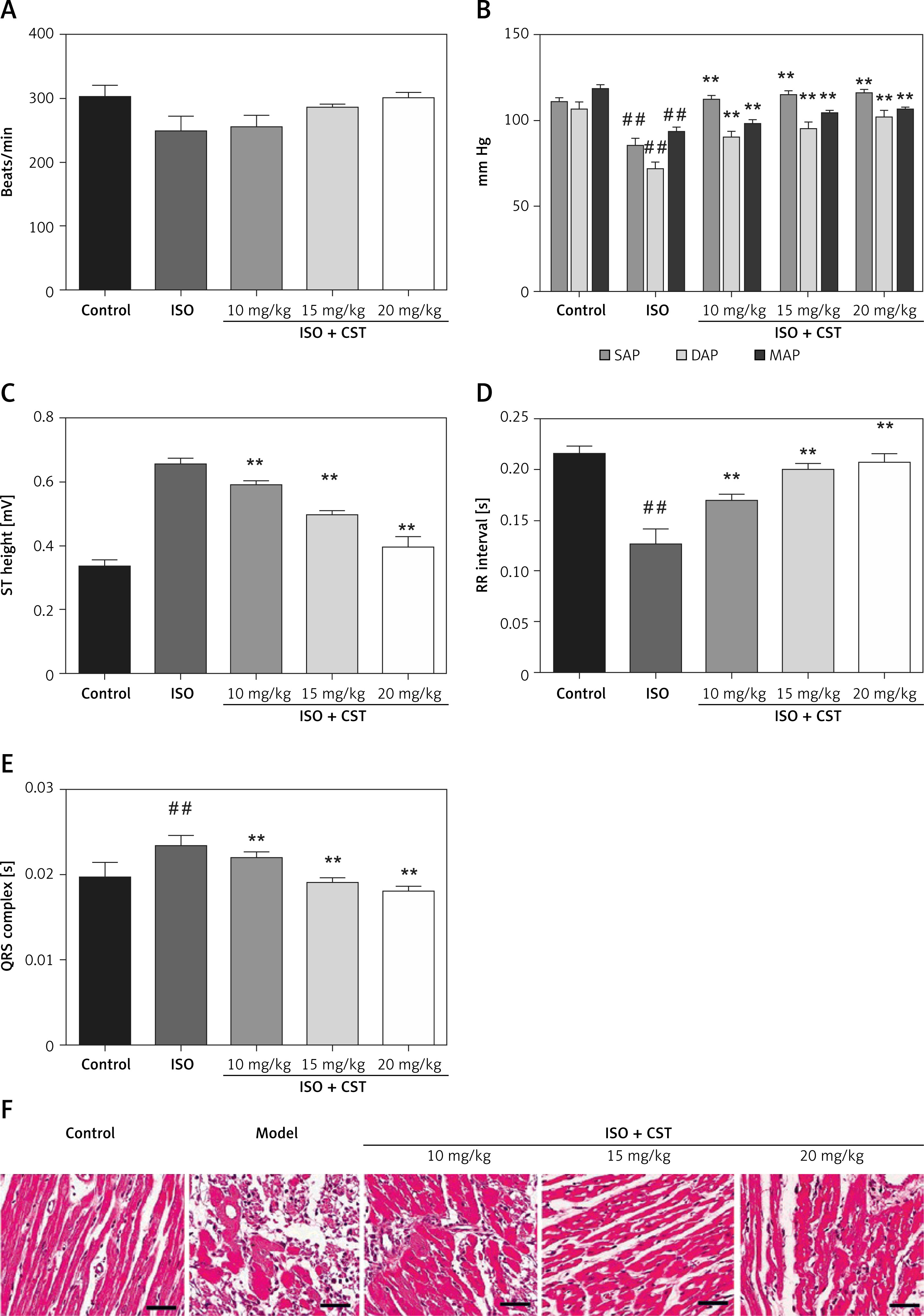
Moreover, CST also causes improvement of heart beat and arterial pressure in a concentration-dependent manner; see Figures 2 A and B, respectively. In ECG analysis as shown in Figures 2 C, D and E, the ISOtreated rats showed amplified heart rate, elevated ST segment, merged T and P waves, flutter waves, decreased RR interval, and widening of QRS complex. In contrast, in the CST treated group, ST segment elevation was reduced together with restoration of the changes in RR interval and QRS complex.
Figure 2
Effect of CST on (A) heart beat and (B) arterial pressure and ECG of rats (C) ST height, (D) RR interval, (E) ORS complex, and (F) cardiac histopathology. Values represent the mean ± SD and are representative of three independent experiments
##p < 0.05 vs. control; *p < 0.05, **p < 0.01 vs. model.
Effect of CST on cardiac histopathology
The effect of CST on the histopathology of cardiac tissue of MRI rats was also investigated to determine its effect at the micro-structural level. As can be seen in Figure 2 F, the control group showed homogeneous and intact histo-architecture with no evidence of cell death or inflammation. However, the model group showed apoptosis of muscle fibers with edema. Necrosis was found to be reduced in a concentration-dependent manner with reduction of edema and myocyte proliferation.
This observation confirmed that CST could mitigate the after-effects of MRI in rats.
Effect of CST on myocardial injury biomarkers
The effect of CST on the serum biomarkers linked with cardiac injury (LDH and CK) was investigated.
As can be seen in Figures 3 A and B, ISO-treated rats showed increased levels of LDH and CK, which were significantly reduced upon administration of CST in CST treated groups.
Figure 3
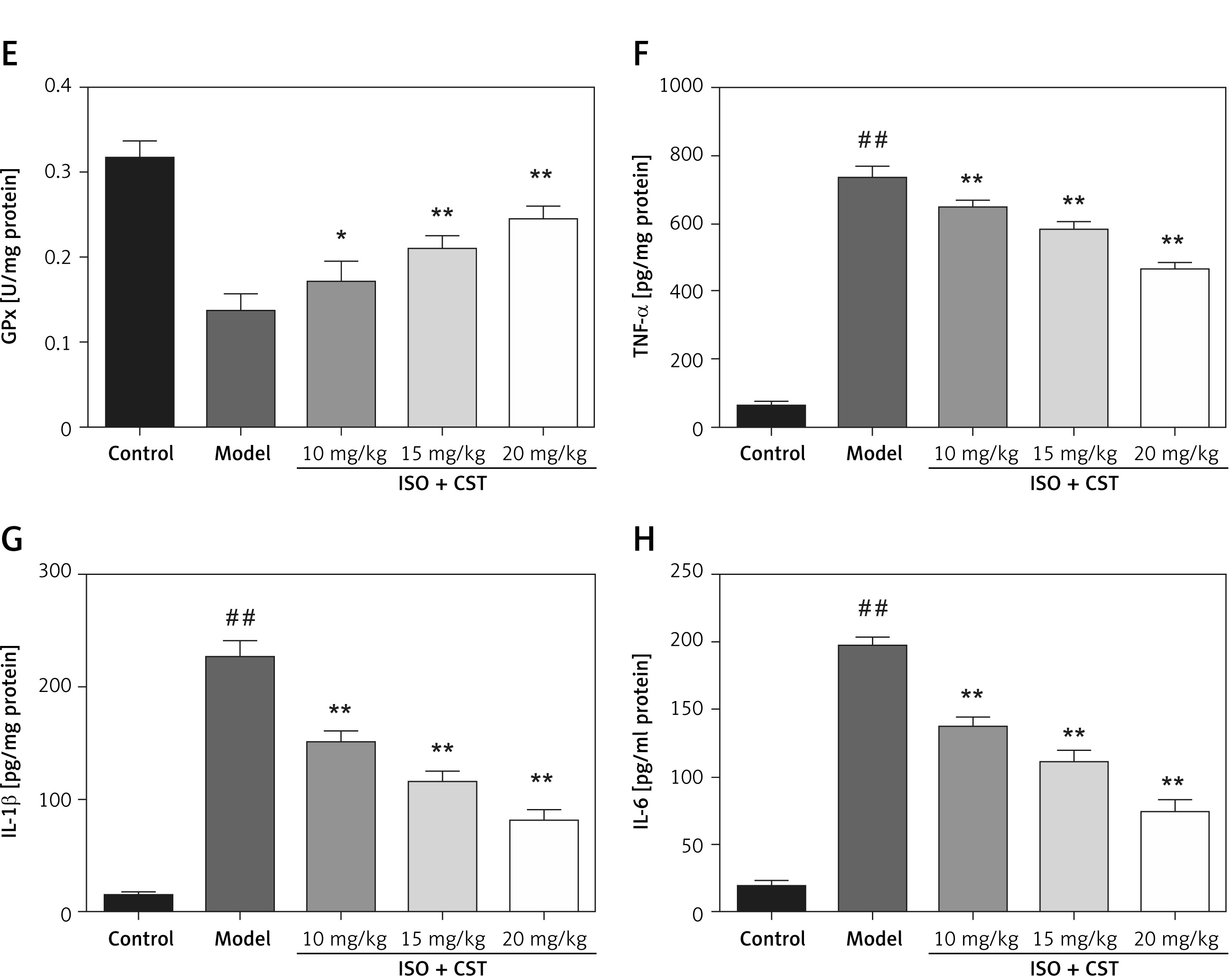
Effect of CST on cardiac injury biomarkers, (A) LDH (B) CK; indices of oxidative stress (C) MDA, (D) SOD. (E) GPx; and inflammatory cytokines (F) TNF-α, (G) IL-1β and (H) IL-6. Values represent the mean ± SD and are representative of three independent experiments
##p < 0.05 vs. control; *p < 0.05, **p < 0.01 vs. model.
Effect of CST on oxidative stress biomarkers
The effect of CST on various biomarkers of oxidative stress was quantified, as presented in Figures 3 C, D and E. The disease model group showed an increased level of MDA with a reduced level of SOD and GPx as compared with the control group.
Moreover, in the CST treated group, the level of these biomarkers of oxidative stress was restored approximately to normal in a concentration dependent manner.
Effect of CST on pro-inflammatory cytokines
As shown in Figures 3 F, G and H, the cytokines (TNF-α, IL-1β and IL-6) were increased in the model group as compared to the control. However, the level of these cytokines was significantly reduced in the CST treated group in a dose-dependent manner. It could suggest that CST had an inhibitory effect on the increased level pro-inflammatory cytokines.
Effect of CST on apoptotic genes
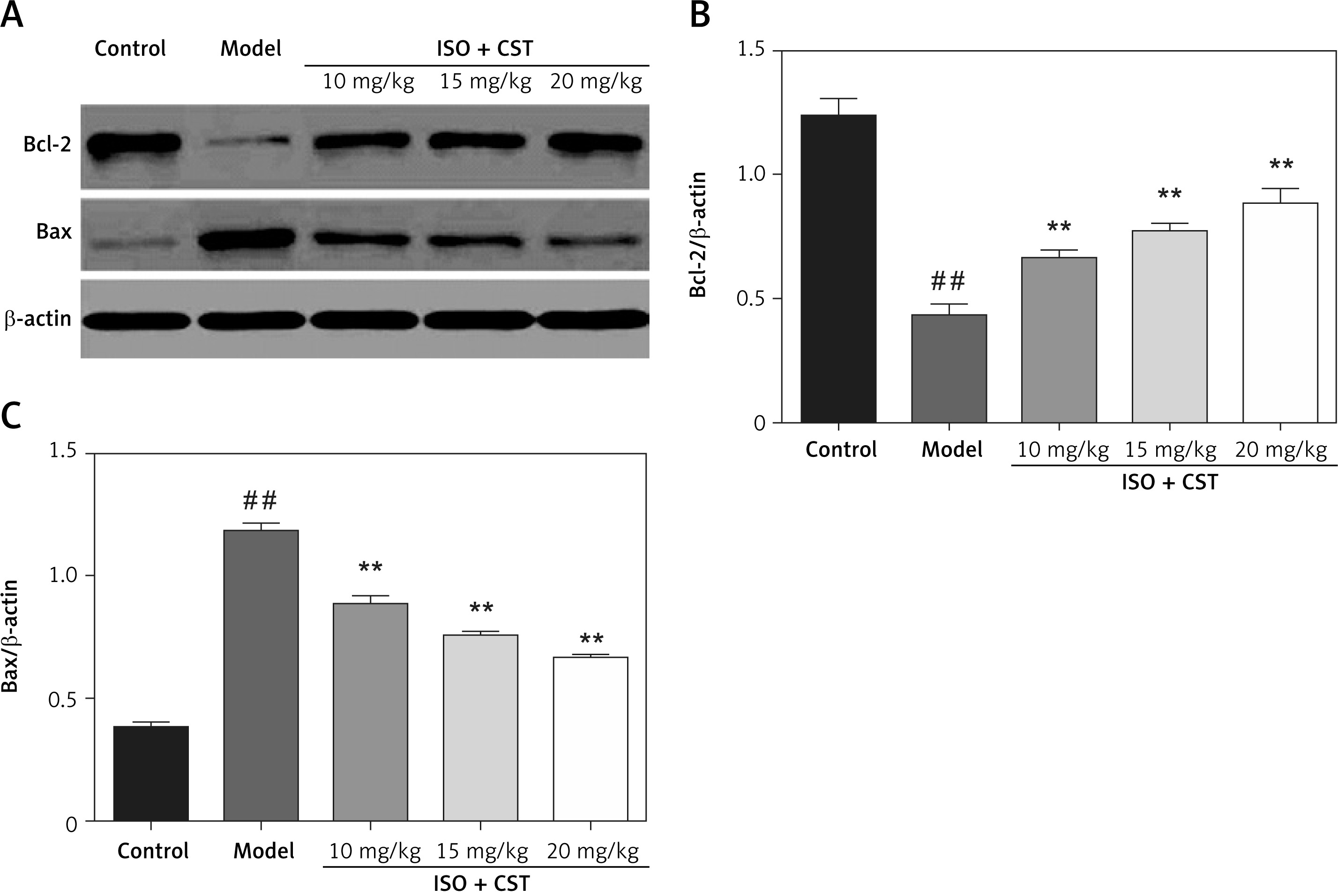
The effect of CST on genes which are known to induce pro-apoptotic and apoptotic action was investigated. As presented in Figure 4, the level of Bcl-2 was found to be decreased with an increase in BCL2-associated X protein (Bax) in the model group as compared to the control. Moreover, the levels of these genes were found to be restored to near normal as compared to the model group. This suggests that CST can inhibit apoptosis of myocardial tissues under MI/R injury.
Effect of CST on NF-κB and iNOS
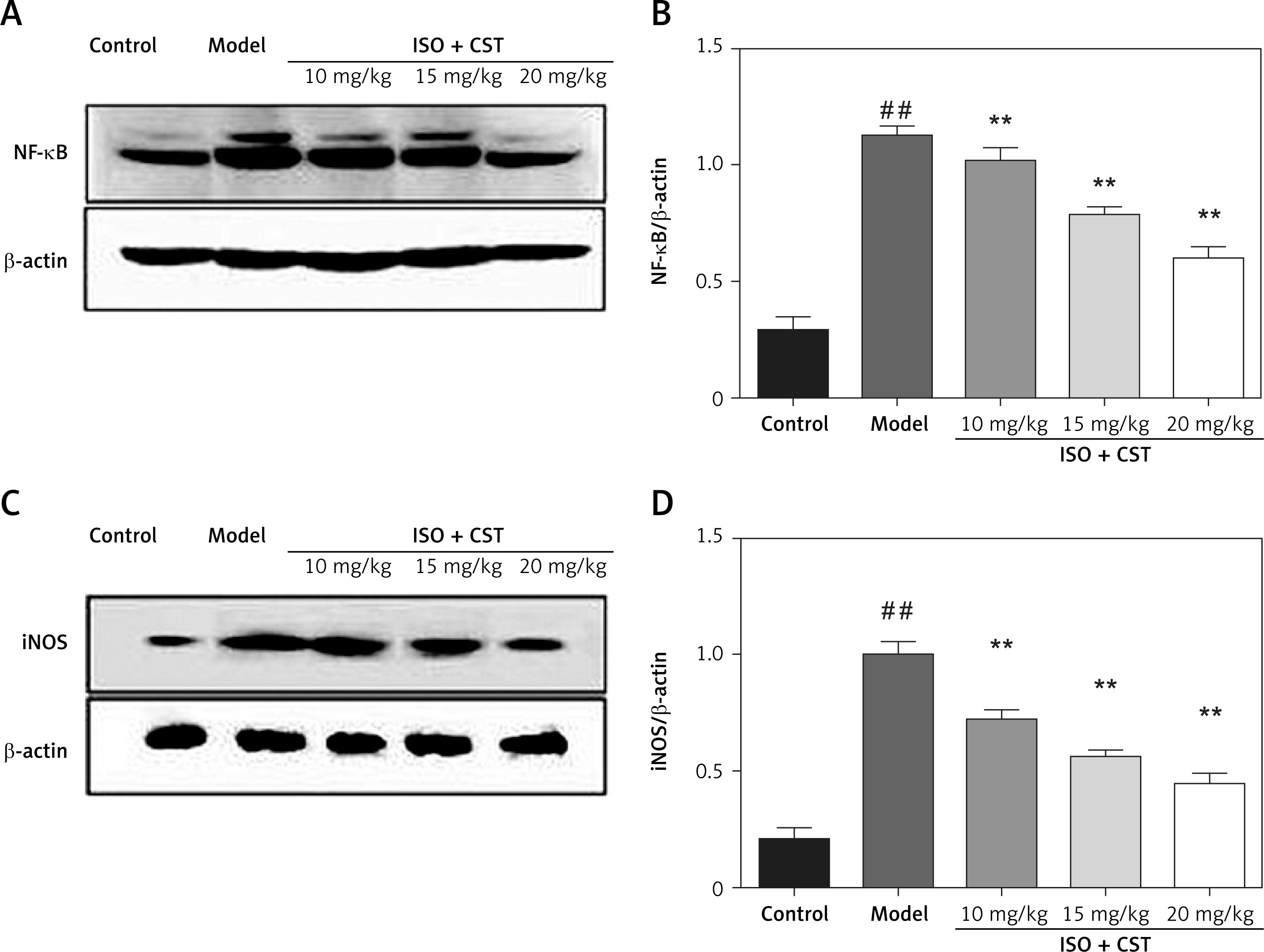
In order to understand the mechanism of action, the effect of CST on NF-κB and inducible nitric oxide synthase (iNOS) was investigated by western blot analysis. As shown in Figure 5, the expression of both NF-κB and iNOS was found to be increased in the model group in comparison to the control. Moreover, the levels of these biomarkers were found to be reduced significantly in the CST treated group in a concentration-dependent manner.
Discussion
Sustained myocardial ischemia–reperfusion induces injury to cardiomyocytes and initiates various forms of cell death that contribute to myocardial infarction. Myocardial ischemia–reperfusion also induces injury to the coronary microcirculation, including capillary rupture and hemorrhage [14]. The present study showed that casticin could effectively mitigate the biochemical alteration in MRI rats. The results suggested that casticin improved the blood supply together with reduction of oxidative stress, inflammation and apoptosis via inhibition of NF-κB and iNOS.
After MI, the key aim of the therapeutics is to restore the blood supply by improving the blood flow [15]. In the present study, it was found that casticin caused improvement in LVSP and ± dp/dtmax with reduction of LVEDP in MRI rats. It also improved the RR interval and QRS complex with reduction of ST segment elevation. Studies have shown the clinical significance of LDH and CK as vital biomarkers to predict cardiac injury. The results suggested that casticin causes significant reduction of both LDH and CK levels in cardiac tissue of rats. The histopathology of rats was also found to be in agreement with the above observation, where casticin causes reduction of the necrosis of myocardial tissue with reduction of infiltration. These results indicated that casticin may have a cardioprotective effect against MI/R injury.
Prompted by the above results, in the next part, we aimed to investigate the probable mechanism underlying the cardioprotective effect of casticin. Studies have shown that oxidative stress has a very deleterious effect on the recovery from MI/R injury [16–20]. It induces production of an excessive amount of reactive oxygen species in the impaired anti-oxidant defense system [21–23]. It has been suggested that these free radicals cause degradation of DNA, lipids, and membranes, which promotes tissue necrosis [21, 24, 25]. Thus agents improving the antioxidant system showed benefit against myocardial ischemia/reperfusion. In the present study, we observed that casticin induced significant reduction of MDA production and restored the level of SOD and GPx in cardiac tissue. Thus, it could be suggested that casticin protects against myocardial damage possibly by restraining the production of free radical via improving the anti-oxidant mechanism.
Studies have shown that oxidative stress promotes inflammation in myocardial tissues [26–30]. It promotes the generation of various pro-inflammatory cytokines regulated via activation of NF-κB [17, 31–34]. NF-κB is an important inflammatory mediator which promotes the transcription of various genes after activation by numerous stimuli responsible for cellular viability, proliferation, apoptosis and the inflammatory response. The activation of NF-κB is generally mediated via ligands of various cytokine receptors, pattern-recognition receptors (PRRs), TNF receptor (TNFR) superfamily members, as well as T-cell receptor (TCR) and B-cell receptor [35–38]. Upon activation NF-κB dissociates from its inhibitory regulator IκB-α and translocates in free form into the nucleus from the cytoplasm [39, 40]. In the present study, we found that casticin causes reduction of NF-κB in myocardial tissues together with iNOS. iNOS is another important mediator found overexpressed in myocardial tissue. A high level of iNOS promotes excessive production of nitric oxide for a prolonged duration. It also induces oxyradical-mediated myocardial damage or acts as a negative inotrope due to myocardial cGMP production [41–44]. Numerous studies have shown the involvement of apoptosis or planned cell death of cardiomyocytes in MI/R injury [45–49]. The Bcl-2 family genes are notably acknowledged as the main regulators of apoptosis. Bcl-2 was shown to inhibit the process of apoptosis, whereas Bax acts as a pro-apoptotic gene; thus the intricate balance between these two genes regulates apoptosis [50]. In the present study, casticin caused reduction of Bax, and increased Bcl-2 in a dose-dependent manner. Thus, the above observations suggest that casticin exerts a cardioprotective effect via inhibition of inflammation and apoptosis.
In conclusion, it could be suggested that casticin protects the heart after MI/R injury in rats via multiple mechanisms, such as reduction of oxidative stress, inflammation, and apoptosis and improvement of hemodynamic status. The present study may serve as a template for further studies on casticin application against MI/R injury.