

Introduction
Chronic kidney disease (CKD) is an increasingly common clinical and public health problem worldwide. It affects approximately 8% to 16% of the general adult population [1]. It is estimated that by 2040, CKD will become the fifth most common cause of death in the world [2]. Treatment of CKD is complex. It requires treatment of the pathophysiology of CKD itself and a multidisciplinary approach to reduce CKD risk factors and treat common complications and comorbidities [3]. In addition to pharmacological interventions, regular physical activity and appropriately selected exercises are considered a basic element of the lifestyle modification strategy in the treatment of CKD [4]. Oxidative stress plays a key role in the development and progression of CKD, causing kidney cell damage, inflammation, and fibrosis [5]. There is a lack of effective therapeutic interventions to slow the progression of CKD [6, 7].
CKD impairs the regulation of muscle protein metabolism, which leads to muscle atrophy [6]. Myostatin is a protein belonging to the transforming growth factor β (TGF-β) family. It is a myokine that is mainly expressed in skeletal muscle [8]. It inhibits the growth of muscle mass and also plays a key role as a mediator of muscle atrophy in CKD. Moreover, it is a muscle growth inhibitor [9]. In an animal model, a 2- to 3-fold increase in muscle myostatin expression has been observed accompanying CKD. Sarcopenia is a serious complication that significantly reduces the quality of life of patients [10]. Therefore, it has been suggested that inhibiting myostatin expression may be a strategy for the treatment of muscle wasting in CKD [8, 11]. Studies indicate that plasma myostatin levels correlate with kidney function, highlighting its potential as a biomarker for assessing kidney health and muscle mass in patients with CKD. MSTN expression is elevated in skeletal muscles of patients with CKD, which may play a role in the pathogenesis of sarcopenia or protein-energy wasting (PEW). The observation suggests that plasma MSTN levels may be correlated with renal function [12]. Low physical activity, persistent chronic inflammation, and the accumulation of uremic toxins observed in patients with CKD are factors that may contribute to the increase in myostatin production [13, 14].
Transcription factors belonging to the forkhead box class O (FOXO) family are integral to numerous cellular functions, thereby contributing significantly to the maintenance of cellular homeostasis [15–17]. The group includes four molecules – FOXO1 (FKHR), FOXO3A (FKHRL1), FOXO4 (AFX1), and FOXO6 – that affect various gene expression mechanisms [18]. There are few studies in the human population, and existing studies on animal models based on the inhibition of the action of individual FOXO proteins emphasize their great importance in physiology [15, 19, 20]. FOXO transcription factors, due to their distribution in tissues and multidirectional actions, may constitute a therapeutic target in many diseases. Further research is necessary on the physiological effects of FOXO proteins and their participation in the pathogenesis of civilization diseases [21–23]. Studies have shown that FOXO3 is involved in the transition from acute to CKD by regulating autophagy and the stress response. Activation of FOXO3 during hypoxia leads to its accumulation in the cell nucleus, where it acts as a transcription factor, promoting the expression of genes related to stress response and cell repair [24]. In turn, FOXO1 is crucial for the regulation of muscle energy homeostasis, playing an important role as a regulator of glucose metabolism in skeletal muscles [24]. In addition, FOXO1 and FOXO2 regulate the transcription of cell cycle genes that control cell proliferation and are involved in cancer, obesity, diabetes, autoimmune diseases, and aging [25].
Patients undergoing renal replacement therapy often have a negative attitude towards physical exercise, which makes cooperation with them in this area extremely difficult. Modern technologies such as virtual reality (VR) are increasingly being incorporated into the rehabilitation process [26, 27]. The incorporation of VR technology into the hemodialysis process represents a groundbreaking approach that has the potential to enhance patients’ motivation for physical activity. By integrating VR into rehabilitation protocols, patients can benefit from real-time feedback and engage in repetitive, functional exercises. Integrating these elements is particularly advantageous during hemodialysis, as it maximizes the efficiency of treatment time, alleviates the tedium associated with the procedure, boosts exercise motivation, and guarantees ongoing medical oversight [28–33]. Increasingly, evidence indicates that physical exercise reduces levels of inflammatory markers (e.g. IL-6, CRP), improves psychological parameters (e.g. reduced anxiety and stress), increases muscle mass and strength, and reduces cardiovascular risk (e.g. reduced blood pressure, improved lipid profile) [13, 14, 34–36].
Further studies, including functional analysis and correlation of myostatin and FOXO protein expression levels with other clinical and metabolic parameters, may contribute to a better understanding of these mechanisms and enable the development of new therapeutic strategies.
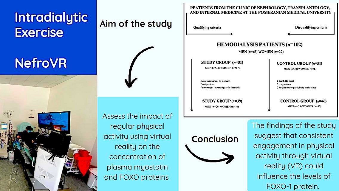
The aim of this study was to comprehensively evaluate the impact of regular physical activity using VR during hemodialysis sessions on plasma concentrations of myostatin and FOXO proteins, with particular emphasis on enhancing training attractiveness and patient motivation.
Material and methods
Inclusion and exclusion criteria
The study was conducted at the Department of Nephrology, Transplantology, and Internal Diseases at Pomeranian Medical University in Szczecin (Poland) between February 2021 and December 2021. The research focused on individuals diagnosed with the fifth stage of CKD who were undergoing renal replacement therapy through hemodialysis at the dialysis facility within the Department of Nephrology, Transplantology, and Internal Diseases at Pomeranian Medical University in Szczecin. Participants in the research program were recruited by a nephrology specialist taking into account the inclusion and exclusion criteria.
The inclusion criteria included: written consent to participate in the study, complete lack of diuresis, undergoing hemodialysis as renal replacement therapy for at least 3 months (three sessions per week), and age over 18 years. The exclusion criteria included: lack of written consent to participate in the study, musculoskeletal disease preventing participation in the study, serious cardiovascular diseases (NYHA III or IV heart failure), acute coronary syndrome in the last 3 months, uncontrolled arterial hypertension, uncorrected visual impairment, poorly controlled diabetes (HbA1c above 8% for 3 months), senile dementia, other neurological or mental disorders preventing consent to the study or understanding the nature of the study and the conditions of participation, malignant tumors, surgery performed in the last month or amputation of the lower limb preventing participation in the study, or epilepsy.
Organization and schedule of the study
The study was conducted after obtaining the consent of the Bioethics Committee (number KB-0012/144/2020), issued on October 5, 2020. Each patient gave written informed consent to participate in this study. Every effort has been made to protect the privacy and anonymity of patients. The study was conducted in accordance with the current version of the Declaration of Helsinki. Financing for the research was provided by the European Regional Development Fund under the grant number RPZP.01.01.00-32-0010/19-10. The costs of publication were covered by the Pomeranian Medical University in Szczecin.
Patients were randomly assigned to the study group or the control group. Researchers and study participants were aware of group assignment. The study group was tasked with engaging in training sessions using the NefroVR system prototype, which occurred three times weekly for a duration of 20 min during hemodialysis (HD) over a period of 3 months. The sessions were conducted during the initial 2 h of HD treatment or until a target ultrafiltration (UF) of 2.5 was reached. In contrast, patients in the control group received standard dialysis care, without additional exercise intervention.
Research tools
The research employed a prototype of the NefroVR system, which comprised various components assembled on a mobile platform equipped with ballast. Key elements of the system included: a central unit that integrates all components and operates specialized software; a rehabilitation rotor featuring a flywheel that enables load adjustment during exercises conducted during hemodialysis; VR goggles designed for the patient to provide an immersive experience; a panoramic display for the patient; a touchscreen interface for medical personnel (such as doctors, nurses, or physiotherapists); and a patient control kit that consists of a digital joystick and buttons. The study had three identical NefroVR system sets available, which were used in rotation by the study group participants.
The system operated based on the concept of audiovisual stimulation, motivating the patient to partake in physical activity through a virtual game that used a rehabilitation rotor for movement. The rotor’s speed influenced the game’s tempo. The rotor was attached to the hemodialysis chair, allowing patients to easily perform the exercise during hemodialysis. Physical activity consisted of performing movements of the lower limbs on the rotor. Healthcare professionals, including physicians and physiotherapists, oversaw the rotor’s revolutions and the flywheel’s resistance, adjusting these parameters according to the patient’s health status to prevent undue stress, such as elevated heart rates or hazardous fluctuations in blood pressure.
In the course of clinical trials, participants were presented with a selection of five mini-games, each with an approximate duration of 20 min. The selection of five mini games was made by a research team consisting of specialists in nephrology, physiotherapy, and new technologies in medicine. The selection criteria included safety of dialysis patients, the possibility of performing exercises in a sitting position, the level of involvement, and intuitive use. Alongside the use of the rotor, patients were able to engage with the game through a joystick and buttons. The extent of interactivity was deliberately constrained to facilitate the introduction of patients to the NefroVR system. Before the study began, all patients received training in use of the VR system. When necessary, medical staff assisted participants in using the device. The correctness of the exercises was assessed by a physiotherapist. His task was to supervise the session, correct any errors in technique and ensure the safety of patients during the exercises. Both the VR device and the games were designed specifically for hemodialysis patients. Their selection took into account the specific needs of this group of patients, such as the ability to perform exercises in a sitting position, safety, and intuitive operation.
The NefroVR research tool has also been used in other studies, where its detailed description is also available [28, 29, 37, 38].
At the outset of the research (E0), participants were requested to fill out a tailored survey questionnaire specifically developed to address the objectives of this study. The questionnaire included inquiries about participants’ demographic characteristics, health conditions, lifestyle choices, and routine behaviors.
Laboratory tests
Two milliliters of blood was collected from study participants. Blood was collected twice: on the day of study initiation (E0) and after 3 months (E3). In both cases, samples were collected before the hemodialysis procedure. In the study participants, venous blood samples were obtained from the dialysis fistula prior to the initiation of the hemodialysis procedure. The samples were collected into test tubes containing EDTA at the dialysis facility located within the Department of Nephrology, Transplantology, and Internal Diseases at the Medical University of Szczecin, specifically on the designated days for cyclical evaluations (E0, E3). Following collection, the blood samples underwent centrifugation at 4000 rpm for 10 min at 4°C, using an MPW-350R centrifuge. The resulting blood plasma was aliquoted into two separate Eppendorf Safe-Lock Tubes (1.0 ml, Eppendorf Quality, colorless), each containing 1.0 ml, and was promptly frozen. The samples were subsequently preserved at –70°C until they were ready for analysis. For laboratory testing, the samples were thawed at room temperature immediately prior to use.
Prior to the commencement of the study, standard plates were prepared in accordance with the guidelines provided by Sun Red Biotechnology Company. Subsequently, biotin-labeled antibodies, the test samples, and streptavidin were added to the plates. The volumes of the materials and reagents used were contingent upon the specific parameter being assessed. The plates underwent incubation for 60 min at 37°C, followed by five washes with a washing buffer. Afterward, Chromogens A and B were added, and the mixture was incubated for an additional 10 min at 37°C before the application of the stopping solution. Absorbance readings were taken at a wavelength of 450 nm, and data analysis was conducted using the Envision program, which was based on a linear calibration curve.
The evaluation of myostatin and FOXO protein levels in plasma was conducted using the commercially available Human MSTN test and the FOXO ELISA Kit from SunRed Biotechnology Company. Myostatin concentrations were reported in nanograms per liter (ng/l), with a sensitivity threshold of 5.113 ng/l and a linearity range extending from 7 to 2000 ng/l. Additionally, myostatin levels were expressed in ng/ml, with a sensitivity of 0.175 ng/ml and a linearity range of 0.2 to 60 ng/ml. The concentrations of FOXO proteins were measured in ng/l, with a sensitivity of 2.827 ng/l and a linearity range from 3 to 900 ng/l.
Statistical analysis
Statistical analyses were performed using Statistica 13 (StatSoft, Inc., Tulsa, OK, USA). All data regarding continuous variables were presented as mean ± standard deviation (± SD) and median, while qualitative variables were presented as number and percentage. The normality of the distribution was tested using the Shapiro-Wilk test. The data used in the study did not meet the assumptions of a normal distribution. The χ2 test or χ2 test with Yates’ correction was used to analyze qualitative data. The Mann-Whitney U test was employed to analyze continuous variables across different groups. The evaluation of laboratory data collected prior to and following the intervention in both the study and control groups was conducted using the Wilcoxon test. A p-value of less than 0.05 was considered statistically significant.
Results
Table I presents the characteristics of the study and control groups. The analysis of data regarding demographics, comorbidities, and habitual behaviors did not reveal statistically significant differences between these groups.
Table I
Characteristics of the study and control groups
| Parameter | Study group (n = 39) | Control group (n = 46) | P-value | |||
|---|---|---|---|---|---|---|
| Age, mean ± SD; Me | 57.56 ±17.61; 63.0 | 62.63 ±15.47; 64.0 | 0.266a | |||
| Sex, n (%) | ||||||
| Male | 29 | 74.36% | 29 | 63.04% | 0.264b | |
| Female | 10 | 25.64% | 17 | 36.96% | ||
| BMI [kg/m2] | 28.23 ±5.75; 28.28 | 27.89 ±5.79; 27.57 | 0.760a | |||
| Professional activity, n (%) | ||||||
| No | 26 | 72.22% | 40 | 88.89% | 0.103b | |
| Yes | 10 | 27.78% | 5 | 11.11% | ||
| Professional activity before the start of hemodialysis, n (%) | ||||||
| No | 10 | 27.78% | 21 | 46.67% | 0.082b | |
| Yes | 26 | 72.22% | 24 | 53.33% | ||
| Type of job, n (%) | ||||||
| Blue-collar | 18 | 47.37% | 18 | 54.55% | 0.829b | |
| White-collar | 11 | 28.95% | 7 | 21.21% | ||
| No job | 9 | 23.68% | 8 | 24.24% | ||
| Currently smoking cigarettes, n (%) | ||||||
| No | 30 | 76.92% | 35 | 76.09% | 0.868b | |
| Yes | 9 | 23.08% | 11 | 23.91% | ||
| Number of cigarettes per day, mean ± SD; Me | 14.44 ±6.13; 15.0 | 14.09 ±7.41; 10.0 | 0.676a | |||
| How many years ago quit smoking, mean ± SD; Me | 9.71 ±10.95; 5.0 | 16.67 ±16.17; 13.0 | 0.520a | |||
| Number of HD per week, mean ± SD; Me | 2.95 ±0.23; 3.0 | 2.98 ±0.15; 3.0 | 0.805a | |||
| Duration of dialysis [min], mean ± SD; Me | 223.85 ±20.47; 240.0 | 216.52 ±28.92; 210.0 | 0.110a | |||
| Concomitant diseases, n (%) | ||||||
| Diabetes | 5 | 14.71% | 13 | 28.89% | 0.224b | |
| Arterial hypertension | 25 | 73.53% | 32 | 71.11% | 0.812b | |
| Epilepsy | 4 | 12% | 3 | 7% | 0.697b | |
| Ophthalmic | 8 | 24% | 15 | 33% | 0.484b | |
| Neurological | 2 | 6% | 3 | 3 | 0.745b | |
| Treatment with another renal replacement therapy | 7 | 20.59% | 8 | 17.78% | 0.979b | |
A total of 102 patients, comprising 65 males and 37 females, were selected for participation in the study. Of these, 85 patients who underwent renal replacement therapy via hemodialysis were included, consisting of 58 males and 27 females, as illustrated in Figure 1. The participants were randomly assigned to two distinct groups: the experimental group, which included 39 participants (mean 57.56 ±17.61), and the control group, consisting of 46 patients (mean 62.63 ±15.47). All patients reported a level of physical fatigue during the exercise, which was assessed at 8–14 on the Borg scale.
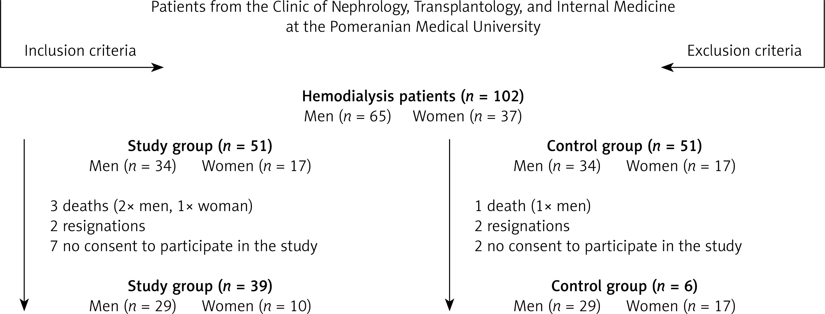
Table II presents a comparison of the values of four laboratory parameters (FOXO-1, FOXO-2, FOXO-3, and myostatin) between the study and control groups. For each parameter, the mean (M), standard deviation (SD), median (Me), lower quartile (Q1), and upper quartile (Q3) are presented for both groups. In measurement 2, a statistically significant difference between the groups was noted (p = 0.041), where the study group showed lower FOXO-1 values compared to the control group. In the remaining parameters (FOXO-2, FOXO-3, and myostatin), no statistically significant differences were found between the groups in any of the measurements (p > 0.05).
Table II
Comparison of laboratory parameters between experimental and control groups
| Variable | Study group | Control group | P-value (statistical power of the test) | ||
|---|---|---|---|---|---|
| M (SD) | Me (Q1–Q3) | M (SD) | Me (Q1–Q3) | ||
| Measurement 1 | |||||
| FOXO-1 | 23.66 (65.07) | 7.49 (3.90–13.18) | 13.36 (11.12) | 8.53 (6.88–18.39) | 0.176 (0.25) |
| FOXO-2 | 4.71 (4.91) | 2.92 (1.63 –5.93) | 4.67 (4.47) | 2.37 (1.80–7.11) | 0.917 (0.05) |
| FOXO-3 | 4.88 (4.92) | 2.66 (1.64–6.44) | 4.74 (6.05) | 2.31 (1.78–5.62) | 0.966 (0.05) |
| Myostatin | 681.40 (590.68) | 565.755 (140.7250–1272.15) | 517.21 (422.23) | 412.13 (250.7– 657.1) | 0.483 (0.31) |
| Measurement 2 | |||||
| FOXO-1 | 10.14 (14.06) | 4.88 (3.47–9.92) | 13.87 (12.34) | 9.03 (6.46–19.43) | 0.041* (0.23) |
| FOXO-2 | 3.48 (3.68) | 2.23 (1.34–3.92) | 5.10 (4.95) | 2.84 (1.82–5.89) | 0.169 (0.50) |
| FOXO-3 | 4.85 (6.23) | 3.06 (1.45–5.90) | 5.26 (4.97) | 3.18 (2.07–6.89) | 0.151 (0.061) |
| Myostatin | 669.18 (659.03) | 416.34 (194.79–1087.70) | 618.51 (602.94) | 404.29 (123.54–1061.42) | 0.522 (0.065) |
Table III presents a comparison of the correlations between two measurements (measurement 1 and measurement 2) of four laboratory parameters (FOXO-1, FOXO-2, FOXO-3, and myostatin) in the study and control groups. In the study group, a statistically significant correlation between measurements 1 and 2 in the FOXO1 measurement was observed (p = 0.045). In the control group, no statistically significant correlation was found (p = 0.434). In both groups, no statistically significant correlations between measurements 1 and 2 were found for the remaining parameters (p > 0.05).
Table III
Comparison of laboratory parameters between baseline and 3-month measurements within study and control groups
| Variable | Study group | Control group | |
|---|---|---|---|
| P-value (statistical power of the test) | P-value (statistical power of the test) | ||
| FOXO1 | Measurement 1 | 0.045* (0.94) | 0.434 (0.35) |
| Measurement 2 | |||
| FOXO2 | Measurement 1 | 0.075 (0.15) | 0.777 (0.09) |
| Measurement 2 | |||
| FOXO3 | Measurement 1 | 0.658 (0.08) | 0.502 (0.21) |
| Measurement 2 | |||
| Myostatin | Measurement 1 | 0.829 (0.89) | 0.875 (0.95) |
| Measurement 2 |
Discussion
In the conducted study assessing the impact of VR exercises performed over a 3-month period on FOXO proteins and myostatin concentrations, a statistically significant difference was observed in FOXO-1 protein levels between the experimental and control groups after 3 months. Additionally, a reduction in FOXO-1 protein concentration was noted in the experimental group following the 3-month exercise intervention. No statistically significant differences were found for the other analyzed parameters. The observed differences between groups may be attributed to the implementation of the exercise intervention using VR technology, which – by increasing patient engagement and diverting attention from the discomfort of the dialysis environment – may have contributed to more consistent and effective participation in physical activity. This, in turn, could have influenced favorable changes in biomarkers such as myostatin and FOXO protein levels. However, we cannot exclude the influence of additional factors, such as individual motivation, fatigue level, or undetected differences in the clinical status of participants.
An increasing number of studies focus on encouraging physical activity among patients undergoing hemodialysis, highlighting its advantages for both mental and physical well-being, which include enhanced laboratory outcomes and improved physical performance. However, motivating these patients to exercise is a challenge due to their poor condition and lack of hope for full recovery. The use of VR technology in dialysis settings may reduce patients’ subjective perception of treatment duration, improve mood, increase engagement in intradialytic exercise, and support adherence to therapeutic recommendations [28, 37–42].
In the study, after a 3-month intervention using VR during physical activity, a statistically significant difference in FOXO-1 protein concentrations was found between the study group and the control group. Patients from the study group who participated in the VR program showed lower FOXO-1 concentrations compared to the control group. A notable decrease in FOXO-1 levels was observed following a three-month intervention with the NefroVR system in the experimental group. The changes observed in FOXO-1 are specific to this protein and may be the result of the body’s specific response to VR-related stimuli. The findings suggest that physical activity, particularly engaging and motivating forms such as those involving VR, may induce changes regulated by FOXO-1. No comparable studies exist in the literature that have evaluated these parameters within a group of patients undergoing hemodialysis. Therefore, the study can be considered pioneering in this field.
Few studies on FOXO expression were found in the literature. For example, an experimental study performed by Reed et al. on an animal model showed that FOXO expression in skeletal muscles increased significantly during the period of cachexia (3.1-fold in the soleus). In response to inflammation, FOXO1 activity increased (1.8-fold in the soleus), and aerobic exercise may slightly reduce its acetylation [43].
The results of our study showed no significant differences in FOXO-2 and FOXO-3 protein concentrations in patients with stage 5 CKD treated with hemodialysis who participated in a regular physical activity program using VR technology. Although previous studies have suggested a significant role of FOXO proteins, especially FOXO3, in the regulation of cellular mechanisms related to the response to oxidative stress, autophagy, and in the transition from acute to CKD, our results did not confirm clear changes in the concentrations of these proteins as a result of physical intervention.
It is important to emphasize that the lack of significant differences in the concentrations of FOXO-2 and FOXO-3 does not diminish the possible health advantages linked to consistent physical activity among individuals with CKD. As other studies show, exercise can improve inflammation, cardiovascular function, and overall quality of life in patients, even if it does not directly affect the expression of specific transcription proteins.
It is worth noting that in our study, FOXO protein concentrations were determined using the ELISA method, while most previous studies focused on the analysis of FOXO gene expression at the mRNA level. Differences in methodological approach may partially explain the lack of significant differences in our results. Determining proteins directly from plasma samples allows for the assessment of their actual concentration in the body, but this does not always reflect changes in their gene expression that may occur at earlier stages of the transcription process [19, 44].
Myostatin, a myokine that negatively regulates muscle growth, is a well-studied factor whose levels may change with physical activity. Previous studies have shown that regular physical exercise can reduce the concentration of myostatin, which helps increase muscle mass and improve muscle strength [37, 42, 45]. However, in the context of our study, in which patients underwent 3 months of VR exercises, no significant changes in myostatin levels were observed. The lack of significant change may be due to several factors. First, the intensity and type of physical activity may not be sufficient to induce changes in myostatin concentrations. While VR can increase motivation to exercise, it can also reduce the intensity of exercise, which is key to inducing significant changes in myostatin levels. Second, a 3-month period could be too short to observe noticeable changes, especially in patients with CKD, whose metabolism and body responses may be slowed. Additionally, individual differences in response to exercise may also influence the results.
The lack of significant changes in the concentrations of the analyzed proteins may be due to several factors. Possible reasons include the relatively short duration of the intervention, the moderate intensity of the exercises, and the large individual variability in the physiological response. Additionally, the results could have been influenced by confounding factors such as nutritional status, chronic inflammation level, or vitamin D level, which were not controlled in this study.
Our study did not include a healthy control group, so we cannot directly compare myostatin and FOXO protein values to the healthy population. Studies suggest that circulating FOXO1 levels in healthy adults are in the tens of ng/ml range. For example, in healthy pregnant women, the mean serum FOXO1 concentration was approximately 29.1 ±3.2 ng/ml [46]. There are no reference data for FOXO2 protein in the literature. It is assumed that circulating FOXO2 levels in healthy individuals are similar to those reported for FOXO3. For example, in a group of healthy adults, the median FOXO3 concentration was approximately 30 ng/ml (range ~5–56 ng/ml) [47]. The reference range for myostatin in healthy adults is approximately 7–32 ng/ml, and its concentration decreases slightly with age [48]. The above values constitute reference points (reference levels) for the healthy population, which enables comparison with the results of patients in the interventional study.
Further research should focus on more intense exercise programs and longer monitoring periods to better understand the effects of VR on FOXO protein expression and myostatin levels. Such studies can provide important information on the long-term effects of VR interventions on the metabolic health and overall health of dialysis patients, which is crucial for the development of effective rehabilitation strategies and motivation for physical activity. Furthermore, to determine whether the observed effect on FOXO-1 concentration results specifically from the use of VR or from the physical activity itself, further studies are needed to compare groups performing identical training with and without VR.
This study had many limitations. First of all, the COVID-19 pandemic limited the study to one dialysis center, which affected the representativeness and sample size. Additionally, the deteriorating health status of patients with CKD and the presence of comorbidities could have significantly influenced the study results. Vitamin D status was not assessed in our study. Since myostatin levels may be dependent on vitamin D, it is necessary to include this measurement in future studies. The degree of fatigue of the patients may have influenced the results, especially in the context of physical activity performed during dialysis. Although fatigue was not assessed quantitatively in our study, the observations of the physiotherapist and the subjective feelings of the patients suggested good exercise tolerance. Nevertheless, this could have been a potential confounding factor. Motivating hemodialysis patients to regularly participate in an exercise program was also a challenge. The study group experienced a reduction in population throughout the project, attributed to various independent factors, including kidney transplantation and participant mortality. The incorporation of VR as a motivational instrument may have enhanced patients’ willingness to engage in the prescribed intervention. Future research should consider expanding both the study and control groups to facilitate a more accurate evaluation of the intervention’s effects and to support a more detailed development of research findings within the cohort of patients suffering from CKD.
In conclusion, the findings of the study suggest that training performed 3 times a week for 3 months through VR could reduce FOXO-1 protein levels. The study is pioneering, as no previous work has been found on these parameters in the context of CKD patients participating in VR programs.

