

Introduction
The importance of enlarged perivascular spaces (EPVS, also known as Virchow–Robin spaces) as an imaging marker of cerebral small vessel disease (CSVD) has been recognized [1]. With age, the number and volume of EPVS gradually increase [2], and more than 80% of people over the age of 70 have enlarged perivascular spaces [3]. Normal perivascular spaces (PVS) are important for maintaining brain health and are thought to function as part of the brain’s perivascular clearance system, serving as a pathway for fluid transport, cerebrospinal fluid-interstitial fluid (CSF-ISF) exchange, and clearance of metabolic wastes from the brain. On MRI, PVS are typically visible as small fluid-filled spaces, whereas enlarged PVS (EPVS) are defined as prominent PVS that meet established radiological criteria. EPVS typically have a diameter of less than 3 mm, although larger spaces may also be observed [4]. EPVS have a smooth and clear boundary on MRI, which is round, oval, or linear, consistent with the course of perforating arteries, and have the same signal as CSF on T2WI, T1WI, and FLAIR sequences [4]. PVS expansion suggests brain microcirculation disorders and brain clearance mechanism dysfunction [5]. EPVS are mainly distributed in the basal ganglia (BG), center semiovale (CS), midbrain, and hippocampus (HP) [6]. At present, visual semi-quantitative methods are often used to assess the severity of EPVS, but the methods are not uniform. The most commonly used method now is the Potter assessment method [7]. The basal ganglia and the center of the semiovale of the cerebral hemisphere on the side with heavier EPVS load were assessed with a score of 0-4, and the midbrain was assessed with a score of 0-1.
Recent years have witnessed a gradual increase in research on EPVS and clinical symptoms, such as cognitive impairment, movement disorders, depressive symptoms, sleep disorders, and stroke [8–13], which impose huge social and economic burdens [14]. EPVS are closely related to age, and population aging is an increasingly important public health concern [15]. Therefore, understanding the clinical significance of a high EPVS burden on MRI has important clinical implications. However, PVS are characterized by an insidious clinical course associated with cerebral small vessel disease, and their imaging appearance can be easily confused with that of lacunes. Some scholars believe that EPVS represent a pathological manifestation of age-related brain tissue loss and are therefore an incidental finding. In addition, current research on neurological dysfunction, such as dementia and stroke, has largely focused on blood vessels and neurons/glial cells. These factors may contribute to EPVS receiving insufficient attention from clinicians [1, 16]. It should still be noted that the relevance and importance of EPVS to neurological disorders is still controversial [3, 17]. For example, Van Sloten [18] found that EPVS were significantly associated with the appearance of depressive symptoms over time. However, Zhang et al. [19] found no significant correlation between EPVS and depressive symptoms in a study of CSVD and post-stroke depression. Similarly, in studies investigating the association between EPVS and sleep disorders, Shuna Yang et al. [20] and Burnett et al. [12] obtained conflicting results. Studies investigating the association between EPVS and cognitive impairment, such as those by Arba et al. [21] and Sim et al. [22], have also obtained inconsistent results. In addition, the mechanisms underlying the development of EPVS in different brain regions and their possible clinical consequences remain controversial [23]. Therefore, we conducted a meta-analysis to investigate the association between EPVS in different brain regions and clinical outcomes.
Methods
Search strategy for the literature
The meta-analysis was reported in accordance with the PRISMA guidelines. We searched PubMed, Embase, Cochrane, OVID, and Web of Science databases from the date of establishment to April 2022 using predefined search terms, and manually screened the reference lists of relevant articles. The study was conducted by three independent investigators, and disagreements were resolved by discussion. The search strategy and keywords are presented in Table I.
Table I
The search strategy of PubMed
Inclusion and exclusion criteria
We aimed to include all papers reporting EPVS related to cognitive impairment, motor impairment, sleep disturbance, depressive symptoms, and stroke. We excluded studies without effect estimates such as odds ratios (ORs), relative risks (RRs), hazard ratios (HRs), and confidence intervals (CI), and excluded case reports, animal studies, and reviews. We also excluded studies where EPVS occur in inflammatory conditions (e.g., multiple sclerosis, lupus) and inherited CSVD (e.g., cerebral autosomal dominant arteriopathy with subcortical infarcts and leukoencephalopathy, CADASIL). Additionally, we excluded nervous system diseases (such as traumatic brain injury, brain tumor, hydrocephalus, encephalitis, secondary and inherited Parkinson’s syndrome) that may cause relevant clinical symptoms of the participant.
Data extraction
We removed duplicate articles, deleted irrelevant articles based on titles and abstracts, and included eligible articles by reading the full text. The extracted data were as follows: first author name, year of publication, country or region of publication, study type, study samples, main clinical symptoms, follow-up time, EPVS location, association outcomes (ORs or RRs or HRs and corresponding 95% CI).
Quality score assessment
The Newcastle-Ottawa Scale (NOS) was used to evaluate the quality of the study [24]. On this scale, a score of 9 is considered high quality, a score of 7–8 is considered moderate quality, and a score below 7 is considered low quality. Two reviewers independently assessed quality, and differences were resolved through discussion with a third reviewer.
Statistical analysis
We performed meta-analyses using Stata software version 15.1. Multivariate-adjusted ORs and 95% CI were plotted, and summary ORs were generated. Inter-study heterogeneity was assessed using the I2 test, with a random-effects model when I2 > 50%, and a random-effects model when I2 < 50% [25]. The z-test was used to assess the significance of the pooled ORs, and p < 0.05 was considered statistically significant. Sensitivity analyses were performed to evaluate the robustness of the results. Potential publication bias was investigated using Begg’s test. Subgroup analyses were performed to explore sources of heterogeneity.
Results
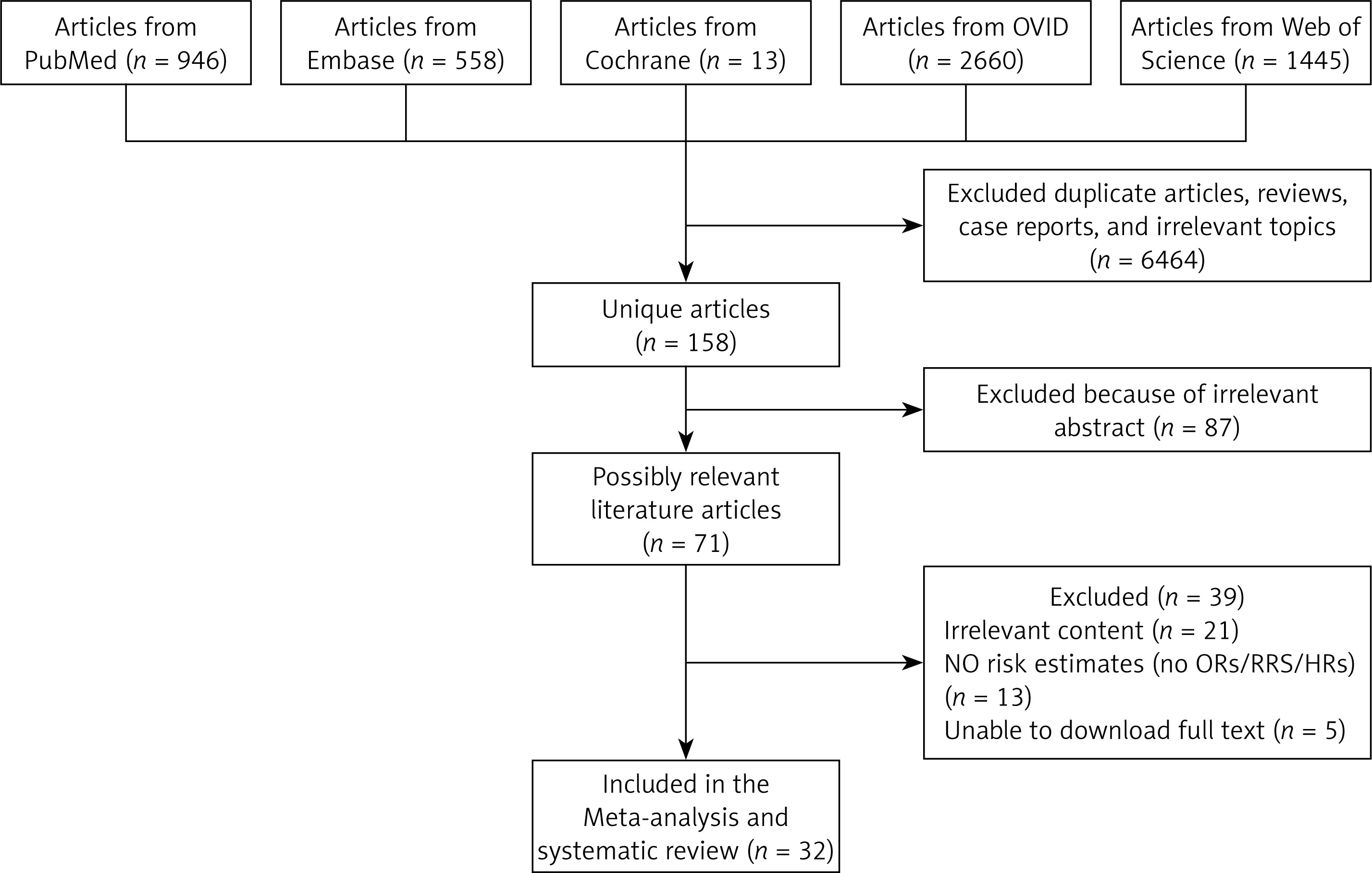
The initial search in PubMed, Embase, Cochrane, OVID, and Web of Science identified 6622 articles. In total, 32 articles (n = 19,987) met our inclusion criteria (Figure 1).
Characteristics of 32 studies included in the meta-analysis
This systematic review and meta-analysis summarized data from 32 studies (n = 19,987), including 14 cross-sectional studies, 11 prospective studies, and 7 retrospective studies. They come from different countries and regions, including China, France, the United Kingdom, the United States, South Korea, Sweden, the Netherlands, etc. In our article, we mainly divide the regions of EPVS into BG, CS, and HP. According to anatomical characteristics, Swiss cheese striatum enlarged perivascular spaces (SCS EPVS) were classified into BG EPVS, and white matter enlarged perivascular spaces (WM EPVS) were classified into CS EPVS.
For cognitive impairment, 12 studies (n = 9205) met our inclusion criteria [12, 21, 22, 26–34]. For movement disorders, 5 studies (n = 1010) met our inclusion criteria [9, 12, 35–37]. For sleep disturbances, 8 studies (n = 1846) met our inclusion criteria [11, 12, 20, 38–42]. For depressive symptoms, 5 studies (n = 3465) met our inclusion criteria [10, 12, 18, 19, 39]. For stroke, 7 studies (n = 6867) met our inclusion criteria [13, 31, 43–47]. The EPVS scoring scale used is mainly the Potter scale, and some studies have used other scales. The included studies mainly used 1.5 T or 3.0 T MRI scanners. Studies with a quality-assessment score of ≥ 7 were included. Basic characteristics and quality assessments of the included studies are shown in Tables II–IV.
Table II
Characteristics of studies included in the meta-analysis
| Study | Year | Total | Study type | Follow-up time | Clinical symptoms | OR (95% CI) | Brain region | Geographic region |
|---|---|---|---|---|---|---|---|---|
| Cognitive impairment | ||||||||
| Zhu et al. [26] | 2010 | 1778 | Prospective | 4 years | Dementia | 5.8 (1.2–28.4) | BG | France |
| 9.8 (1.7–55.3) | WM | |||||||
| Burnett et al. [12] | 2014 | 79 | Retrospective | / | Dementia | 1.57 (0.48–5.13) | SCS | Minnesota |
| Yao et al. [27] | 2014 | 1818 | Prospective | 6.12 years | Dementia | 0.95 (0.50–1.80) | HP | Dijon |
| Arba et al. [28] | 2016 | 234 | Retrospective | / | PSCI | 2.66 (1.82–3.87) | BG | VISTA |
| 2.08 (1.42–3.05) | CS | |||||||
| Arba et al. [21] | 2016 | 430 | Retrospective | / | Cognitive disorder | 2.52 (1.90–3.35) | BG | VISTA |
| 1.67 (1.25–2.18) | CS | |||||||
| Riba-Llena et al. [29] | 2016 | 733 | Prospective | 8 years | MCI | 1.88 (1.03–3.41) | BG | Barcelona |
| Shams et al. [30] | 2017 | 1504 | Cross-sectional | / | Vascular dementia | 11.1 (1.1–112.2) | BG | Sweden |
| 1.3 (0.6–2.9) | CS | |||||||
| Yilmaz et al. [31] | 2018 | 1651 | Prospective | 7.2 years | Dementia | 0.97 (0.35–2.72) | BG | Netherlands |
| Xiong et al. [32] | 2018 | 158 | Cross-sectional | / | Dementia | 1.283 (0.967–1.703) | CS | Massachusetts |
| Banerjee et al. [33] | 2019 | 117 | Prospective | 1 year | Cognitive impairment | 2.60 (0.74–9.19) | BG | UK |
| 1.83 (1.06–3.15) | CS | |||||||
| Sim et al. [22] | 2020 | 109 | Cross-sectional | / | Memory function | 2.142 (–0.515–4.799) | HP | South Korea |
| Paradise et al. [34] | 2021 | 414 | Prospective | 6 years | Dementia | 3.25(1.14–9.30) | BG | Sydney |
| 1.77(0.58–5.39) | CS | |||||||
| Motor disorder | ||||||||
| Wan et al. [36] | 2018 | 137 | Cross-sectional | / | PIGD | 2.97 (0.98–9.03) | CS | China |
| Lioutas et al. [35] | 2018 | 175 | Retrospective | / | Functional independence | 1.4 (0.34–5.77) | BG | USA |
| Del Brutto et al. [37] | 2019 | 288 | Cross-sectional | / | risk of falls | 3.36 (1.85–6.09) | BG | Atahualpa |
| Heiland et al. [9] | 2021 | 331 | Prospective | 9 years | Walking speed limitation | 2.13 (1.04–4.36) | / | Sweden |
| Burnett et al. [12] | 2014 | 79 | Retrospective | / | Any movement disorder | 1.47 (0.43–5.04) | SCS | Minnesota |
| Sleep disturbance | ||||||||
| Song et al. [38] | 2017 | 170 | Cross-sectional | / | Moderate-to-severe (AHI ≥ 15) | 3.64 (1.02–13.01) | / | South Korea |
| Wang et al. [42] | 2020 | 106 | Cross-sectional | / | ArI | 2.108 (1.032–4.017) | / | China |
| Ramirez et al. [11] | 2021 | 153 | Cross-sectional | / | Daytime Dysfunction | 5.31 (1.38–22.26) | BG | ONDRI |
| Del Brutto et al. [39] | 2019 | 338 | Cross-sectional | / | Sleep quality | 1.68 (1.01–2.79) | BG | Atahualpa |
| Del Brutto et al. [40] | 2020 | 146 | Cross-sectional | / | PLMS | 1.36 (0.56–3.33) | BG | Atahualpa |
| Burnett et al. [12] | 2014 | 79 | Retrospective | / | Any sleep disorder | 1.37 (0.37–4.97) | SCS | Minnesota |
| Aribisala et al. [41] | 2020 | 457 | Cross-sectional | / | Interrupted sleep | 1.84 (1.08–3.15) | BG | Scotland |
| Yang et al. [20] | 2022 | 398 | Retrospective | / | Poor sleep quality | 2.125 (1.113–4.058) | BG | China |
| 1.882 (1.005–3.527) | WM | |||||||
| Depressive symptoms | ||||||||
| Van Sloten et al. [18] | 2015 | 1949 | Prospective | 5 years | Depression | 3.44 (1.71–6.91) | / | Reykjavik |
| Liang et al. [10] | 2018 | 725 | Cross-sectional | / | PSD | 1.49 (1.04–2.13) | CS | China |
| Zhang et al. [19] | 2016 | 374 | Cross-sectional | / | PSD | 1.557 (0.958–2.530) | / | China |
| Del Brutto et al. [39] | 2019 | 338 | Cross-sectional | / | Depression | 1.359 (0.608–3.039) | BG | Atahualpa |
| Burnett et al. [12] | 2014 | 79 | Retrospective | / | Depression | 1.57 (0.48–5.13) | SCS | Minnesota |
| Stroke | ||||||||
| Yilmaz et al. [31] | 2018 | 1651 | Prospective | 7.2 years | Stroke | 1.80 (0.71–4.59) | BG | Netherlands |
| Charidimou et al. [13] | 2013 | 121 | Retrospective | / | ICH | 3.58 (1.70–7.54) | BG | UK and Belgium |
| 0.74 (0.35–1.57) | CS | |||||||
| Xu et al. [46] | 2021 | 167 | Cross-sectional | / | Large hematoma volume | 0.395 (0.147–1.064) | / | China |
| Zhang et al. [45] | 2020 | 1204 | Prospective | 3 years | Stroke | 3.34 (0.78–14.37) | / | China |
| Gutierrez et al. [43] | 2017 | 1228 | Prospective | 9 ±2 years | Stroke | 0.9 (0.60–1.61) | / | Manhattan |
| Lau et al. [44] | 2017 | 2002 | Prospective | 42 ±23 months | Ischemic stroke | 1.82 (1.18–2.80) | BG | UK and China |
| Intracerebral hemorrhage | 2.58 (0.97–6.89) | BG | ||||||
| Recurrent stroke | 1.94 (1.31–2.89) | BG | ||||||
| Ischemic stroke | 0.83 (0.54–1.28) | CS | ||||||
| Intracerebral hemorrhage | 1.36 (0.51–3.59) | CS | ||||||
| Recurrent stroke | 0.89 (0.60– 1.33) | CS | ||||||
| Song et al. [47] | 2021 | 494 | Retrospective | / | HT | 0.638 (0.454–0.897) | BG | China |
| 0.690 (0.502–0.949) | CS | |||||||
[i] PSCI – post-stroke cognitive impairment, VISTA – Virtual International Stroke Trial Archive, MCI – mild cognitive impairment, PIGD – postural instability and gait disability, AHI – apnea-hypopnea index, ArI – arousal index, PSD – poststroke depression, HT – hemorrhagic transformation, HR – hazard ratio, OR – odds ratio, BG – basal ganglia, CS – centrum semiovale, WM – white matter, SCS – Swiss cheese striatum, HP – hippocampus, ICH – intracerebral hemorrhage, PLMS – periodic limb movements during sleep, ONDRI – Ontario Neurodegenerative Disease Research Initiative.
Table III
Eight questions of included study quality were evaluated according to Newcastle-Ottawa Scale (NOS) evaluation criteria
Table IV
Quality assessment of the included studies using eight criteria
| Study | Year | 1 | 2 | 3 | 4 | 5 | 6 | 7 | 8 | Total |
|---|---|---|---|---|---|---|---|---|---|---|
| Prospective | ||||||||||
| Zhu et al. [26] | 2010 | Y | N | Y | Y | Y | Y | Y | Y | 8 |
| Yao et al. [27] | 2014 | Y | N | Y | Y | Y | Y | Y | Y | 8 |
| Van Sloten et al. [18] | 2015 | Y | Y | Y | Y | Y | N | Y | Y | 8 |
| Riba-Llena et al. [29] | 2016 | Y | Y | Y | N | Y | N | Y | Y | 7 |
| Gutierrez et al. [43] | 2017 | Y | N | Y | Y | Y | N | Y | Y | 7 |
| Lau et al. [44] | 2017 | Y | N | Y | Y | Y | Y | Y | Y | 8 |
| Yilmaz et al. [31] | 2018 | Y | Y | Y | N | Y | Y | Y | Y | 8 |
| Banerjee et al. [33] | 2019 | Y | Y | Y | Y | N | Y | Y | Y | 7 |
| Zhang et al. [45] | 2020 | Y | N | Y | Y | Y | Y | Y | Y | 8 |
| Heiland et al. [9] | 2021 | Y | Y | Y | N | Y | N | Y | Y | 7 |
| Paradise et al. [34] | 2021 | Y | Y | Y | N | Y | Y | Y | Y | 8 |
| Retrospective or cross-sectional | ||||||||||
| Charidimou et al. [13] | 2013 | Y | Y | Y | Y | N | Y | Y | Y | 7 |
| Burnett et al. [12] | 2014 | Y | Y | Y | Y | N | Y | Y | Y | 7 |
| Arba et al. [28] | 2016 | Y | Y | Y | Y | Y | N | Y | Y | 8 |
| Arba et al. [21] | 2016 | Y | N | Y | Y | Y | N | Y | Y | 7 |
| Song et al. [38] | 2016 | Y | Y | Y | Y | Y | N | Y | Y | 8 |
| Zhang et al. [19] | 2016 | Y | Y | Y | Y | N | Y | Y | Y | 7 |
| Shams et al. [30] | 2017 | Y | Y | Y | Y | N | Y | Y | Y | 7 |
| Xiong et al. [32] | 2018 | Y | Y | Y | Y | N | Y | Y | Y | 7 |
| Wan et al. [36] | 2018 | Y | Y | Y | Y | Y | N | Y | Y | 8 |
| Lioutas et al. [35] | 2018 | Y | Y | Y | Y | Y | N | Y | Y | 8 |
| Liang et al. [10] | 2018 | Y | Y | Y | Y | N | Y | Y | Y | 7 |
| Del Brutto et al. [37] | 2019 | Y | Y | Y | Y | N | Y | Y | Y | 7 |
| Del Brutto et al. [39] | 2019 | Y | Y | Y | Y | N | Y | Y | Y | 7 |
| Del Brutto et al. [40] | 2019 | Y | N | Y | Y | Y | Y | Y | Y | 8 |
| Aribisala et al. [41] | 2019 | Y | Y | Y | Y | Y | N | Y | Y | 8 |
| Sim et al. [22] | 2020 | Y | Y | Y | Y | Y | N | Y | Y | 8 |
| Wang et al. [42] | 2020 | Y | N | Y | Y | Y | N | Y | Y | 7 |
| Ramirez et al. [11] | 2021 | Y | Y | Y | Y | Y | N | Y | Y | 8 |
| Xu et al. [46] | 2021 | Y | N | Y | Y | Y | Y | Y | Y | 8 |
| Song et al. [47] | 2021 | Y | Y | Y | Y | N | Y | Y | Y | 7 |
| Yang et al. [20] | 2022 | Y | Y | Y | Y | Y | N | Y | Y | 8 |
Correlation between EPVS and clinical symptoms
Cognitive impairment
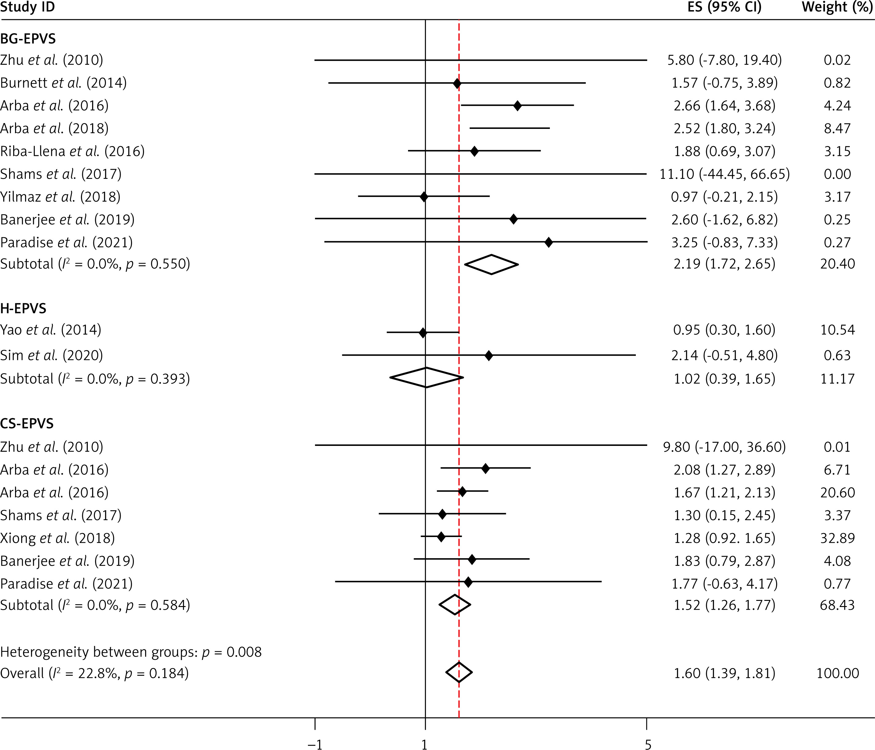
Twelve studies (n = 9205) reported on associations between cognitive impairment and EPVS in the BG, CS, or hippocampus. EPVS was significantly associated with cognitive impairment (OR = 1.60, 95% CI: 1.39–1.81, I2 = 22.8%, p = 0.184), with the strongest association observed for BG (OR = 2.19, 95% CI: 1.72–2.65, I2 = 0.0%, p = 0.550), followed by CS (OR = 1.52, 95% CI: 1.26–1.77, I2 = 0.0%, p = 0.584). However, hippocampal EPVS were not significantly associated with cognitive impairment (OR = 1.02, 95%CI: 0.39–1.65, I2 = 0.0%, p = 0.393) (Table II, Figure 2).
Motor impairment
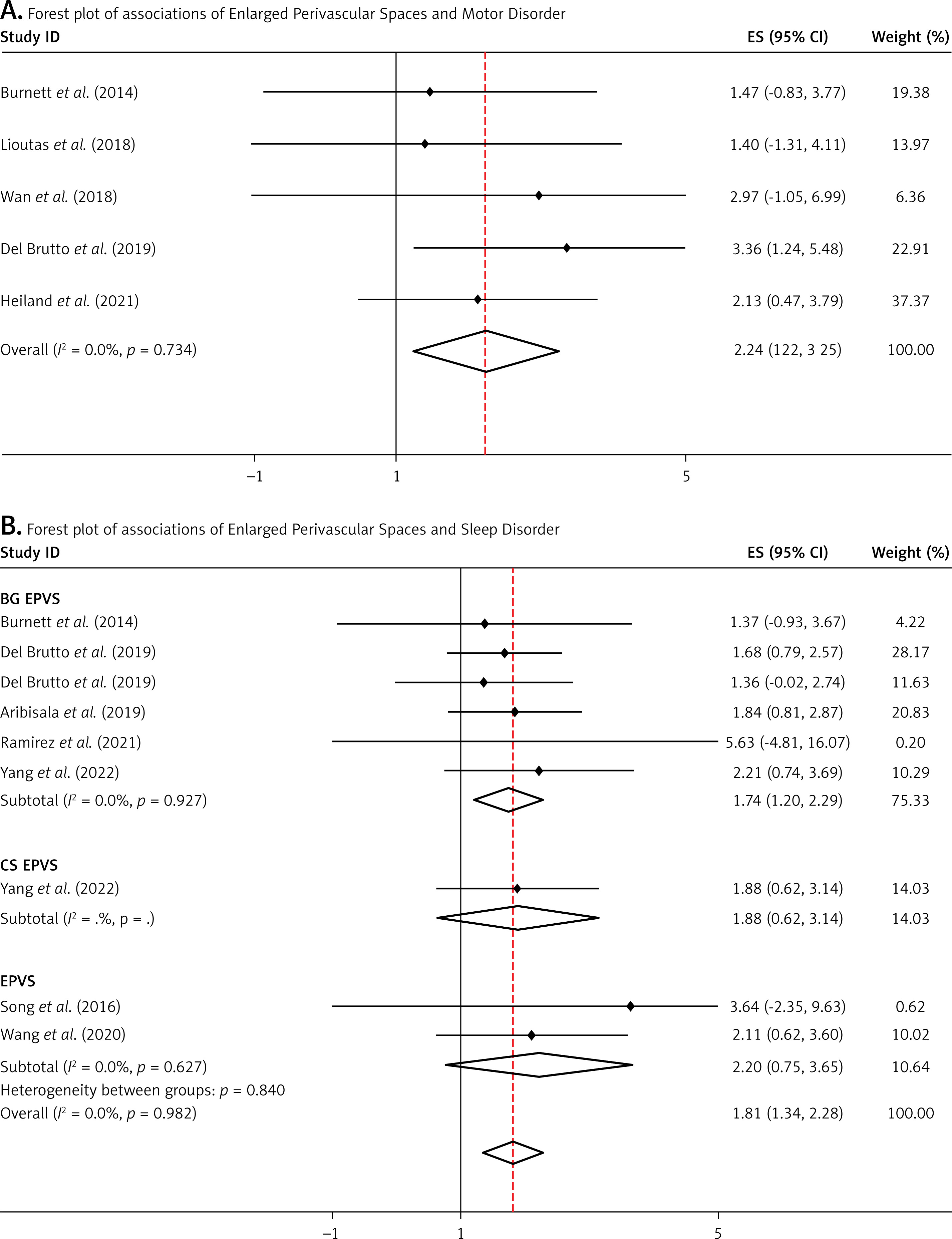
We found a significant association in 946 participants from five studies of EPVS with motor impairment (OR = 2.24, 95% CI: 1.22–3.25, I2 = 0.0%, p = 0.734) (Table II, Figure 3).
Sleep disturbance
In 1846 participants from eight studies, EPVS was significantly associated with sleep disturbance (OR = 1.81, 95% CI: 1.34–2.28, I2 = 0.0%, p = 0.982), especially in BG EPVS (OR = 1.74, 95% CI: 1.20–2.29, I2 = 0.0%, p = 0.927). There were insufficient data to compare CS EPVS and sleep disturbance (Table II, Figure 3).
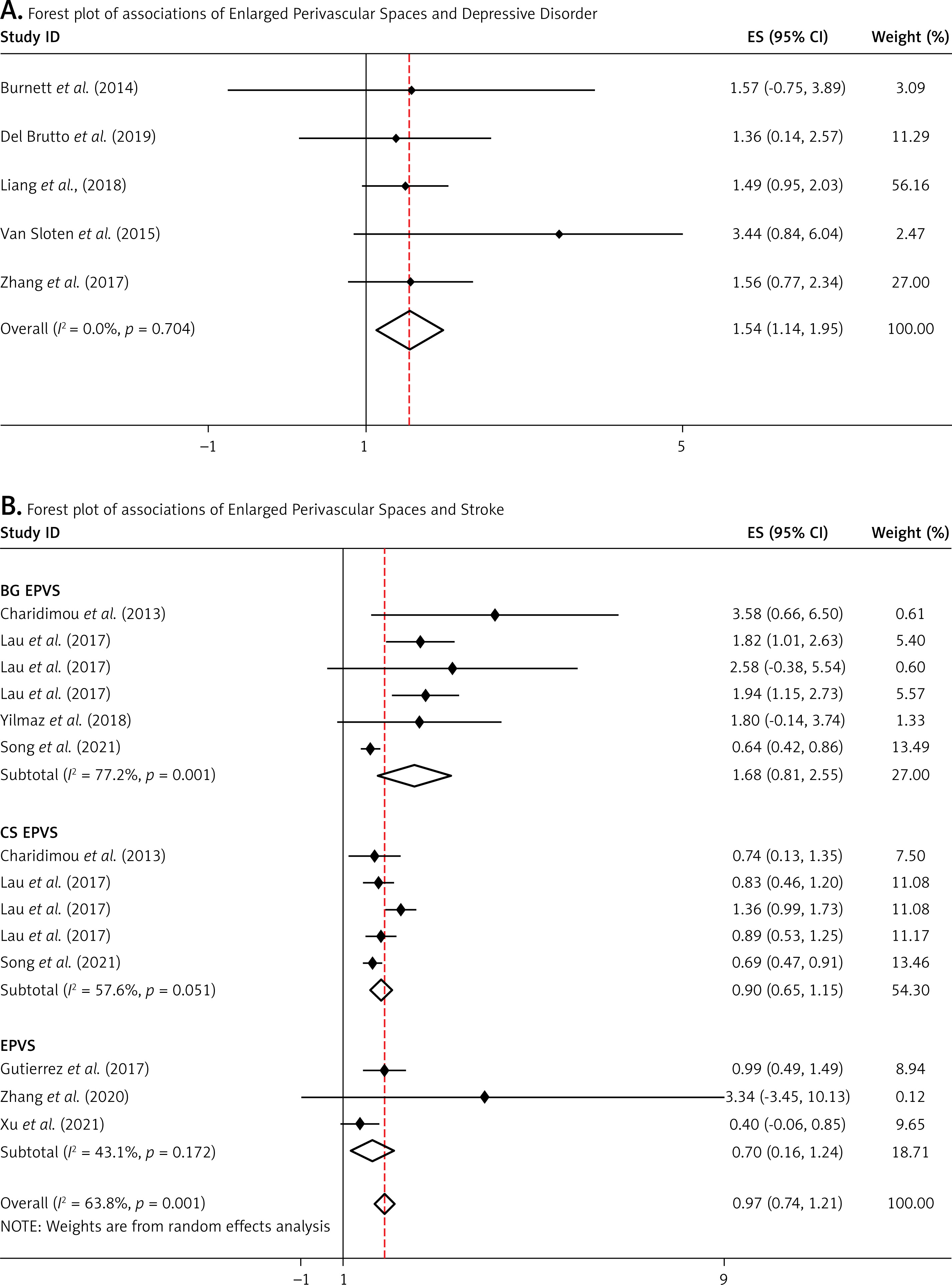
Sensitivity analyses and publication bias
The purpose of the sensitivity analyses was to detect whether any of the included studies would affect the primary outcome of the meta-analysis. The OR values did not change significantly when either study was excluded (Supplementary Figures S1–S5 show the results of the sensitivity analyses). Begg’s test based on the OR value and SE showed no publication bias (Table V).
Discussion
We investigated the correlation between EPVS and clinical symptoms using meta-analysis. Since BG EPVS and CS EPVS have different formation mechanisms, we performed a subgroup analysis to explore whether there are differences in their impact on clinical symptoms. Our findings showed that EPVS were associated with cognitive impairment, motor impairment, sleep disturbance, and depressive symptoms, but not with stroke.
In recent years, many studies have shown that EPVS are associated with the occurrence of cognitive impairment [8]. Impaired cortical cholinergic pathways can lead to cognitive impairment, and a high burden of EPVS may impair the cortical cholinergic pathways, thereby contributing to cognitive decline [48]. However, these findings remain controversial. For example, a prospective study by Yilmaz et al. [31] showed that EPVS were not associated with the risk of dementia. This finding may have been influenced by the small number of participants with EPVS included in the study. Our findings also show that BG EPVS were more strongly associated with cognitive impairment than CS EPVS, which may be related to their different anatomical characteristics and formation mechanisms. Studies have shown that BG EPVS are more strongly associated with age and hypertension [17], and CS EPVS are more strongly related to cerebral amyloid angiopathy (CAA) [30]. In light of our findings, we speculate that vascular risk factors such as age and hypertension may have a greater influence on cognitive impairment than CAA; however, further clinical studies are needed to investigate this hypothesis.
Several recent human studies have reported an association between sleep and EPVS. The perivascular space is a critical pathway for lymphatic flow and an important fluid waste removal system that may be associated with compensatory changes in human sleep physiology [49, 50]. Larger PVS volumes have been reported to be associated with periodic limb movements during sleep [51] and lower sleep quality [52]. These observations reflect the effect of sleep deprivation on the structure of the perivascular space. However, one study found that the association between sleep disturbance and EPVS disappeared after adjusting for relevant confounders [39]. After subgroup analysis, we found that BG EPVS were more significantly associated with sleep disturbance. There are few studies on CS EPVS and sleep disturbance, which may be related to the anatomical structure of cerebrospinal fluid drainage. BG EPVS can communicate directly with the basal cisterns, while CS EPVS communicate with subarachnoid CSF at certain locations. This may result in a greater role for BG EPVS in CSF drainage than CS EPVS, potentially contributing to the stronger association between BG EPVS and sleep disturbances [53, 54].
Our results show that EPVS were associated with motor impairment. Unfortunately, we did not perform a subgroup analysis due to the small number of eligible studies. A prospective study with a 9-year follow-up showed that the highest tertile of EPVS burden was associated with a 2.13-fold higher risk of motor impairment (HR = 2.13; 95% CI: 1.04–4.36), suggesting that motor impairment may be associated with the burden and progression of EPVS [9]. However, the study by Pinter et al. [55] showed that white matter hyperintensities (WMH) were a significant predictor of gait and balance impairment, whereas EPVS was not significantly associated with movement impairment. This discrepancy may partly reflect differences in study populations, as the study mainly included non-disabled, community-dwelling older adults with lower overall CSVD scores, potentially attenuating the observed association.
Our findings suggest that EPVS are associated with depressive symptoms. Depression in later life is associated with cerebrovascular disease, and treatment resistance may be associated with vascular lesions in the white matter and basal ganglia [56]. Common sites of EPVS overlap with emotion regulation pathways (such as frontal-subcortical loops, cingulate-cortical loops, and cortico-basal ganglia loops), potentially disrupting neural circuits involved in emotion regulation and contributing to depressive symptoms [57, 58]. A cross-sectional study by Patankar et al. [56] also showed that EPVS were associated with an increased risk of depression and a poorer response to antidepressants, which was consistent with our overall findings. However, Zhang et al. [19] did not find a clear association between EPVS and poststroke depression (PSD). These differences may be related to differences in study populations and designs, particularly in the assessment of PSD and EPVS. Future research should address these issues.
At present, the evidence regarding the association between EPVS and stroke is inconsistent, and our results show that there was no statistically significant association between EPVS and stroke. The study of Francis et al. also found no significant association between the two [17]. However, some studies have shown that CS EPVS are associated with lobar intracerebral hemorrhage (p = 0.041), and BG EPVS are associated with non-CAA-induced intracerebral hemorrhage (OR = 3.58, 95% CI: 1.70–7.54) [13]. Since few studies have examined the association between EPVS and stroke, we did not perform separate analyses of ischemic and hemorrhagic stroke. Given the different pathophysiological mechanisms and clinical management of these conditions, the associations of EPVS with ischemic and hemorrhagic stroke should be evaluated separately in future studies.
Our results have important implications for understanding the relationship between EPVS and clinical symptoms, but several limitations remain. The sources of heterogeneity may include the following: (1) differences in study populations; (2) differences in study designs; (3) differences in sample sizes; (4) differences in EPVS detection and evaluation methods. Future studies should adopt more consistent, objective, and quicker methods to quantify EPVS. The potential of automated methods to assess EPVS burden has recently been recognized. Boespflug et al. [59] developed a method for fully automatic identification and segmentation of EPVS based on multimodal MRI images. However, this technique requires multiple MRI sequences, which may increase scanning time. Dubost et al. [6] developed a method for automated identification and quantitative assessment of EPVS using only T2-weighted MRI.
Second, while most studies used ORs, a few used HRs [9, 32, 43–45]. In meta-analyses, pooling HRs with ORs, although numerically similar to each other, may not be methodologically appropriate and may affect the validity of the pooled estimates. Although the sensitivity analysis indicated that our results were robust, the inclusion of different effect measures remains a methodological limitation. In addition, due to the limited number of studies, we could not explore the relationship between EPVS in different regions and movement disorder and depressive symptoms, and high-quality cohort studies require longer follow-up. Although our results did not show statistically significant evidence of publication bias, our findings require further validation in future large prospective studies and meta-analyses using standardized EPVS classification criteria and statistical methods.
In conclusion, our results provide evidence for an association between EPVS and clinical symptoms, indicating that EPVS are associated with an increased risk of cognitive impairment, motor impairment, sleep disturbances, and depressive symptoms, but not with a significantly increased risk of stroke. In subgroup analyses, we found that BG EPVS were more strongly associated with cognitive impairment and sleep disturbance, and CS EPVS were more strongly associated with depressive symptoms. Therefore, the presence of EPVS may warrant attention to associated clinical symptoms and risk factors, and further research is needed to determine whether EPVS can be used to identify individuals at increased risk of these outcomes and to investigate potential preventive and therapeutic strategies. Furthermore, future research should continue to optimize automated EPVS quantification methods, including improvements in algorithms, MRI acquisition sequences, and post-processing techniques.

