

Introduction
In the last 10 years, significant advancements have been made in understanding the human microbiota and its role in various diseases [1]. Gut microbiota is the collection of bacteria, fungi, viruses, archaea, and parasites in the gastrointestinal tract (GIT), producing a diverse ecosystem of about 1014 microorganisms. Our eating habits are directly connected with our microbiota composition. As an illustration, epidemiological evidence on immigrants suggests that there is a potential fourfold increase in obesity risk within 15 years of emigrating to the U.S. compared to populations remaining in their birth country. This can be furthermore accompanied by a decrease in their gut microbial diversity and function [2, 3]. It has been shown that environmental factors (e.g., diet, household cohabitation) greatly outweigh heritable genetic contributions to the composition and function of gut microbiota [4]. Furthermore, Rothschild et al. constructed a microbiome-association index that mimics heritability statistics [4, 5]. The most significant described associations were between the gut microbiome and host phenotypes for body mass index (BMI), waist-to-hip ratio, fasting glucose levels, glycemic status, high-density lipoprotein (HDL) cholesterol levels, and monthly lactose consumption [4, 5]. If the homeostasis of the gut microbiota, which acts almost like an endocrine organ, is disturbed, dysbiosis can contribute to the development of various diseases [6, 7].
It comes as no surprise that some of these potential diseases include cardiovascular diseases (CVD), chronic kidney disease, type 2 diabetes mellitus, non-alcoholic fatty liver disease, and even certain types of cancer [1, 8–10]. The term dysbiosis denotes a change in the composition of the gut microbiota. Reasons for such a change are manifold and can range from exposure to several factors (diet, increased stress, antibiotic usage). Ilya Ilyich Mechnikov (also written as Élie Metchnikoff) coined the term dysbiosis at the beginning of the 20th century. Together with Paul Ehrlich, they were awarded in 1908 the Nobel Prize in Physiology or Medicine “in recognition of their work on immunity”. Dysbiosis might offer an explanation as to why certain individuals are more susceptible to develop specific diseases. Moreover, it has recently been recognized that dysbiosis increases the risk of developing atherosclerosis and hypertension [1, 9, 11].
Two authors (P.S. and K.S.) performed an electronic bibliographic search of the PubMed and Cochrane databases. The databases were primarily searched using the keywords/MeSH terms “cardiovascular diseases”, “gastrointestinal microbiome” and “dysbiosis” with various subheadings, taking into account the latest findings (last 5 years) with exceptions when citing older original findings. The initial search resulted in 231 entries that were further screened by applying additional filters and eligibility criteria (full text, books and documents, clinical trial, meta-analysis, randomized controlled trial, systematic review, in the last 5 years). This alongside the exclusion criteria (exclusion of studies, chapters and articles with similar findings published as different bibliographic units) narrowed the final result to 28 articles. During analysis of these articles, any articles cited therein that were thematically relevant were also included. The inquiry was performed without time restriction at the Research Department of the University Clinical Center Maribor (from 2020–2021).
Diagnostic procedures for determining composition of the gastrointestinal microbiota
The composition of the microbiota, its diversity and potential significance in maintaining homeostasis of epithelial cell function, prevention of pathogenic microorganism growth and production of different substances as well as ingredients can be determined using various methods, which differ in resolution [11–13]. These methods may be employed to compare and specify the microbiota composition between samples, determine the specific microorganisms, their intercellular relationships and dependencies as well as their role in metabolism based on their genetic information [14]. Some approaches and the corresponding terms are shown in Table I [15, 16].
Table I
Methods for gut microbiota analysis and their definitions
| Areas | Name | Principle | Method | Pros | Cons |
|---|---|---|---|---|---|
| Composition | Biomarker profiling | DNA | NGS | Cost-effective; semi-quantitative | No functional information |
| Metagenomics | DNA | NGS | Strain-level resolution | Expensive Computationally intensive | |
| Productivity | Metabolomics | Metabolites | LG/GC – MS | Semi-quantitative Targeted or untargeted | Origin or metabolite unclear |
| Function | Metatranscriptomics | RNA | NGS | Host and microbial gene transcripts | Samples require RNA preservation; host genes may predominate signal |
| Metaproteomics | Proteins | LG/GC – MS | Semi-quantitative | Origin of proteins unclear | |
| Term | Definition (based on Marchesi and Ravel [15]) | ||||
| Microbiome | The term represents all microorganisms (bacteria, viruses, etc.), their genomes (i.e., genes), and the surrounding environmental conditions | ||||
| Metagenomics | Metagenomics is the process used to characterize the metagenome (DNA from a group of species) and gain information on the potential function of the microbiota | ||||
| Metabolomics | This term describes systematic identification and quantification of small molecule metabolic products of any given strain or single tissue | ||||
| Metabonomics | Is a subset of metabolomics and describes the approach used to measure metabolic changes, with respect to time, due to an intervention | ||||
| Metatranscriptomics | The analysis of the suite of expressed RNAs (meta-RNAs), which provides information on the regulation and expression profiles of complex microbiomes | ||||
| Metaproteomics | Large-scale characterization of the entire protein complement of environmental or clinical samples at a given point in time | ||||
[i] NGS – next-generation sequencing, LG/GC – liquid/gas chromatography, MS – mass spectrometry. First part adapted from Durack and Lynch [16].
An accurate representation of the human microbiota composition, as well as its characterization, was one of the main goals of the quite recent Metagenomics of the Human Intestinal Tract project [17, 18]. Methods for defining the microbiota composition can be divided into traditional and molecular. Commonly known traditional methods are “the counting of cells on a specific culture medium” and the “most probable number” [14]. Culturing methods have certain important drawbacks and restrictions, namely: a large amount of laboratory work, limited culturing possibility, and range (only 30% of the intestinal microbiota) [14]. It has to be stressed that successful growth can be observed during cultivation only in 0.01–10% of the cells in the microbiological sample. Most molecular techniques use the ribosomal 16S and 18S RNA (rRNA), which function as phylogenetic markers for the taxonomic classification of organisms and are preserved in all bacteria, archaebacteria, and eukaryotes. Some molecular methods include:
quantitative polymerase chain reaction (qPCR) – amplification and quantification of 16S rRNA, which enables the phylogenetic identification of microbiota;
denaturing gradient gel electrophoresis – analysis of microbial communities by the sequence-specific separation of PCR-amplified 16S rRNA fragments using a linear gradient of denaturants or temperature;
terminal restriction fragment length polymorphism (T-RFLP) – the amplification is performed with one or both the primers having their 5′ end labeled with a fluorescent molecule with a subsequent restriction of 16S rRNA products with enzymes and gel electrophoresis separation;
automated method of ribosomal intergenic spacer analysis (ARISA) – PCR multiplication of a region between the 16S and 23S RNA regions, with subsequent fragment separation via capillary electrophoresis;
fluorescence in situ hybridization (FISH) – hybridization of oligonucleotides marked with a fluorescent molecule with 16S genes with subsequent measurement of fluorescence via a flow cytometer;
DNA-microarray – hybridization of oligonucleotide probes, marked with a fluorescent molecule, with complementary oligonucleotides and subsequent measurement of fluorescence with a laser;
sequencing of cloned 16S rRNA genes – cloning of all the 16S rRNA products, Sanger sequencing and capillary electrophoresis;
sequencing of 16S rRNA products – also known as deep sequencing of 16S rRNA products;
shotgun metagenomics sequencing of the whole microbiome;
shotgun metatranscriptomics sequencing for determining gene expression of the microbiota.
All of the mentioned methods have their advantages as well as drawbacks, which become evident when determining phylogenetic differences or considering accurate identification, accessibility, and of course price.
Mechanisms of microbiota activity
One of the major risk factors for CVD is atherosclerosis. Its pathophysiological basis is the accumulation of cholesterol, followed by an immune response that leads to the formation of plaques [1, 11, 19]. Gut dysbiosis can, via modulation of the inflammatory response as well as production of microbial metabolites, accelerate this process [20–22]:
gut dysbiosis and atherosclerosis: The GIT acts as a barrier; any changes in its permeability can lead to complications. These changes are associated with the reduced expression of tight junction proteins (e.g., zonula occludens-1, claudin-1 and occludin) and an imbalance between epithelial cell death and regeneration [1, 21, 22]. What follows is the translocation of bacteria, which stimulate, via the recognition of their pathogen-associated molecular patterns (PAMPs), an immune response and inflammation. What is more, lipopolysaccharide (LPS) and peptidoglycan, which are part of the cell wall, have also been described as risk contributing factors.
Humans: The correlation between LPS and CVD risk was first proposed in 1999, based on measurements of plasma endotoxin levels [21]. The correlation of endotoxemia and CVD burden has been confirmed in some studies. Cani et al. reported the correlation between dysbiosis and the suppressed expression of tight junction proteins, which in turn leads to the above-described cascade and translocation of LPS into the blood [23]. On the pathophysiological level, it has been proposed that the (gut dysbiosis-derived) LPS might act as a modulator of toll-like receptors (TLRs), which are mostly present on immune sentinel cells, which are responsible for the immune system’s defense mechanisms. The upregulation of these proteins has been associated with inflammatory activation, which in turn promoted the process of atherosclerosis. The bacterial cell wall component peptidoglycan (PG) can apparently also impair the intestinal epithelial barrier via an inflammatory response. This has been demonstrated in patients with over-representation of genes for PG synthesis. Furthermore, this polymer might be responsible for more vulnerable plaques in sclerotic arteries [11, 19]. Inflammatory processes can also be stimulated by other PAMPs (CpG oligodeoxynucleotides flagellin, lipopeptides, etc.). All in all, the scientific results in the last years have confirmed the correlation of the gut microbiota and atherosclerosis risk [1, 11, 24];
gut microbial metabolites in atherosclerosis: in the metabolism of intestinal bacteria, different metabolites are produced that show involvement in the pathophysiology of atherosclerosis (Figure 1).
Figure 1
Microbial metabolites. Depiction of different microbial metabolites that trigger specific pathophysiological mechanisms in the development of cardiovascular diseases
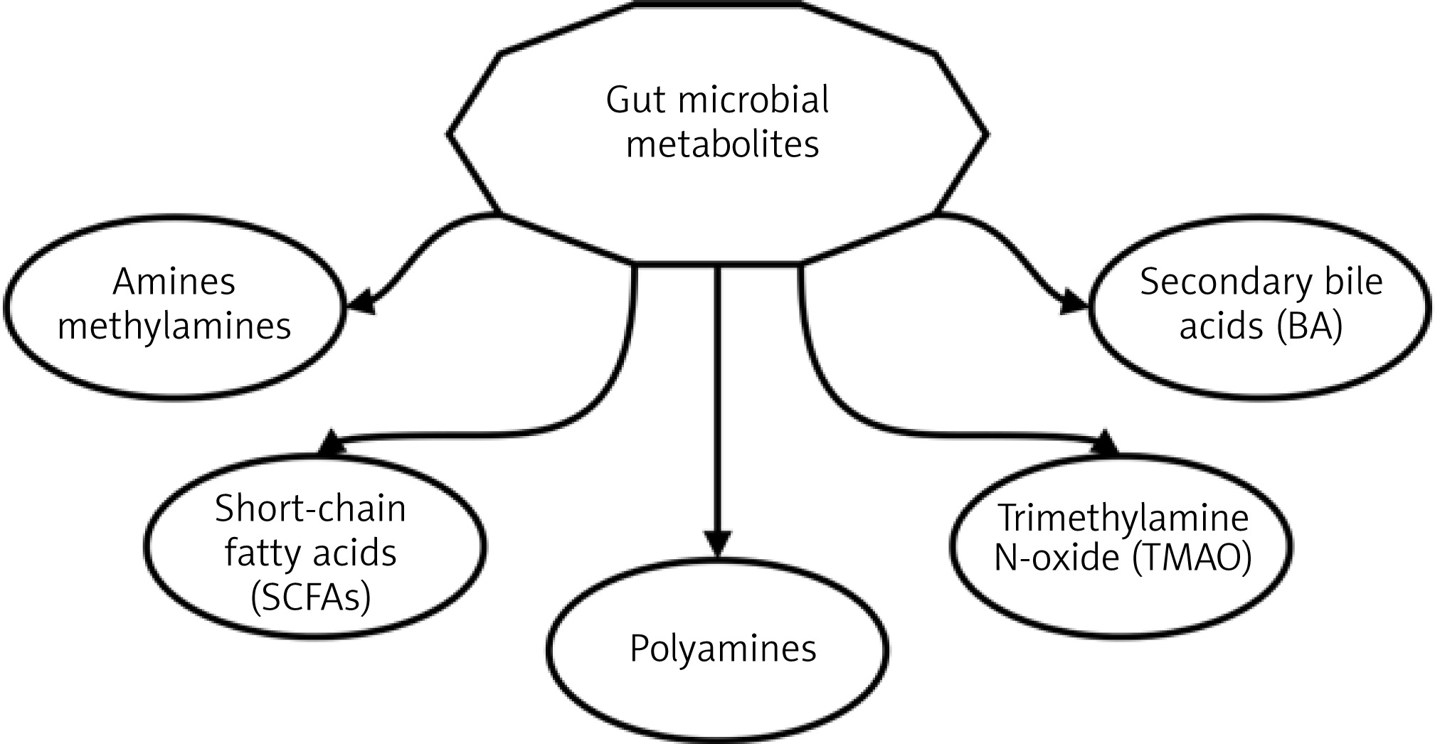
Short-chain fatty acids (SCFAs) play a significant role in the development of metabolic diseases. Bacteria can via the use of choline-specific and carnitine-specific trimethylamine (TMA) lyases form TMA, which in turn is, after absorption, transferred to the liver. Through further metabolic processes (flavin-containing monooxygenases) the TMA is converted into trimethylamine N-oxide (TMAO) [11, 22]. TMAO has according to the literature a variety of different mechanism which all promote atherosclerosis (cholesterol influx, cholesterol efflux inhibition, bile acid (BA) pathway blockade, excessive activation of platelets) [1, 11]. According to researchers, TMAO could represent, in addition to the role of a biomarker for CVD and atherosclerosis, a potential therapeutic target in the future;
gut microbiota and hypertension: in 1982, Honour et al. demonstrated that blood pressure could be elevated by the use of antibiotic treatment [20].
Animals: A study of Tang et al. from 2015 in spontaneously hypertensive rats confirmed that altering gut microbiota (e.g., decreasing/increasing) can influence the regulation of blood pressure. They specifically stressed the increase in the Firmicutes/Bacteroidetes ratio [11].
Humans: Current evidence, even though it might not yet be complete, has elucidated and shown the importance of SCFAs and oxidized low-density lipoprotein (ox-LDL) in hypertension. The microbiota of a person is very specific and stable throughout the adult life span, despite the fact that 90% of them are dominated by representatives of only two bacterial phyla, Firmicutes and Bacteroidetes. Bacteria of these two phyla are good structural polysaccharides and SCFAs producers (e.g., butyrate, acetate, propionate). They are crucial for the homeostasis of the gut microbiome and host immunity [1, 11, 13]. It is interesting that different bacteria form different types of SCFAs. The study by Gomez-Arango et al. showed that in obese pregnant women an increase in butyrate-producing bacteria (Lachnospiraceae, Ruminococcaceae, and Acidaminococcaceae families) is associated with lower blood pressure [22]. SCFAs can stimulate host G-protein-coupled receptor (GPCR)-regulated pathways to affect renin secretion and therefore blood pressure. From a physiological standpoint, the blood pressure regulatory mechanisms are primarily dependent on vasoconstriction and vasodilation. Another fascinating mechanism is low-density lipoprotein (LDL) oxidation by bacteria, which causes excessive vasoconstriction. This is also promoted by pro-inflammatory cytokine expression, which causes oxidative stress and stimulates this process [1]. All in all, higher levels of oxLDL can lead to a vasodilator/vasoconstrictor production disequilibrium;
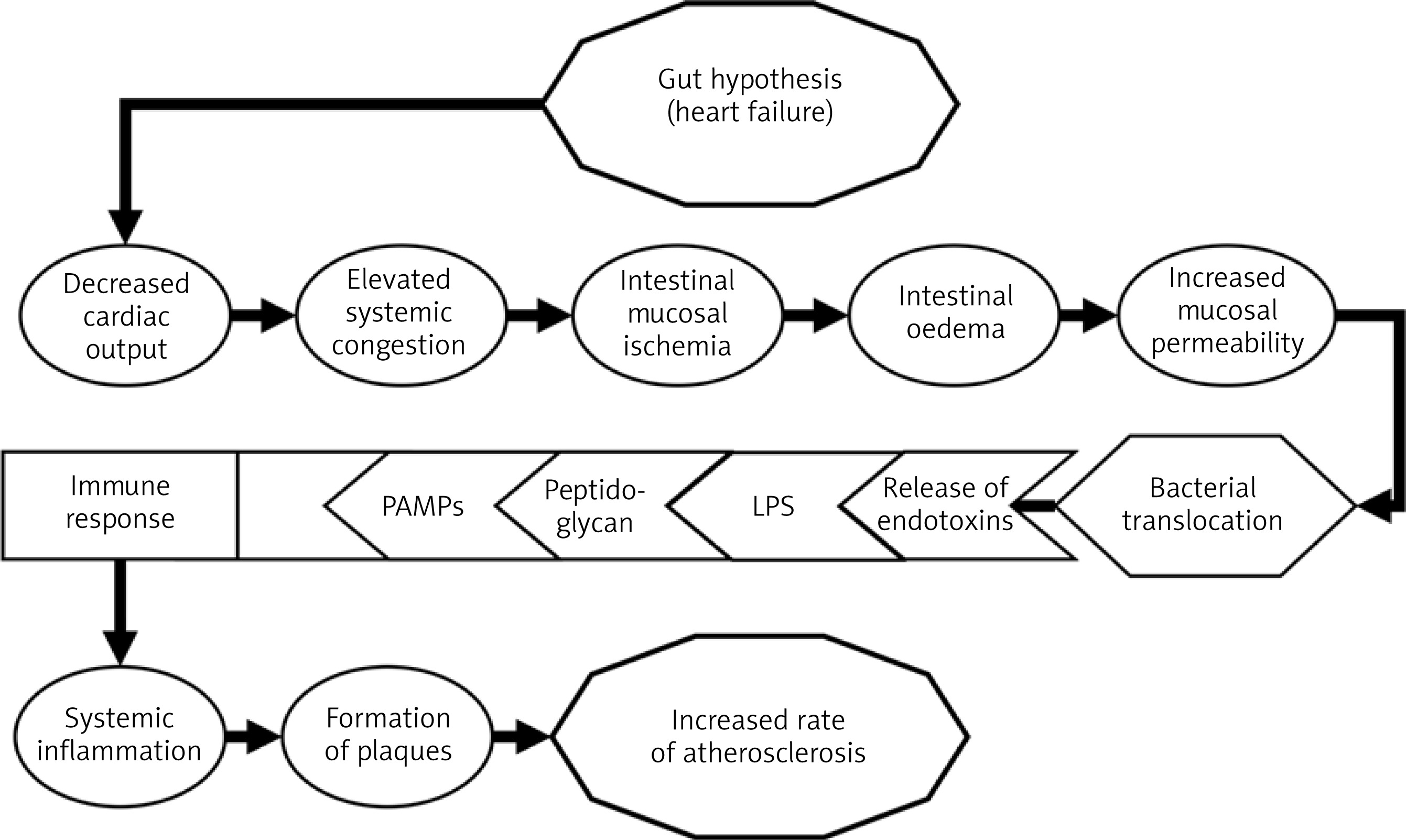
gut microbiota and heart failure: with a growing body of scientific evidence the link between the gut in the pathogenesis of heart failure, the so-called “gut hypothesis of heart failure,” is becoming more and more plausible [23, 25, 26]. The pathophysiological events are shown in Figure 2.
Humans: In a fascinating study by Niebauer et al. it was shown that heart failure patients who had accompanying peripheral edema exhibited increased concentrations of plasma inflammatory markers (endotoxin, cytokines) in comparison with those without edema [11, 21, 23]. When patients received diuretic treatment (short-term), serum concentrations of endotoxin, but not cytokines, decreased. Furthermore, in a different study, higher serum concentrations of immunoglobulin A – anti-lipopolysaccharide were seen in individuals with heart failure and a lower intestinal blood flow. Surprisingly the microbiota was different in these individuals in comparison with the control group [11]. Moreover, studies have also shown that TMAO levels were elevated in patients with heart failure in comparison with the control group. TMAO levels exhibited a remarkably strong adverse prognostic value in a cohort of stable patients with heart failure;
gut microbiota and myocardial infarction: Studies showed that atherosclerotic plaques (especially vulnerable/instable ones) can contain bacterial DNA.
Humans: In those specific individuals the bacterial species found in the plaques was then also found in the GIT [11, 25, 26]. This means that the composition of one’s microbiota might be a reason for an increased rate as well as instability of plaque formation.
Animals: Lam et al. studied the impact of gut microbiota composition and the severity of myocardial infarction in rats [11, 27]. The authors reported that the levels of leptin and other catabolic amino acid metabolites as well as the myocardial infarct size were lower when rats were given broad-spectrum antibiotics [27]. What is more, administration of Lactobacillus plantarum showed a significant reduction in infarct size and improved left ventricular function after a myocardial infarction in rodents. A different study showed that the addition of Lactobacillus rhamnosus GR-1 attenuated left ventricular hypertrophy and heart failure after experimental myocardial infarction [28].
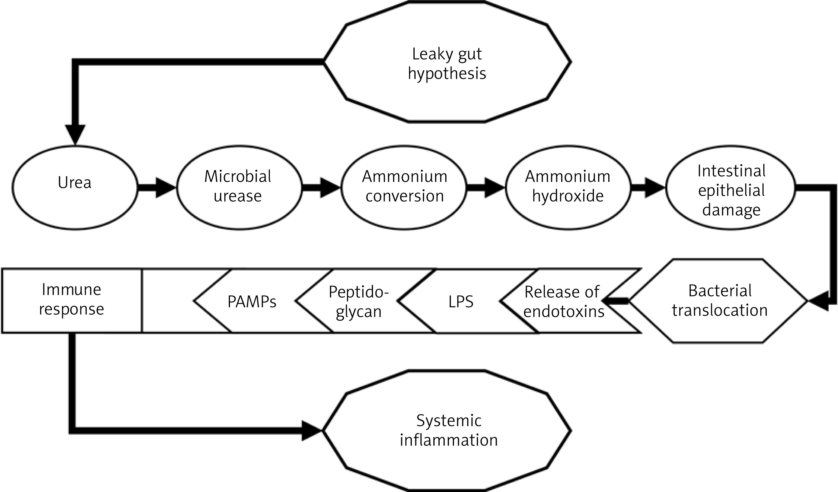
gut microbiota and chronic kidney disease: CVD and kidney diseases are closely interrelated (e.g., cardiorenal syndrome). Patients with chronic kidney disease (CKD) have a greater risk of CVD complications as well as an increased mortality rate; therefore, in many research environments, there are attempts to identify the most appropriate biomarkers of potential complications [29]. Studies have confirmed that patients with CKD have a distinctly different composition of gut microbiota. In CKD an influx of circulating urea and other uremic toxins into the gut lumen occurs and induces the so-called “leaky gut” [11, 30–34]. The pathophysiological mechanism is shown in Figure 3.
Humans: Recently, the DNA of gut microbiota has been detected in the plasma of CKD patients on chronic hemodialysis using bacterial 16S rDNA amplification and DNA pyrosequencing. Moreover, the levels of the bacterial DNA correlated with increased plasma inflammatory marker levels. Poorly dialyzable protein-bound uremic toxins such as indoxyl sulfate and p-cresyl sulfate are associated with poor cardiovascular outcomes [11, 32]. TMAO has been known to accumulate in the plasma of patients with CKD, and higher TMAO levels were associated with higher mortality and progressive loss of kidney function [33, 34], which has to an extent also been proven by the data from the Framingham Heart Study [35].
Research has also been focused on links between dysbiosis and obesity, type 2 diabetes, and dyslipidemia. Obesity has been linked to a higher ratio of Firmicutes to Bacteroidetes. Type 2 diabetes was associated with a reduction in butyrate-producing bacteria and an increase of Lactobacillus spp. [1, 10, 11, 19, 24]. The gut microbiota is able to regulate BA metabolism via its own enzymes, e.g., bacterial bile-salt hydrolase (BSH). This is essential for the formation of secondary BAs. The decrease of mentioned BSH activity in a dysbiotic ecosystem leads to a variety of pro-atherosclerotic effects. Specifically, dysbiosis can lead to impaired cholesterol elimination and dyslipidemia by modulating hepatic and/or systemic lipid metabolism, as well as glucose metabolism [1, 11, 19, 24].
Clinical relevance of gut microbiota
Over the last decade, knowledge about the relationship between dysbiosis and the pathogenesis of CVD has rapidly accumulated [36–39]. Some of the lessons are new opportunities for early, targeted action, and at the same time, many research questions are being raised about the relationship between “what is the cause and what is the consequence” and therapeutic options. Research on dysbiosis in some groups of patients is surprising. Typical signs of disrupted microbiota are reduced diversity, a decrease in anti-inflammatory species such as Faecalibacterium prausnitzii and an increase in various members of the Enterobacteriaceae [40, 41]. Mahnic et al. have confirmed that bacterial and fungal alterations of the gut microbiota, which are often reported to be disease-specific, such as a decrease of Faecalibacterium and an increase in Escherichia coli, Enterococcus and Candida, are often found in a broader population of hospitalized patients with different diseases and also in healthy controls [41]. Furthermore, the authors noted a prominent correlation between levels of C-reactive protein and the abundance of Enterococcus. Although gut dysbiosis is often perceived as random, the research group described two different types in which the severity of the disorder was correlated with specific microbial patterns, the degree of inflammation and, to some extent, the use of antibiotics [41]. Specifically, the clinical examples of gut microbiota interventions for CVD can be divided into multiple groups: 1) dietary interventions; 2) exercise; 3) pro-, pre-, antibiotics; 4) fecal transplantation; 5) TMAO reduction 6) other (e.g., nanomedical approaches) [7, 42].
Modulating the gut microbiota with the help of dietary changes has been shown to be a promising intervention for lowering the risk for coronary diseases [43, 44] as well as general atherosclerosis [19]. A Mediterranean diet intervention has been reported to alter the gut microbiome in older people and thus reduce frailty and improving health status [45]. Furthermore, in animal models, a high-fiber diet has been associated with lower blood pressure, lower cardiac hypertrophy and a lower degree of fibrosis [46]. Even trace elements as zinc have been shown to have a significant impact on the homeostasis of the microbiota [47]. The habitual diet of a person is considered a key driver in establishing this core microbial profile [48]. Acute dietary interventions in humans lead to transient microbial shifts (e.g., days to weeks) [49]. Moreover, for quite some time, gluten-free diet (GFD) plans have been trending in the general unaffected population as a healthy diet change, despite being primarily aimed at those with gluten-related disorders (e.g., celiac disease, gluten allergy) [50]. Many studies have evaluated the impact of such a dietary change [51–54]. Some of the commonly reported changes include a reduction in Eubacterium hallii, Anaerostipes hadrus, and Bifidobacterium and an increase in Enterobacteriaceae and E. coli [55]. It has been reported that the effects of GFD, while reducing bacterial richness, strongly depend on the subject’s health as well as disease status (e.g., celiac disease, healthy) [55]. Reports on this matter differ based on study population, geographical diversity as well as the individual characteristics of patients. Recent studies reported that in normal subjects the diet had deleterious effects [54, 55] and that the opposite was the case in patients with celiac disease [55, 56]. Furthermore, as stated by Lebwohl et al., avoidance of gluten in healthy subjects may result in reduced consumption of beneficial whole grains, which may affect cardiovascular risk [57]. It has to be stressed that the results from the effect of GFD on health and the gut microbiota cannot be extrapolated from one population (or region) to others, nor are they universally applicable [58]. This statement applies to all dietary interventions in any other dysbiosis-associated gastrointestinal disease (e.g., inflammatory bowel disease). Such alterations should not be applied lightly. Nevertheless, it is generally considered that irregular eating habits, such as skipping breakfast, having dinner late, and late-night eating, contribute to obesity and other metabolic disorders [59].
Exercise is of the utmost importance for a heathy human being. Not only does it lower the risk for CVD and improve long-term survival in patients with preexisting heart conditions [60], it has also been shown that regular exercise promotes a healthy gut microbiota while protecting the permeability and function of the gut barrier [61]. Several studies indicate that exercise leads to an increase in the number of health-promoting bacterial species [62–64]. For example, in active women, a higher abundance of Faecalibacterium prausnitzii, Roseburia hominis and Akkermansia muciniphila was demonstrated [64]. However, according to Allen et al. [65], the effects of exercise on the gut microbiota depend on the continuity of exercise and are therefore reversible.
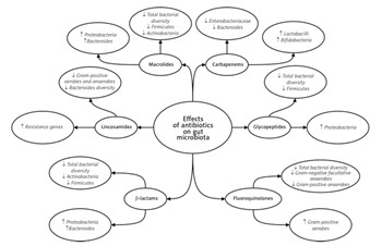
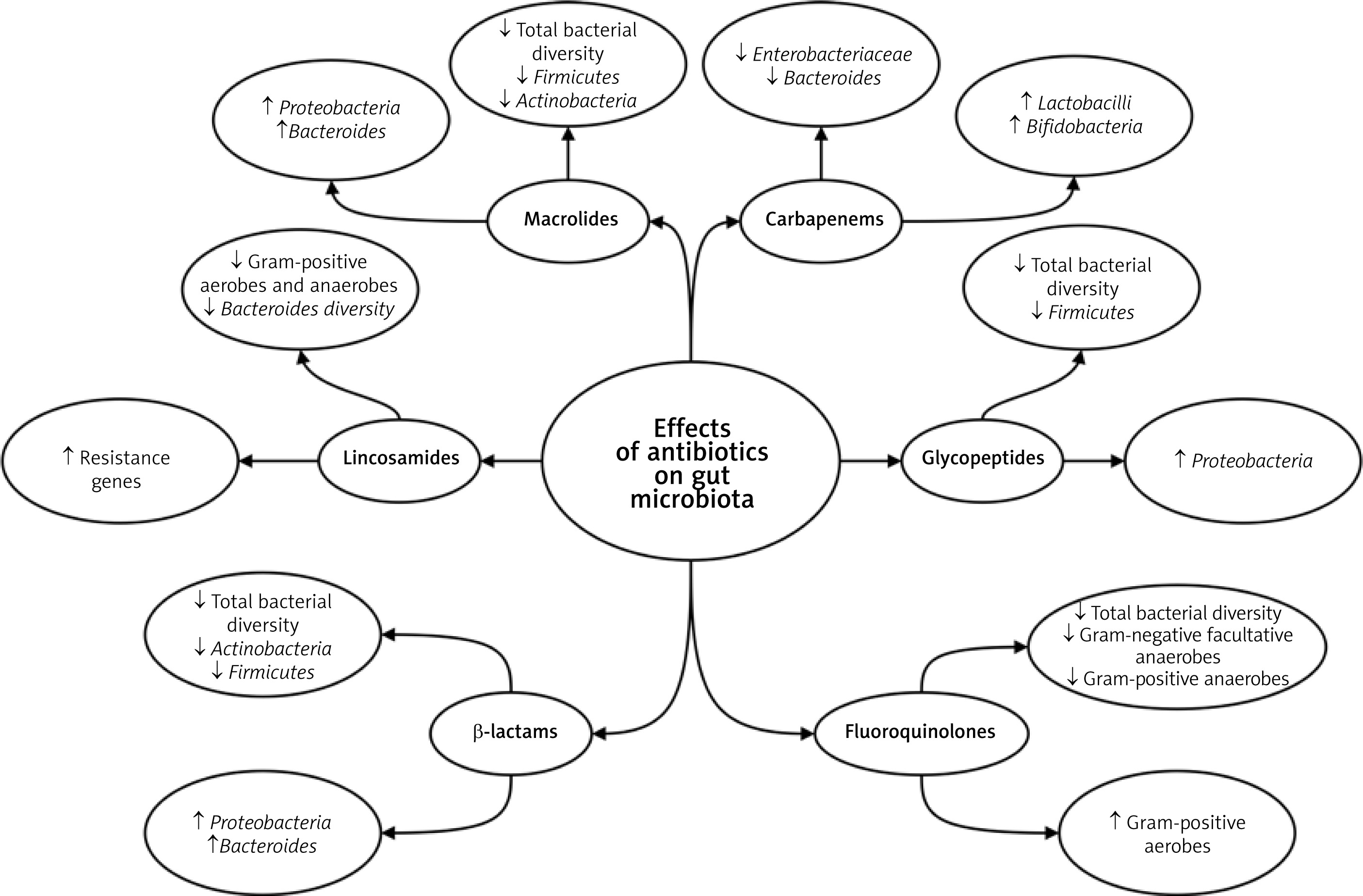
Other modalities of microbiota modulation include probiotics [66], prebiotics [67], postbiotics, and antibiotics [68]. Probiotics are live microorganisms administered to reestablish an intestinal ecological balance, through a variety of different mechanisms [68], which also include immunomodulation of the host and inhibition of bacterial toxin production. Therapy with probiotics has shown promise in patients with impaired cardiac function [68, 69] and has been associated with a protective effect against colorectal cancer [67]. The by-products of probiotic cultures are called postbiotics. These, despite only recently receiving attention, have been shown exhibit positive effects (e.g., suppression of colonic inflammation and restoration of gut barrier integrity) [67]. However, the exact identity of the postbiotics and the molecular mechanisms are not yet fully understood [67]. Moreover, prebiotics have been reported to beneficially modify lipid metabolism [70]. The use of antibiotics to specifically alter the microbiota is, due to a wide range of potential side effects, still debatable. Antibiotic administration presents the most aggressive means to manipulate gut microbiota composition. Negative effects include the depletion of bacterial diversity, altered gene expression and metabolism, selection for intrinsically resistant bacteria, etc. [71, 72]. That is why antibiotics have also been referred to as deep modulators of the gut microbiota (Figure 4) [72, 73]. Some examples of modulation include studies on obesity [74], insulin resistance, diabetes [75], and myocardial infarction (mentioned previously] [27]. Mouse models showed that the effects of antibiotic treatment towards weight appears to depend on several factors (e.g., drug dosage, timing of exposure) [74]. The results depending on dosage showed either a tendency to become underweight [76] or overweight [75]. This has been explained as selective dysbiosis. At the same time, certain antibiotics showed in obese mice antidiabetic effects [77]. Another study provided evidence that early life treatment of mice with vancomycin was beneficial in preventing the onset of diabetes by an increase in health-promoting bacteria [78]. In human studies, antibiotic exposure during infancy was linked to being a risk factor for becoming overweight later in childhood [79]. These results still need to be validated by additional studies [80].
Figure 4
Effects of antibiotics on microbiota composition. Depiction of the overall changes of specific antibiotic groups in the microbiota. Adapted from Bhalodi et al. [72] and Ianiro et al. [73]
Fecal microbiota transplantation has already therapeutically confirmed the importance of a healthy gut microbiota in certain patients. This form of treatment is several decades old and is still an important intervention [81]. It has even been shown that this method might improve insulin resistance [82]. Due to the negative effect of TMAO, probiotics as well as other pharmacological interventions in the form of TMAO reduction inhibitors can be used to inhibit or block specific microbial metabolic pathways. In mice the treatment with a TMA-lyase inhibitor has shown promise by improving hemodynamical parameters [83]. However, further studies will have to be performed to fully determine the safety profiles and possible consequences of such therapies.
Conclusions
Microbiota and dysbiosis represent areas of research interest that will most certainly change some of the established methods of treatment in the future. These changes show great promise in the field of cardiovascular diseases. The present article has discussed different aspects of dysbiosis, its pathophysiological pathways and its effects on cardiovascular health as well as possible promising interventions. All of the presented methods alter the microbial composition in different ways (e.g., suppression of TMA, increase in beneficial cultures) and may lead to positive changes that help prevent and/or reduce deleterious effects of atherosclerosis, hypertension, heart failure, obesity as well as diabetes. The presented changes have in certain cases still only been reported in animal models and should therefore not be directly extrapolated to humans. Furthermore, although we can change the composition of the microbiota, unfortunately at the present moment we cannot fully predict the long-term effects of our actions or offer universal guidelines for all interventions.

