

Introduction
Ultrasonography plays an important role in evaluation of many diseases in the pediatric population. The noninvasiveness of the method allows for its wide use in children. It is a first-line diagnostic test for detecting lymphadenopathy, benign and malignant abnormalities of the thyroid gland, abdominal structures or testes in boys. Also congenital anomalies, mainly of the kidney and urinary tract, can be found [1–3].
Regardless of the type of the disease, early diagnosis is essential for successful treatment, especially in the case of cancer. Although cancer is not common in the pediatric population, it remains one of the major causes of death in children, as was shown in studies conducted on huge pediatric cancer registries [4, 5]. The effects of anticancer treatment depend largely on the time of diagnosis. Early detection of neoplastic lesion is essential for early diagnosis and successful treatment and allows morbidity and mortality to be minimized. Although it is well known, still many tumors in pediatric population are detected in rather advanced stages (III or IV) [6]. A possible reason may be delayed diagnosis and thus delayed treatment [7, 8]. However, since childhood cancer is a rare disease, the cost-effectiveness of populational ultrasound screening would be low. Therefore, searching for a spectrum of abnormalities in children would be more justified.
The Ultrasound Screening Program “No to Cancer in Children” was introduced by the Ronald McDonald House Charities (RMHC) Chapter in Poland in 2006 to detect neoplastic pathologies and possible developmental disorders in children with no or few symptoms to allow early diagnosis. The program was also intended for children from areas with poor access to general practitioners and ultrasound examinations. The introduction of the protocol was proposed by the national consultant in pediatric oncology and pediatric radiologists. The main goal of the program was to detect neoplastic lesions, as well as any abnormalities in the neck, abdomen, pelvis and testes in boys at earlies stage, before the onset of clinical symptoms. Another goal was to reach places with difficult access to health care.
Material and methods
In the years 2006-2019, 67 594 children, 34 892 boys and 32 702 girls aged from 9 months to 6 years were examined for the detection of early neoplastic changes. Ultrasound examinations were performed involving the neck, the abdominal cavity, the pelvis and scrotum. In total, 188 660 ultrasound examinations were performed, including 67 169 cervical scans, 67 448 abdominal scans, 18 822 female pelvic scans and 34 648 scrotal scans. Parents of children at a particular age were invited to participate in the study voluntarily. Informed consent was received from all parents. The children were scanned in a special mobile ambulance – a Ronald McDonald Care Mobile – equipped with two high-tech ultrasound devices with two probes: convex C 3–7 MHz and linear L 5–9 MHz. Two experienced pediatric radiologists performed examinations simultaneously, and overall fifteen pediatric radiologists took part in the project. All scans were performed according to standardized protocols.
Results
In total, 18 544 various abnormalities were detected. In the case of the neck they were found in 7542 children, which represents nearly 11.2% of all patients. In 3842 cases (5.7%) further diagnostics was required, which included medical examinations or follow-up imaging studies. These were mainly suggested in the case of enlarged lymph nodes or thyroid gland lesions. Among these changes some were suspected of oncological origin and then specialist consultations were advised. It was decided in 1644 patients (2.4%). According to results of this management no malignancy in the neck area was found. The occurrence frequency of the main neck abnormalities is included in Table I.
Table I
Abnormalities of the neck (N = 67 169)
Changes in abdominal ultrasound were found in 4528 cases (6.7%), with 2332 (3.2%) urinary tract abnormalities (Tables II and III). Further proceedings were required in 2636 patients (3.9%), including oncologic consultation in 1175 cases (1.7%). These mainly included focal and diffuse changes in the parenchymal organs and enlarged abdominal lymph nodes. In those patients 8 malignant tumors were detected: 1 hepatoblastoma, 6 nephroblastomas and neurogenic tumor of the rib (Tables II and III).
Table II
Abdominal abnormalities (N = 67 448)
Table III
Urinary tract abnormalities (N = 67 448)
Abnormalities of the male reproductive system were detected in 6474 boys (18.5%), out of which 3949 patients were sent for further diagnostics (11.4%), 496 (1.4%) with oncological suspicion. These included focal testis lesions, abnormal testis echostructure, microlithiasis and hypoplasia of the testis. Four teratomas were detected and confirmed by histological examinations. However, also patients with undescended and mobile testicles are more likely to have scrotal malignancy and should be observed in scrotal ultrasound (Table IV).
Table IV
Reproductive system abnormalities
Abnormalities of the female reproductive system were quite rare, found in 246 girls (1.3%). Only a few girls (0.002%) with enlarged inguinal lymph nodes were referred for further consultations, with no malignancy on diagnosis (Table IV).
Summarizing the above, in 3315 out of 67 594 children (4.9%) oncological processes were suspected. Twelve tumors were detected, which represent 0.02% of the examined population. All tumors were confirmed in other studies, finally by histological examinations.
Other common pathologies of the neck concerned thyroid cyst, inhomogeneous thyroid echostructure and hypoplasia of the thyroid gland (Table I). In the case of the abdominal cavity it was enlargement of the liver, spleen and mesenteric lymph nodes (Table II). Dilation of the collecting systems of the kidneys was the most frequent urinary tract pathology. Anatomic variants such as duplication of the collecting systems were also common (Table III). In the scrotal ultrasound mobile, undescended and hydrocele testes were dominant (Table IV).
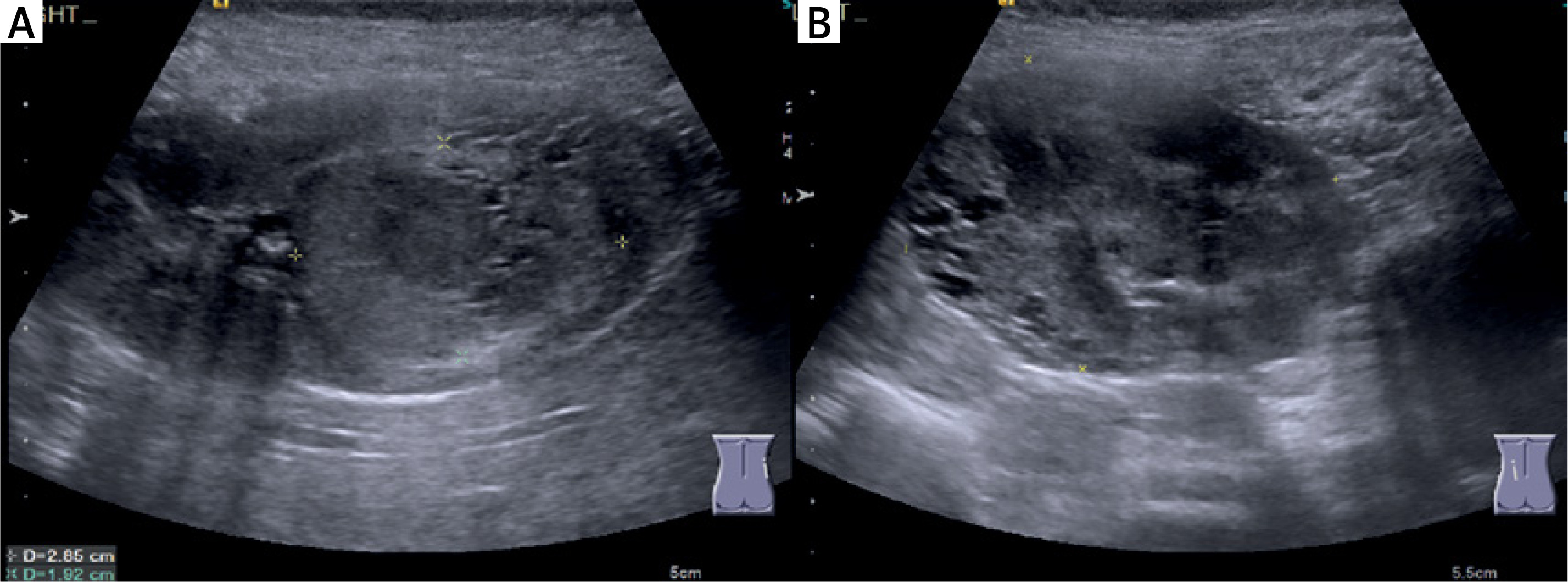
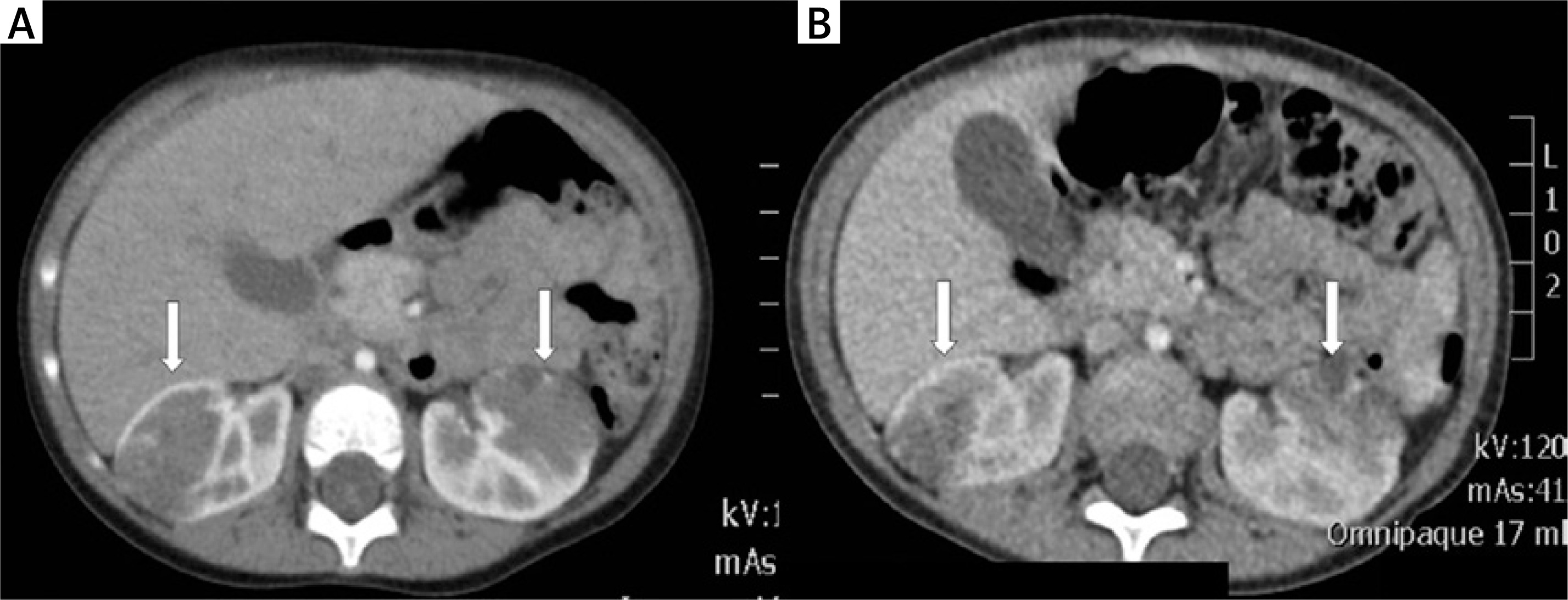
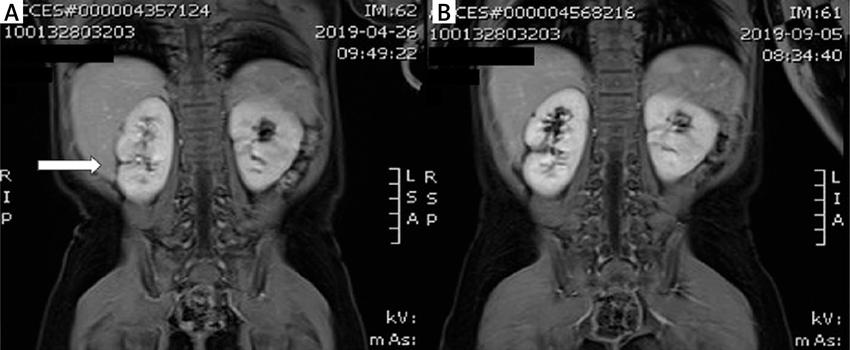
Case report. Screening ultrasound of the abdomen was performed in a girl aged almost 2 years, with no clinical symptoms. Bilateral kidney tumors were found in an early stage with diameter under 30 mm on the right side and 40 mm on the left (Figure 1). Nephroblastoma was suspected and confirmed in computed tomography (CT) examination. In the preoperative CT study, after a month of chemotherapy the tumors showed a slight decrease in volume (Figure 2). Then preserving kidneys surgery was performed. Follow-up magnetic resonance imaging (MRI) study of the abdomen after 6 and 11 months of observation showed only postoperative changes in both kidneys, with no recurrence (Figure 3). The standard observation is continued.
Figure 1
Ultrasound of the right kidney (A) and left kidney (B) in 2-year-old patient. Bilateral kidney tumors with diameter under 30 mm in the right kidney and 40 mm in the left (calipers)
Discussion
Epidemiological studies showed higher incidence of cancers such as lymphomas, central nervous system tumors and solid tumors in children and adolescents during the last decades in Europe [4–9]. According to studies by Steliarova-Foucher et al., the incidence increased by about 1% per year for all cancer types [7, 9]. Although epidemiological studies have not identified clear risk factors for childhood cancer so far, early diagnosis is essential to improve treatment results for childhood cancer [10]. Also lack of specific manifestations in the early stage of many tumors usually delays its diagnosis and prevents treatment in the early stage [11]. Ultrasound screening examinations have been tried several times in the diagnosis of children, mainly in the assessment of the urinary tract [12, 13]. Despite promising results that showed changes in approximately 25% of the examined children, these studies have not been introduced to the entire population. Another attempt was made by the RMHC Chapter in Poland, which purchased a special mobile ambulance equipped with two ultrasound rooms for pediatric ultrasound examinations [14]. These studies were co-financed with local self-governments. The goal of the ultrasound screening program was to identify abnormalities in the early stage, without clinical symptoms, to allow implementation of further procedures in the earliest phase. Ultrasound was used due to its non-invasiveness, which is especially important in pediatric patients. The population of children under 6 years of life was selected for examinations. The experience of pediatricians to date shows that at this age some of the childhood cancers appear, which are usually detected at higher, symptomatic stages. In addition, children at this age are quite difficult to examine and cannot precisely report their symptoms. During the ultrasound examination, the child usually lies on his back; restless children can be held by parents. In the abdominal cavity, mainly parenchymatous organs such as the liver, pancreas, spleen, kidneys and organs containing fluid such as the gall bladder and urinary bladder are assessed. In girls with full bladder, reproductive organs can also be assessed. Within the neck the thyroid gland, salivary glands and lymph nodes and in boys additionally testicles, epididymis and spermatic cord are assessed. In most cases, due to the physiological image of organs, a small area of examination and good, high-resolution images, it is a fast examination, lasting only a few minutes. Therefore, it can be carried out even for restless children. During the examination, parents are kept informed about the physiology and possible pathology of the observed structures and after the examination they receive a report, with suggestions what to do if abnormalities are found. These can be referrals for check-ups, a family doctor or specialist consultations. In 5% of the examined children oncological processes were suspected, mostly in patients with focal lesions and enlarged lymph nodes. Twelve malignant tumors were detected, which represent 0.02% of the examined population. Peak incidence during the earliest years of life features embryonic tumors such as hepatoblastoma, nephroblastoma, or neuroblastoma. Out of these tumors the highest incidence rate is 9.3 for neuroblastoma, slightly lower for nephroblastoma (7.4) and lowest for hepatoblastoma (1.3) [15]. Based on the number of examined patients in this program and detected Wilms tumors, it gives an incidence rate of 9, which is higher than the incidence rate of Wilms tumor of the whole population. The incidence rate of hepatoblastoma (1.5) was similar to the populational index, in contrast to the detection of neuroblastomas, which did not appear in the children examined in the program. The example of a patient with bilateral Wilms tumor, described in the case report, is very important. Finding these changes at an earlier stage allowed for a saving treatment and leaving both kidneys. So far, no recurrence has occurred during the observation.
Moreover, during the study, a large number of other abnormalities were found, with various degree of clinical significance (Tables I–IV). Such findings provide valuable information about the frequency of certain diseases in the whole population. In the neck examination inhomogeneous thyroid echostructure and hypoplasia of the thyroid gland were observed and may indicate potential thyroid dysfunction. The most common urinary tract pathology was related to dilation of collecting systems of the kidneys, reflecting potential risk of urinary tract infections and post-inflammatory nephropathy. Renal congenital defects may give no symptoms for a long time. A significant number of abnormalities were related to scrotal pathologies. Undescended testes and microlithiasis can affect testis growth and are proven conditions predisposing to cancer occurrence [14, 16].
In summary, pediatric screening examinations enable the early identification of childhood malignancies as well as developmental abnormalities, and therefore the early initiation of interventions whose effectiveness, especially in oncology, is crucial and evidence based. The higher rate of Wilms tumors detected in our screening program compared to the rate of diagnosed Wilms tumors in the whole population proves the sense of abdominopelvic screening ultrasound in children. Due to the oncological orientation of the program, all suspicions of malignancies are referred to the oncologists cooperating with the foundation; hence the diagnosis of these changes is really fast. However, there were too few patients diagnosed with cancer to analyze whether the program had an impact on reducing the time to diagnosis in the pediatric population.
We identified in the study a relatively high percentage of benign urinary tract pathologies which developing silently in asymptomatic children could have led to the more advanced stages of disease, leading to irreversible morphological and functional changes. Similarly, the high percentage of abnormalities in scrotal ultrasound supports the necessity of screening in boys up to 6 years, which thanks to early diagnosis and treatment can be crucial for their fertility in adulthood. In these areas, we see the possibilities of using our research to develop specialized procedures to accelerate the treatment of these patients. On the other hand, the high percentage of enlarged lymph nodes in this age group is a kind of physiology due to the activity of the immune system, vaccinations and frequent infections. The lack of symptoms of lymph node malignancy and the low percentage of other pathologies suggest that cervical ultrasound screening would probably not play a significant role in the population. The issue of costs is very important, and reducing the amount of areas of interest could partially reduce these costs. Selection of patients to participate in the program may constitute another limitation. The idea of the program was to assess healthy children, but parents of children with pathologies recognized earlier could have had more motivation to participate in the studies.
In conclusion, ultrasound screening has proved to be very useful for the early identification of cancerous as well as precancerous lesions, but due to the small percentage of the population covered by the screening, not all neoplasms were represented in sufficient numbers to draw definitive conclusions. Nevertheless, it is worth continuing this research even for the reason of effective early detection of other anomalies. However, limiting the research to the abdominal cavity and testes in boys should be considered. The ideal would be the possibility of examining these children at least once a year, as non-invasive ultrasound examination can be used repeatedly.

