

Introduction
Clostridioides difficile, an anaerobic, spore-forming, Gram-positive bacillus, is considered one of the most important pathogens responsible for the occurrence of intestinal infection in hospitals [1]. Clostridioides difficile-induced enteritis (CDE) manifests as abdominal pain, diarrhea, and bloody stools. Complications such as toxic megacolon, intestinal perforation, and septic shock can occur in severe cases [2]. CDE is a major global health problem imposing a serious socioeconomic burden. According to recent estimates by the Centers for Disease Control, Clostridioides difficile infection is reported in approximately 500,000 Americans each year, with the estimated annual economic burden in the United States being $796 million [3]. Hospitalized patients with CDE, irrespective of age or comorbidity, are 2.74-fold more likely to die during their hospital stay than all other hospitalized patients [4]. Despite the establishment of national guidelines, the adoption of well-recognized preventive strategies has not resulted in the eradication of Clostridioides difficile infection [5].
Vitamin D is associated with bone health and has been recently shown to regulate the innate and adaptive immune systems [6, 7]. Therefore, we speculate that vitamin D status, as measured by serum 25-hydroxyvitamin D levels, may play an important role in the susceptibility to nosocomial infections, such as Clostridioides difficile infection [8]. Several clinical studies have shown that vitamin D deficiency may increase the risk of Clostridioides difficile infection. However, this conclusion remains controversial and requires further investigation [9].
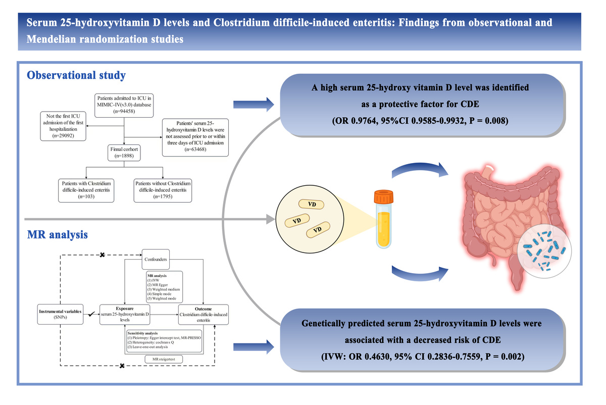
In this study, we integrated observational and Mendelian randomization (MR) approaches to comprehensively investigate the association and potential causal relationship between serum 25-hydroxyvitamin D levels and the risk of CDE. For observational analysis, data were obtained from MIMIC-IV, which is a large database containing high-quality clinical data of patients admitted to intensive care units at a healthcare center [10]. This observational study provided essential epidemiological insights for evaluating the relationship between serum 25-hydroxyvitamin D levels and CDE. MR serves as a robust instrument in epidemiological research. In MR analysis, genetic variations are used to evaluate the causal relationship between risk factors and specific diseases. This technique effectively mitigates issues such as confounding effects and reverse causality inherent in observational studies as well as concerns related to representativeness and feasibility inherent in randomized clinical trials (RCTs) [11, 12]. In this study, MR analysis was used to investigate the causal relationship between serum 25-hydroxyvitamin D levels and the risk of CDE. The findings may provide a reference for the prevention and treatment of CDE in clinical settings.
Material and methods
Observational study
Database
Data were retrieved from the Medical Information Mart for Intensive Care-IV (MIMIC-IV) (version 3.0) database. A researcher from our team completed the Collaborative Training Program (Renli Wang, certificate number: 1797679) to access the database. All patients in the database are anonymous; therefore, informed consent was not required in this study.
Study population
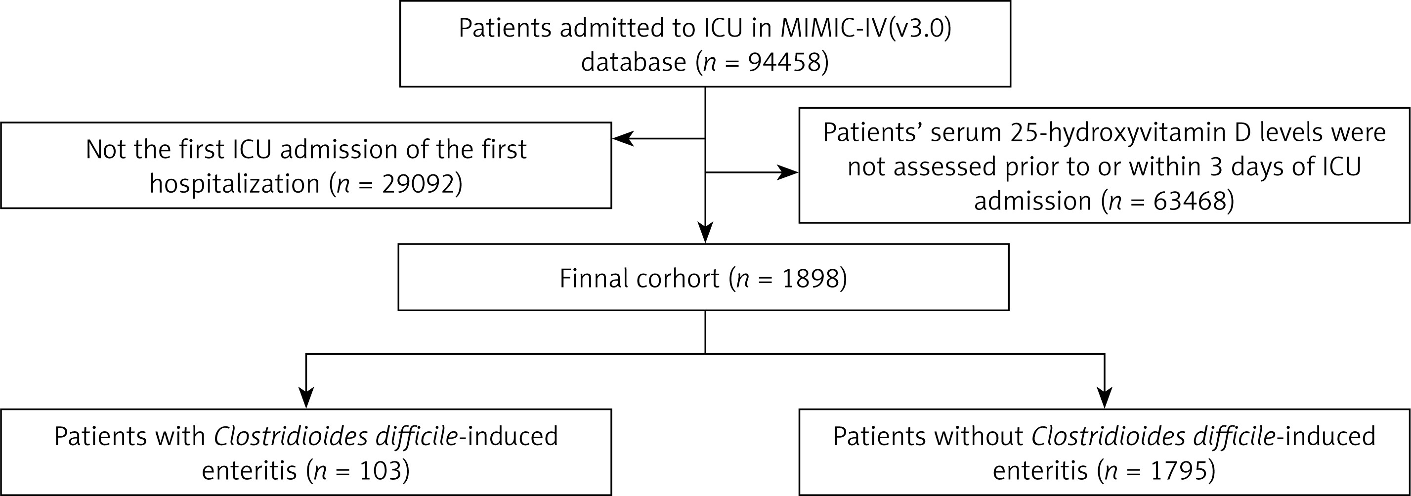
Patients who met the following criteria were included in this study: (1) intensive care unit (ICU) admission during hospitalization, (2) first ICU admission on first hospitalization, and (3) measurement of serum 25-hydroxyvitamin D levels prior to or within 3 days of ICU admission. The cohort selection process is shown in Figure 1.
Selection of variables
Structured Query Language (SQL) based on PostgreSQL tools (version 9.6) was used for data extraction. Patients were categorized into CDE and non-CDE groups. Data regarding the following covariates were collected: (1) demographic characteristics such as age, sex, and race; (2) comorbidities such as congestive heart failure, chronic pulmonary disease, severe liver disease, renal disease, cerebrovascular disease, rheumatic disease, cancer, and diabetes and the Charlson Comorbidity Index; (3) disease severity scores such as the Sequential Organ Failure Assessment (SOFA) score and Simplified Acute Physiology Score II (SAPS II) (in cases of multiple measurements, only the worst value was considered); (4) use of drugs such as antibiotics (clindamycin, fluoroquinolones, and third-generation cephalosporins) and proton pump inhibitors (PPIs); and (5) serum 25-hydroxyvitamin D levels (in cases of multiple measurements, only the minimum value was considered).
Statistical analysis
Baseline characteristics were expressed as the mean (standard deviation) and number (percentage) for continuous and categorical variables, respectively. As appropriate, the t-test, chi-square (χ2) test, or Wilcoxon rank-sum test was used to compare the characteristics of patients between the CDE and non-CDE groups.
Univariate and multivariate logistic regression analyses were used to identify independent risk factors for CDE. Variables that had a p-value of < 0.05 on univariate analysis and were considered potential risk factors were included in the multivariate logistic regression model through backward stepwise selection using Wald’s method. Multicollinearity was assessed using the variance inflation factor (VIF), and variables showing multicollinearity were removed from the multivariate model. A VIF value of < 4 indicated the absence of multicollinearity.
Restricted cubic spline (RCS) analysis was used to evaluate the relationship between serum 25-hydroxyvitamin D levels and the risk of CDE. RCS analysis does not require the assumption of a linear relationship between covariates and outcomes, which provides greater flexibility for fitting data [13]. In this study, RCS analysis was performed based on the multivariate logistic regression model.
R (version 4.2.1) software was used for statistical analysis. A p-value of < 0.05 was considered statistically significant.
Mendelian randomization
Study design
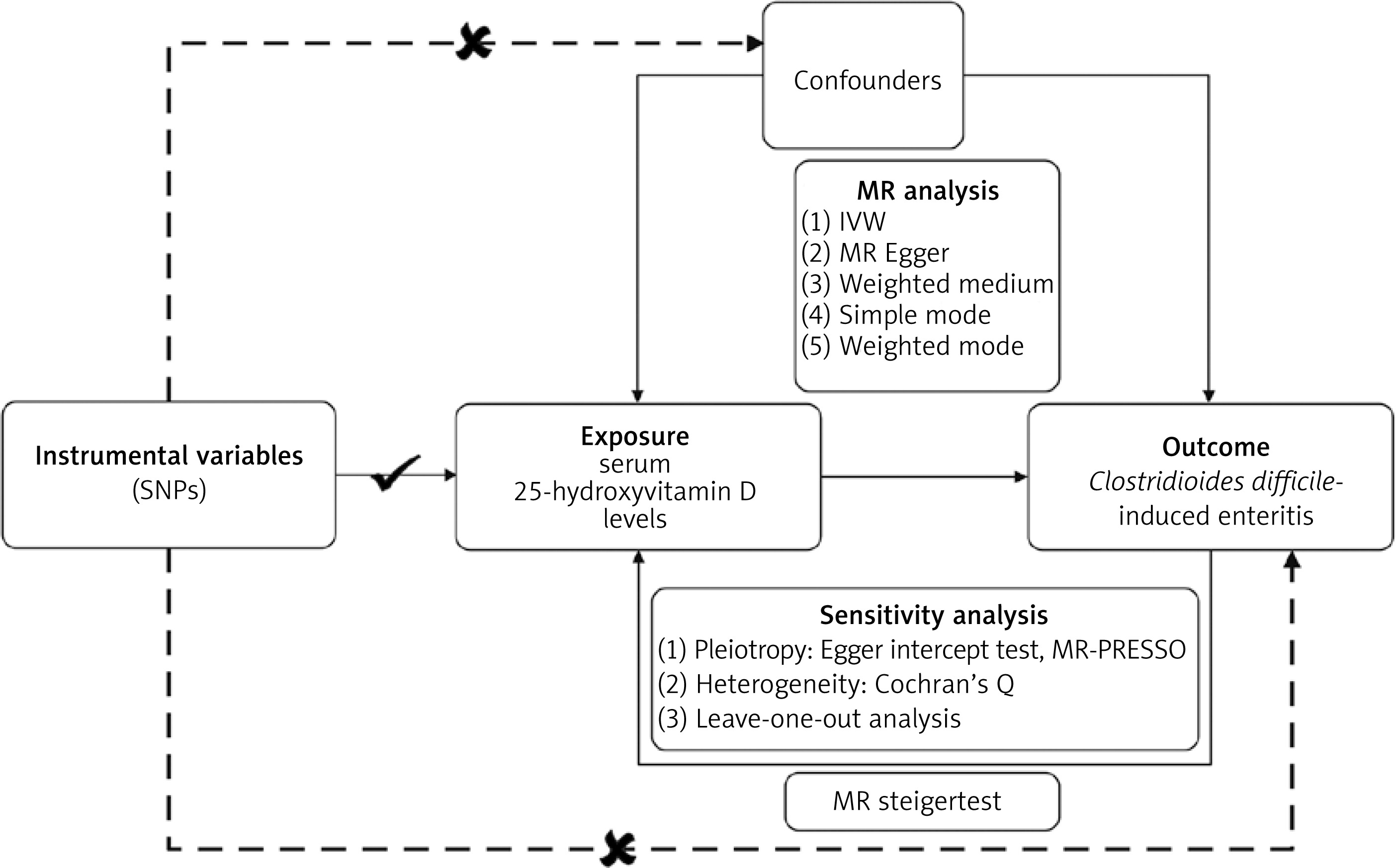
Two-sample MR analysis was used to evaluate the causal effects of serum 25-hydroxyvitamin D levels on the risk of CDE using summary statistics from genome-wide association studies (GWASs). The validity of the MR approach relies on the following three key assumptions: (1) genetic variants exhibit a strong association with the exposure; (2) genetic variants are unrelated to potential confounders; and (3) genetic variants affect the outcome exclusively through their impact on the exposure. In this study, serum 25-hydroxyvitamin D levels and the risk of CDE were selected as the exposure and outcome, respectively, for MR analysis. A flowchart depicting the protocol of MR analysis is presented in Figure 2.
Data source
GWAS summary statistics for serum 25-hydroxyvitamin D levels were derived from UK Biobank (UKB), which had a sample size of 79,366 and was accessed at https://gwas.mrcieu.ac.uk/ (ID: ebi-a-GCST005367). GWAS summary data for CDE were obtained from the FinnGen consortium (ID: finngen_R11_C_DIFFICILE_ENTEROCOLITIS), which included 450,673 participants. All participants in both databases were of European descent. Ethical approval and consent for the use of summary statistics were obtained from the original publication.
Selection of instrumental variables
Single nucleotide polymorphisms (SNPs) with genome-wide significance (p < 5 × 10⁻⁸) were selected as instrumental variables (IVs) to ensure a strong correlation between the genetic variations and the exposure. To prevent linkage disequilibrium (LD) bias, the LD threshold was set at R2 < 0.001, with a window size of 10,000 kb [14]. The F statistic (F=22/SE2) was used to evaluate the strength of each SNP. An F value of > 10 indicated a strong association. SNPs associated with confounders were detected and removed using the LDlink database (https://ldlink.nih.gov/?tab=ldtrait). Data harmonization was achieved by removing palindromic SNPs to prevent unintended bias [15].
MR estimates and sensitivity analysis
Inverse variance weighting (IVW), the most frequently used and effective MR method, was selected as the primary method for MR analysis. This method assumes that all SNPs are valid IVs and hence provides estimates with the highest statistical power. Four other MR methods, namely, MR-Egger, weighted median, simple mode, and weighted mode, were used to verify the reliability of the results of IVW [16]. Furthermore, the MR Steiger directionality test was used to examine the direction of causality between the exposure and the outcome [17].
Horizontal pleiotropy was detected using the MR-Egger intercept test and funnel plot. The presence of horizontal pleiotropy was confirmed when the MR-Egger intercept significantly deviated from 0 (p < 0.05) or when asymmetry was observed in the funnel plot [18]. The Cochran Q statistic was used to assess heterogeneity among causal estimates, with a p-value of < 0.05 indicating the presence of heterogeneity [19]. The MR-PRESSO method was used to detect outliers with potential horizontal pleiotropy. Any detected outliers were excluded to yield unbiased causal estimates through outlier-corrected MR analysis [20]. Subsequently, a leave-one-out analysis was performed, wherein each SNP was sequentially removed to detect pleiotropy attributed to the individual SNPs.
All MR analyses were performed using the “TwoSampleMR” and “MR-PRESSO” packages in R (version 4.2.1). A p-value of < 0.05 indicated statistical significance.
Results
Baseline characteristics of participants in the observational analysis
A total of 1898 participants were included in the observational analysis. Of these participants, 103 were included in the CDE group, whereas 1795 were included in the non-CDE group. The baseline characteristics of the two groups are shown in Table I. The CDE group had lower serum 25-hydroxyvitamin D levels than the non-CDE group (20.36 [13.00] versus 23.91 [13.90], p = 0.011). In addition, some other variables exhibited significant differences between the two groups.
Table I
Baseline characteristics of the study population
Association between serum 25-hydroxyvitamin D levels and the risk of CDE
We calculated the odds ratios (ORs) and confidence intervals (CIs) for all variables included in this study. As shown in Table II, univariate logistic regression analysis showed that severe liver disease (OR = 1.5923; 95% CI: 1.0217–2.4311; p = 0.035), renal disease (OR = 2.4303; 95% CI: 1.6248–3.6272; p < 0.001), higher SOFA scores (OR = 1.0679; 95% CI: 1.0210–1.1162; p = 0.004), higher SAPS II scores (OR = 1.0180; 95% CI: 1.0045–1.0314; p = 0.008), antibiotic use (OR = 2.3952; 95% CI: 1.5177–3.9276; p < 0.001), PPI use (OR = 1.8498; 95% CI: 1.1560–3.0923; p = 0.014), and lower serum 25-hydroxyvitamin D levels (OR = 0.9781; 95% CI: 0.9608–0.9943; p = 0.012) were associated with the risk of CDE. Subsequent multivariate logistic regression analysis showed that only renal disease (OR = 2.6457; 95% CI: 1.7419–4.0136; p < 0.001), antibiotic use (OR = 2.1975; 95% CI: 1.3614–3.6716; p = 0.002), and lower serum 25-hydroxyvitamin D levels (OR = 0.9764; 95% CI: 0.9585–0.9932; p = 0.008) were independently associated with an increased risk of CDE. The RCS curve shown in Figure 3 demonstrates a linear relationship between serum 25-hydroxyvitamin D levels and the incidence of CDE (p for non-linear = 0.165, p for overall < 0.001). Lower serum 25-hydroxyvitamin D levels were associated with a higher risk of CDE. Notably, the risk of CDE significantly increased (OR > 1) when serum 25-hydroxyvitamin D levels decreased below 20.92 ng/ml.
Figure 3
RCS curve of the relationship between serum 25-hydroxyvitamin D levels and the incidence of Clostridioides difficile-induced enteritis (CDE)
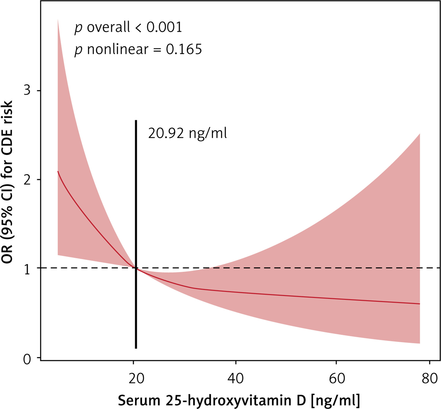
Table II
Univariate and multivariate analyses of prognostic variables
Main MR analysis
According to the screening criteria for IVs, 7 SNPs were eventually included in the MR analysis. Detailed information regarding the included SNPs is presented in Supplementary Table SI. The results of IVW showed that serum 25-hydroxyvitamin D levels were significantly associated with the risk of CDE (OR = 0.4630; 95% CI: 0.2836–0.7559; p = 0.002). Furthermore, the β-values of the other four MR methods were consistent in direction with that of the IVW method, indicating directionally consistency across MR methods (Table III, Supplementary Figures S1, S2). In addition, the Steiger test suggested the absence of a reverse causal relationship between serum 25-hydroxyvitamin D levels and the risk of CDE (Supplementary Table SII).
Table III
Results of Mendelian randomization
Sensitivity analysis for MR
Cochran’s Q test revealed no heterogeneity among the IVs (p of IVW = 0.751, Table III). Additionally, no horizontal pleiotropy was detected (p of Egger intercept = 0.602, Table III). Funnel plots indicated good symmetry, and the MR-PRESSO method revealed no outliers (Supplementary Figure S3). Furthermore, leave-one-out analysis corroborated that the effect estimates were not driven by any single anomalous SNP (Supplementary Figure S4). These findings collectively validated the reliability and validity of the results of MR.
Discussion
In this study, we comprehensively investigated the relationship between serum 25-hydroxyvitamin D levels and the risk of CDE using both real-world observational and MR analyses. In the observational analysis, after adjustments were made for factors such as demographic characteristics, comorbidities, disease severity score, and drug use, low serum 25-hydroxyvitamin D levels were associated with increased susceptibility to CDE. Furthermore, two-sample MR analysis consistently showed that genetically predicted low serum 25-hydroxyvitamin D levels were associated with an increased risk of CDE; however, the reverse relationship (CDE → lower serum 25-hydroxyvitamin D levels) was unlikely to be causal.
CDE is a major health issue in hospitals and long-term care facilities, especially for patients in intensive care units (ICUs) [21]. Low 25-hydroxyvitamin D concentrations are common, and the prevalence of severe vitamin D deficiency (< 25 nmol/l) ranges from 5% to 50% based on the location and population characteristics [22, 23]. Vitamin D supplementation is a simple, safe, and inexpensive measure that is effective in correcting vitamin D deficiency [24]. Therefore, understanding the relationship between serum 25-hydroxyvitamin D levels and the risk of CDE may help prevent, manage, and treat CDE accurately, which is of great clinical significance.
Over the past few decades, several studies have investigated the relationship between serum 25-hydroxyvitamin D levels and CDE. Ananthakrishnan et al. reported that higher concentrations of 25-hydroxyvitamin D were associated with a lower risk of CDE in patients with inflammatory bowel disease [25]. Sahay et al. found that 25-hydroxyvitamin D concentrations of > 20 ng/ml had a protective effect against community-acquired Clostridioides difficile infection [26]. In contrast, Quraishi et al. did not find an association between 25-hydroxyvitamin D concentrations and the risk of hospital-acquired CDE [27]. Despite these controversial findings, vitamin D is usually considered a protective factor in CDE, which is consistent with the results of this study [28]. Existing studies have predominantly focused on American populations and have not adequately addressed the potential limitations associated with observational studies. However, in this study, we analyzed the high-quality clinical data from the MIMIC-IV, UKB, and FinnGen databases and used MR to investigate the nature of the relationship between 25-hydroxyvitamin D levels and the risk of CDE. Therefore, the results of this study may be more reliable. Furthermore, we found that a serum 25-hydroxyvitamin D level of < 20.92 ng/ml was an independent risk factor for CDE. This finding has important implications for clinical practice.
Although this study supports a causal relationship between serum 25-hydroxyvitamin D levels and the risk of CDE, the underlying mechanisms remain unclear. The immunomodulatory effects of vitamin D may be an important consideration in the care of patients with CDE. The active form of vitamin D can activate macrophages by upregulating the expression of cathelicidin and B-defensin 2 in neutrophils, monocytes, natural killer cells, and epithelial cells. These antimicrobial peptides are effective against both Gram-positive and Gram-negative bacteria, fungi, and mycobacteria at various sites, including the gastrointestinal system [29, 30]. Furthermore, vitamin D has been shown to have a protective effect on maintaining the structural integrity of the intestinal lining. Specifically, activation of the vitamin D receptor increases the expression of several intracellular proteins that form tight junctions between epithelial cells of the gut [31]. Therefore, owing to the decreased expression of antimicrobial peptides and the loss of epithelial barrier integrity, patients with vitamin D deficiency may experience impaired immune function, which contributes to the increased risk of developing CDE [32]. In addition, preliminary evidence suggests that vitamin D affects the pathogenesis of Clostridioides difficile infection by reducing the expression of NLRP3 (NLRP3 normally produces IL-1B, an important mediator of inflammatory responses that can trigger apoptosis). The reduced expression of NLRP3 may help prevent macrophage death caused by Clostridium difficile toxins A and B, limit inflammation, and minimize colon damage [33]. However, the specific underlying mechanisms warrant further investigation.
This study has several limitations that should be acknowledged. First, the MIMIC-IV database does not provide all the necessary information. For instance, because the timing of the diagnosis of CDE could not be established, it is possible that vitamin D status was assessed after the occurrence of CDE in some patients. This limitation may influence the finding that low serum 25-hydroxyvitamin D levels are a risk factor for CDE. Second, this study was observational, and serum 25-hydroxyvitamin D levels were not monitored in the general population; consequently, many patients were excluded owing to the lack of data on serum 25-hydroxyvitamin D assessment. This exclusion significantly reduced the sample size of this study. Third, despite our attempts to significantly reduce the bias through multivariable adjustment, estimation bias was unavoidable because complex confounding factors found during actual clinical treatment could not be considered owing to the retrospective nature of this study. Fourth, all participants in GWASs were of European descent; therefore, the findings of this study cannot be generalized to other populations and regions. Fifth, although the LDlink database was used to identify potential confounders in MR analysis, some unaccounted confounders might have affected the reliability of the results. Sixth and last, follow-up vitamin D levels were not available; therefore, we could not determine whether patients with vitamin D deficiency were adequately treated with replacement therapy.
In conclusion, this study provides epidemiologic and genetic evidence indicating that low serum 25-hydroxyvitamin D levels are associated with an increased risk of CDE. Therefore, serum 25-hydroxyvitamin D levels may warrant close monitoring in patients at high risk for Clostridioides difficile infection. Prospective studies are needed to investigate the role of vitamin D deficiency in the development of CDE and the impact of vitamin D supplementation on the risk of CDE.

