

Introduction
Idiopathic pulmonary fibrosis (IPF) is a chronic, progressive interstitial lung disease of unknown cause, characterized by gradual fibrosis of the lung tissue, leading to a progressive loss of lung function and, ultimately, death [1]. The disease predominantly affects older men, with a median survival time of only 3 to 5 years after diagnosis [2]. The incidence of IPF has steadily increased over the years [3]. A recent study on Medicare beneficiaries in the United States reported a rise from 202 cases per 100,000 in 2001 to 495 cases in 2011 among those aged 65 and older [4]. Currently, lung transplantation remains the only definitive treatment for IPF. Nintedanib and pirfenidone are the main FDA-approved drugs used in clinical practice to slow the progression of IPF. The complexity of IPF and the unclear etiology contribute to the significant challenges in its treatment [5–7].
Repeated injury to the alveolar epithelium leads to fibrosis. Factors such as genetic predispositions, environmental influences, immune dysregulation, and microbial elements all act as triggers for alveolar damage in IPF [8]. Among microbial factors, viruses are the most extensively studied, with chronic viral infections – such as those caused by herpes simplex virus (HSV), Epstein-Barr virus (EBV), and cytomegalovirus (CMV) – being implicated in the onset or progression of IPF. A meta-analysis of studies from 10 countries, involving 1,287 participants, revealed that viral infections were significantly associated with the risk of developing IPF, but were not associated with exacerbation of the disease. All the analyzed viruses, including EBV, CMV, human herpesvirus 7 (HHV-7), and human herpesvirus 8 (HHV-8), were associated with an increased risk of developing IPF [9]. Herpesviruses, particularly, have been the subject of substantial research. They are categorized into three subfamilies based on their cellular targets and sites of latency: (i) α herpesviruses, such as HSV; (ii) β herpesviruses, such as CMV; and (iii) γ herpesviruses, such as EBV [10]. Once a herpesvirus infects a host, it establishes lifelong latency in the nuclei of host cells, persisting in an episomal form, and can periodically reactivate to produce infectious virions, leading to symptomatic recurrence [11]. The study by Lok et al. [12] in animal models demonstrated that herpesviruses do not directly induce pulmonary fibrosis, but viral replication can accelerate the fibrotic process in the presence of pre-existing lung injury.
Further evidence supporting the link between herpesvirus infections and IPF comes from a study showing elevated titers of HSV immunoglobulin G (IgG), CMV IgG, and EBV capsid antigen in IPF patients compared to healthy controls [13]. Additionally, high viral loads of EBV and CMV have been detected in the alveolar epithelial cells of IPF patients. Interestingly, EBV antigens have been associated with markers of endoplasmic reticulum (ER) stress, suggesting a mechanistic link between latent viral infections and the development of IPF [14]. However, the causal relationship between these chronic viral infections and IPF remains to be determined. In fact, current observational studies in this field have several limitations, such as small sample sizes, residual confounding, detection bias, and reverse causality [15, 16].
In this study, we aimed to analyze the potential causal relationship between genetically predicted antibody responses to herpesvirus antigens and IPF. Mendelian randomization (MR) is a commonly used epidemiological approach for causal inference that leverages summary-level data from genome-wide association studies (GWAS) to evaluate potential causal effects of exposures on outcomes [17]. Since alleles are randomly allocated during meiosis, genetic variants serve as instrumental variables for exposures, reducing – but not eliminating – the influence of confounding and reverse causation that affect observational studies [18, 19]. Routine two-sample MR methods employ multiple complementary approaches, including inverse variance weighting (IVW), MR-Egger, weighted median, and sensitivity analyses, to assess the robustness of results. In contrast to conventional MR, generalized summary-data-based MR (GSMR) offers several advantages: it corrects for biases using linkage disequilibrium (LD) matrices and employs the heterogeneity in dependent instruments (HEIDI) outlier test to identify and remove pleiotropic single nucleotide polymorphisms (SNPs), thereby improving the accuracy and reliability of causal inferences [20]. Given the lack of conclusive epidemiological evidence on the potential associations between genetically predicted antibody responses to herpesvirus antigens and IPF risk. We applied both bidirectional two-sample MR and GSMR methods to evaluate associations between genetically predicted antibody responses to three herpesvirus (HSV, CMV, EBV) and the risk of IPF.
Material and methods
Data sources
This study used large publicly available GWAS databases. To minimize bias due to population stratification, we included study samples of European ancestry. We obtained the summary GWAS data for twelve antibodies against herpesvirus from the study by Butler-Laporte et al. [21]. Antibody titers were based on log-transformed units. This study comprehensively covers various phenotypes associated with antibody responses to herpesvirus antigens (HSV, CMV, EBV).
We accessed summary-level data for three HSV-related antibodies from the MRC-IEU UK Biobank and the IEU OpenGWAS project, including GWAS summary statistics for anti-HSV-1 IgG levels (GWAS ID: ieu-b-4906, n = 683), HSV-1 IgG1 antibody levels (GWAS ID: ebi-a-GCST90006918, n = 6,199), and HSV-2 IgG2 antibody levels (GWAS ID: ebi-a-GCST90006920, n = 1,382). Similarly, we obtained GWAS summary data for four CMV-related antibodies, including anti-CMV IgG levels, CMV pp28 antibody levels, CMV pp52 antibody levels, and CMV pp150 antibody levels. These data came from GWAS analyses involving 5,010, 5,087, 5,681, and 5,136 participants, respectively, and were available in GWAS databases under the GWAS IDs ieu-b-4900, ebi-a-GCST90006894, ebi-a-GCST90006895, and ebi-a-GCST90006896. We also accessed five sets of GWAS summary statistics for EBV-related antibodies, including anti-EBV IgG levels (GWAS ID: ieu-b-4901, n = 5,010), EBV early antigen-D (EA-D) antibody levels (GWAS ID: ebi-a-GCST90006898, n = 7,763), EBV nuclear antigen-1 (EBNA-1) antibody levels (GWAS ID: ebi-a-GCST90006899, n = 7,972), EBV viral capsid antigen (VCA) p18 antibody levels (GWAS ID: ebi-a-GCST90006900, n = 8,518), and EBV Z EBV replication activator (ZEBRA) antibody levels (GWAS ID: ebi-a-GCST90006901, n = 8,191).
We sourced the GWAS summary data for IPF from the FinnGen cohort (https://r7.finngen.fi/), which comprised 1,514 IPF cases and 306,063 controls. All cases met the International Classification of Diseases (ICD-10) criteria. Detailed information on the selected datasets are provided in Table I and Supplementary Table SI.
Table I
Baseline characteristics of participants
| Exposure/outcome | Phenotype | GWAS ID | Data source | Sample size* | Population |
|---|---|---|---|---|---|
| Antibodies against HSV | Anti-HSV-1 IgG levels | ieu-b-4906 | IEU OpenGWAS | 683 | European |
| HSV-1 IgG1 antibody levels | ebi-a-GCST90006918 | UK Biobank cohort | 6,199 | European | |
| HSV-2 IgG2 antibody levels | ebi-a-GCST90006920 | UK Biobank cohort | 1,382 | European | |
| Antibodies against CMV | Anti-CMV IgG levels | ieu-b-4900 | IEU OpenGWAS | 5,010 | European |
| CMV pp28 antibody levels | ebi-a-GCST90006894 | UK Biobank cohort | 5,087 | European | |
| CMV pp52 antibody levels | ebi-a-GCST90006895 | UK Biobank cohort | 5,681 | European | |
| CMV pp150 antibody levels | ebi-a-GCST90006896 | UK Biobank cohort | 5,136 | European | |
| Antibodies against EBV | Anti-EBV IgG levels | ieu-b-4901 | IEU OpenGWAS | 5,010 | European |
| EBV EA-D antibody levels | ebi-a-GCST90006898 | UK Biobank cohort | 7,763 | European | |
| EBV EBNA-1 antibody levels | ebi-a-GCST90006899 | UK Biobank cohort | 7,972 | European | |
| EBV VCA p18 antibody levels | ebi-a-GCST90006900 | UK Biobank cohort | 8,518 | European | |
| EBV ZEBRA antibody levels | ebi-a-GCST90006901 | UK Biobank cohort | 8,191 | European | |
| IPF | – | Idiopathic pulmonary fibrosis | FinnGen cohort | 1,514/306,063 | European |
* Sample size shown as a total number for quantitative traits and cases/controls for binary traits. EBV – Epstein-Barr virus, CMV – cytomegalovirus, HSV – herpes simplex virus, EA – EBV early antigen, EBNA-1 – EBV nuclear antigen-1, IgG – immunoglobulin G, EBV VCA p18 – EBV viral capsid antigen p18, EBV ZEBRA – EBV Z EBV replication activator, IPF – idiopathic pulmonary fibrosis.
This study used GWAS summary datasets for bidirectional MR analysis. We sourced all datasets from previously published publicly available GWAS studies that had obtained ethical approval and participant consent. The summary statistics were publicly available for download, fully de-identified, and could be accessed without any restriction.
Selection of instrumental variables
The following criteria were applied to select instrumental variables (IVs): (1) SNPs reaching GWAS significance threshold (p < 5 × 10⁻⁶) were included as IVs for herpesvirus-related antibodies, except for the anti-EBV IgG levels, for which the GWAS significance threshold was set at p < 5 × 10⁻⁵ to obtain a sufficient number of SNPs, as fewer than three genome-wide significant SNPs were available under the primary threshold. For reverse causal analyses, we used the same threshold (p < 5 × 10⁻⁶) to select SNPs associated with IPF. (2) To ensure independence and minimize the impact of LD, we clumped the GWAS significant SNPs from each split’s GWAS with a clumping window of 10,000 kb and an r2 threshold of 0.001. The reference panel used for LD estimation was the 1000 Genomes phase 3 European population. (3) We also excluded SNPs with a minimum allele frequency (MAF) less than 0.01 because the impact of these SNPs was not stable. Palindromic SNPs with intermediate allele frequencies were removed to avoid strand ambiguity. All non-matching alleles were aligned, and the signs of the beta estimates were flipped [22]. Then, the F-statistic was calculated for the selected SNPs, and strong instruments were defined as those with an F-statistic > 10 [23]. The summary of instrument counts, F-statistics, and related information for each antibody exposure is presented in Supplementary Tables SII–SV.
MR analyses
GSMR as the main analysis
We employed GSMR as the main analysis method. GSMR is a flexible approach that uses summary-level GWAS data from independent studies to conduct MR analysis. Multiple near-independent genetic instruments were selected to test causal relationships between risk factors (or phenotypes) and disease outcomes [24]. The HEIDI-outlier test was used to identify and exclude genetic variants exhibiting pleiotropy, thereby reducing bias in the causal effect estimates.
To select valid and independent IVs, we employed the clumping algorithm to identify genome-wide significant SNPs for each trait (r2 threshold = 0.05, p-value threshold = 5 × 10⁻⁶), using the 1000 Genomes phase 3 European samples as the reference for LD estimation. To address potential pleiotropy, the HEIDI-outlier test was implemented with a p-value threshold of 0.01 to filter SNPs exhibiting horizontal pleiotropy effects on both exposure and outcome traits [24].
Two-sample MR as supplemental analysis
We applied three additional MR methods as supplemental analyses for the antibody-disease associations examined in the main analysis-GSMR: the IVW, MR-Egger, and weighted median methods. Cochran’s Q statistic was used to quantify heterogeneity among the IVs. A statistically significant Cochran’s Q (p < 0.05) indicated heterogeneity among the IVs [25]. MR pleiotropy residual sum and outliers (MR-PRESSO) tests and the MR-Egger intercept were used to detect outliers and horizontal pleiotropy [26]. A significant intercept in the MR-Egger (p < 0.05) indicated the presence of pleiotropy. Leave-one-out sensitivity analyses were conducted to verify the robustness of the causal effect estimates.
Statistical analysis
MR analyses were conducted using the “TwoSampleMR” and “gsmr” packages in R version 4.3.0 (http://r-project.org/). Results were considered significant if the estimates from the four methods (GSMR, IVW, MR-Egger, and weighted median) were directionally consistent, GSMR result was significant (p < 0.05), and there was no significant heterogeneity or pleiotropy.
Results
Effect of HSV-related antibody traits on IPF
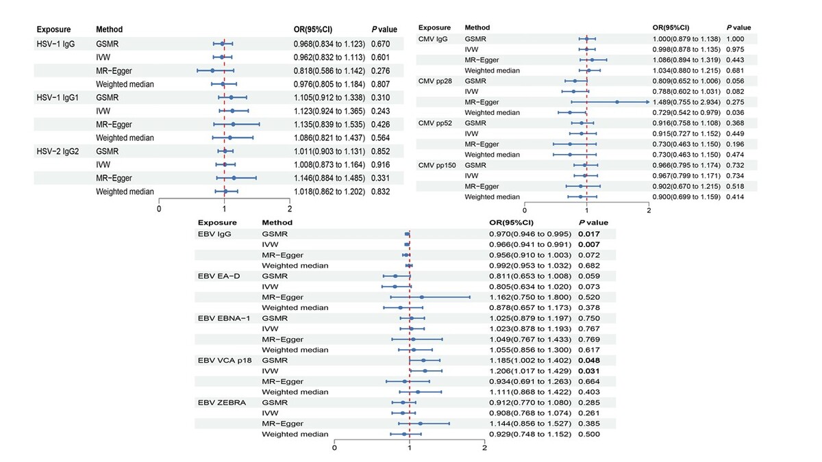
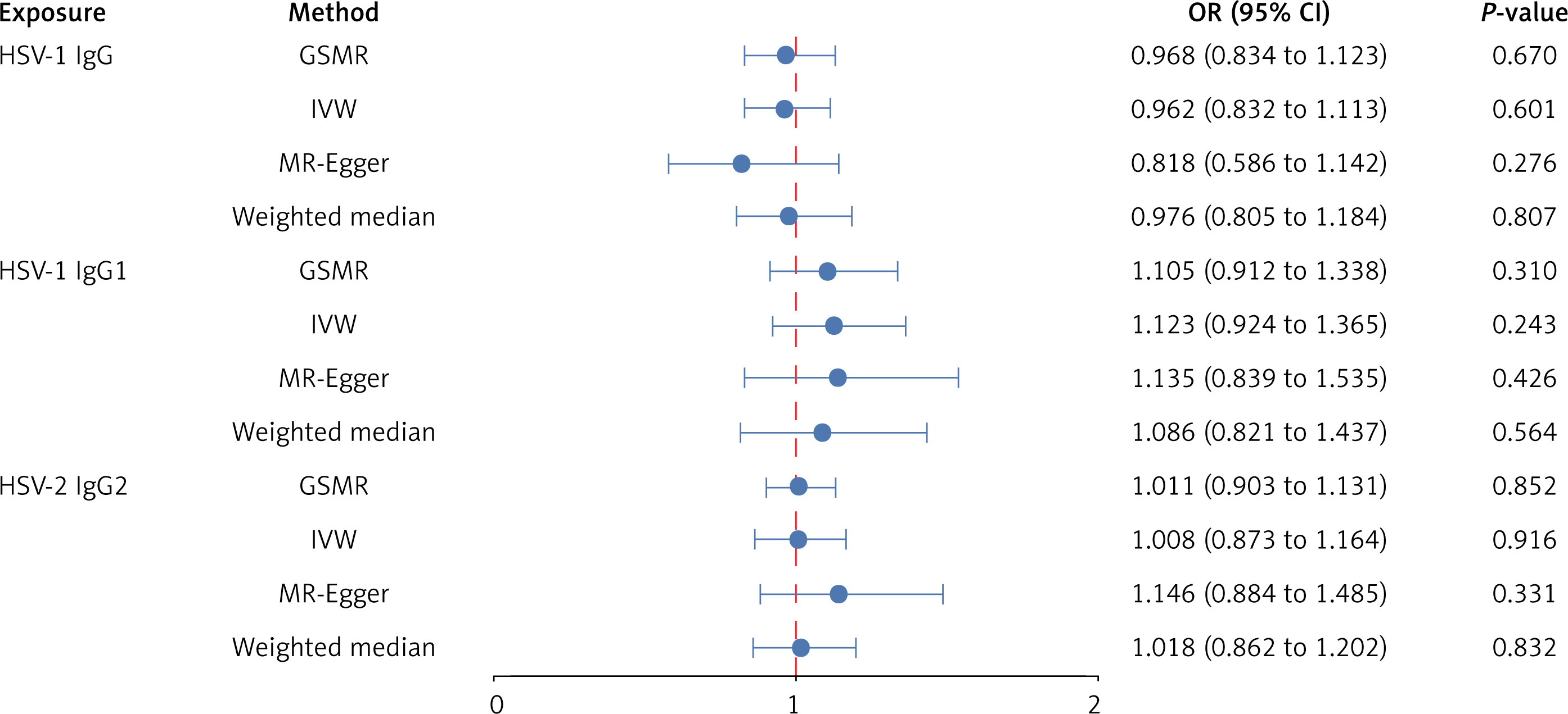
We first investigated the potential causal effects of genetically predicted HSV-related antibody traits on IPF (Figure 1, Supplementary Tables SVI–SVIII, and Supplementary Figures S1–S3). In GSMR analyses, the results did not support a causal effect of antibody levels targeting HSV-1 IgG1 (OR = 1.105, 95% CI: 0.912–1.338, p = 0.310), HSV-2 IgG2 (OR = 1.011, 95% CI: 0.903–1.131, p = 0.852), or anti-HSV-1 IgG (OR = 0.968, 95% CI: 0.834–1.123, p = 0.670) on IPF. Likewise, in analyses using three additional MR methods (IVW, MR-Egger, and weighted median), the results did not support a causal relationship between HSV-related antibody traits and IPF.
Effect of CMV-related antibody traits on IPF
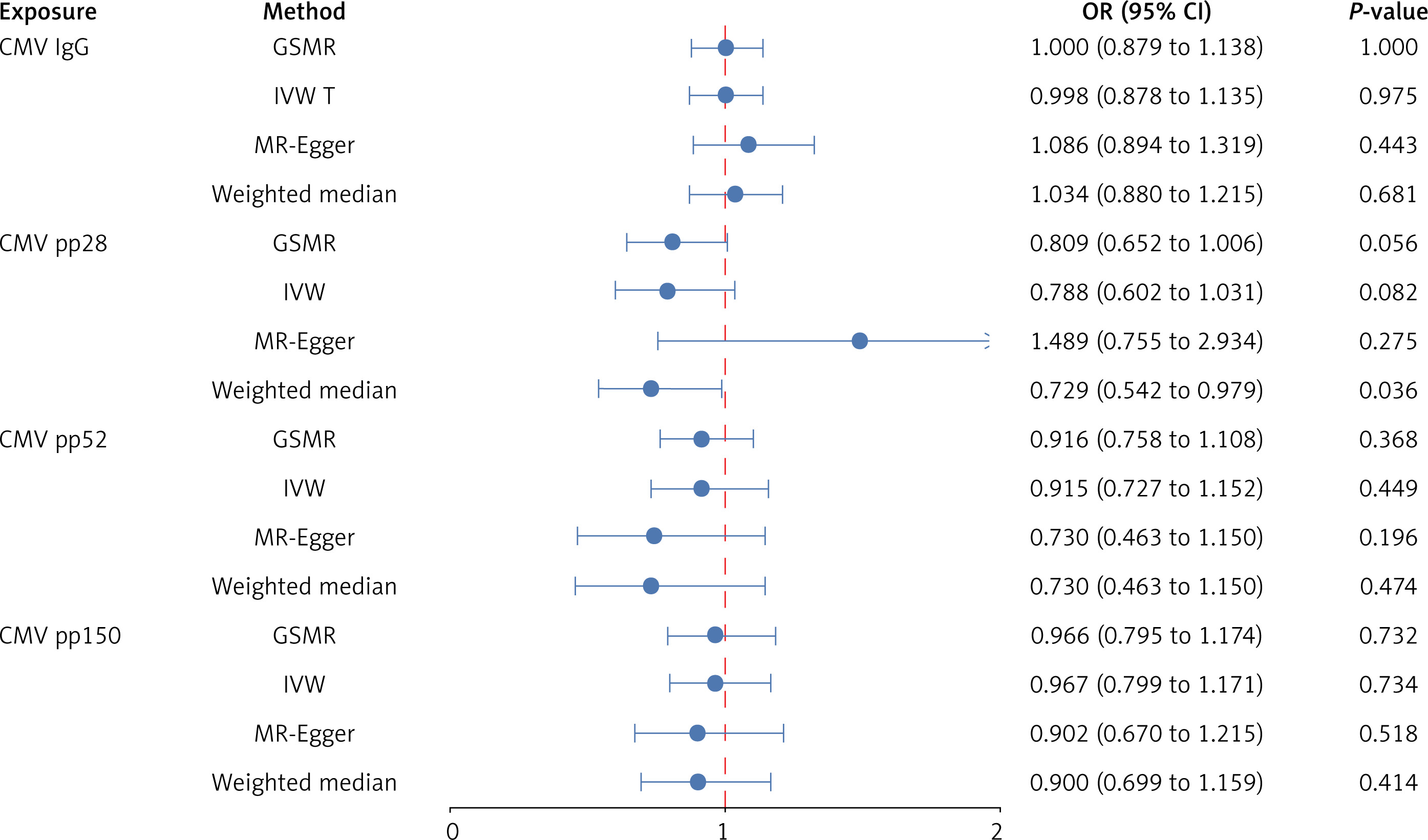
In our main analyses, we assessed the relationships between four antibodies against CMV and IPF using GSMR. In GSMR, the results did not support causal effects of antibody levels against CMV pp28 (OR = 0.809, 95% CI: 0.652–1.006, p = 0.056), CMV pp52 (OR = 0.916, 95% CI: 0.758–1.108, p = 0.368), CMV pp150 (OR = 0.966, 95% CI: 0.795–1.174, p = 0.732), or anti-CMV IgG (OR = 1.000, 95% CI: 0.879–1.138, p = 1.000) on IPF (Figure 2, Supplementary Tables SVI–SVIII, and Supplementary Figures S4–S7).
Effect of EBV-related antibody traits on IPF
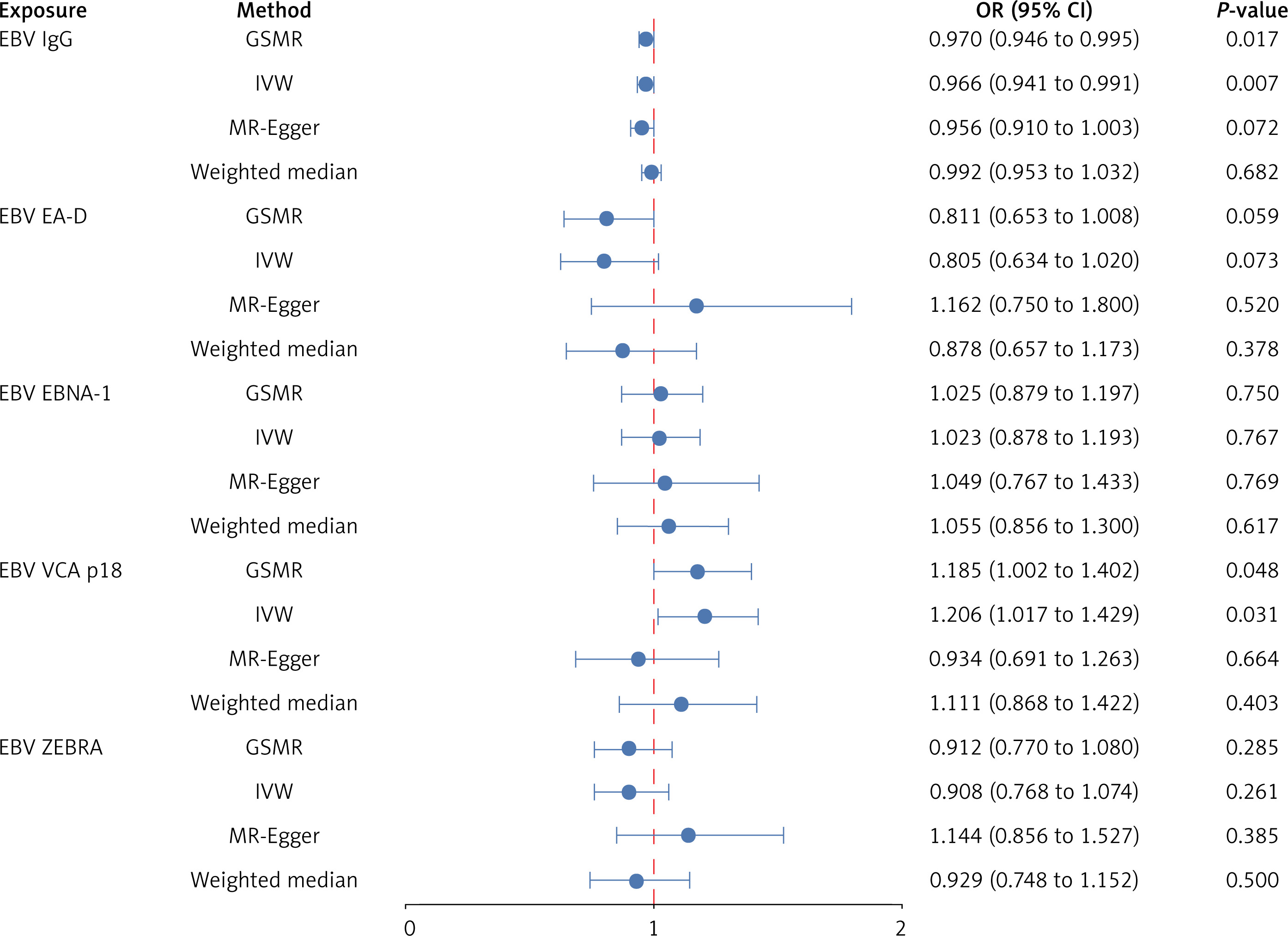
We then examined whether genetically elevated levels of five antibodies against EBV showed potential causal associations with IPF (Figure 3, Supplementary Figures S8–S12). In GSMR, genetically increased antibody levels against EBV VCA p18 were positively associated with IPF (OR = 1.185, 95% CI: 1.002–1.402, p = 0.048). In IVW, the association remained significant (OR = 1.206, 95% CI: 1.017–1.429, p = 0.031). However, in GSMR, anti-EBV IgG levels were negatively associated with IPF (OR = 0.970, 95% CI: 0.946–0.995, p = 0.007). In IVW, the association remained significant, confirming the protective effect of anti-EBV IgG levels on IPF (OR = 0.966, 95% CI: 0.941–0.991, p = 0.007). There was no other evidence to support a causal relationship between the other EBV antibody levels and IPF.
Figure 3
Forest plot of the causal association between Epstein-Barr virus-related antibody traits and idiopathic pulmonary fibrosis
EBV – Epstein-Barr virus, OR – odds ratio, CI – confidence interval, GSMR – generalized summary data based Mendelian randomization, IVW – inverse variance weighted.
No heterogeneity was found with the Cochran’s Q for EBV VCA p18 antibody levels (p = 0.549) (Table II). The calculated p-value of Egger intercept for EBV VCA p18 antibody levels was 0.061 (Table II), indicating no potential directional horizontal pleiotropy in the MR analysis.
Table II
Pleiotropic and heterogeneous results of five antibodies against EBV and IPF
Effect of IPF on herpesvirus-related antibody traits
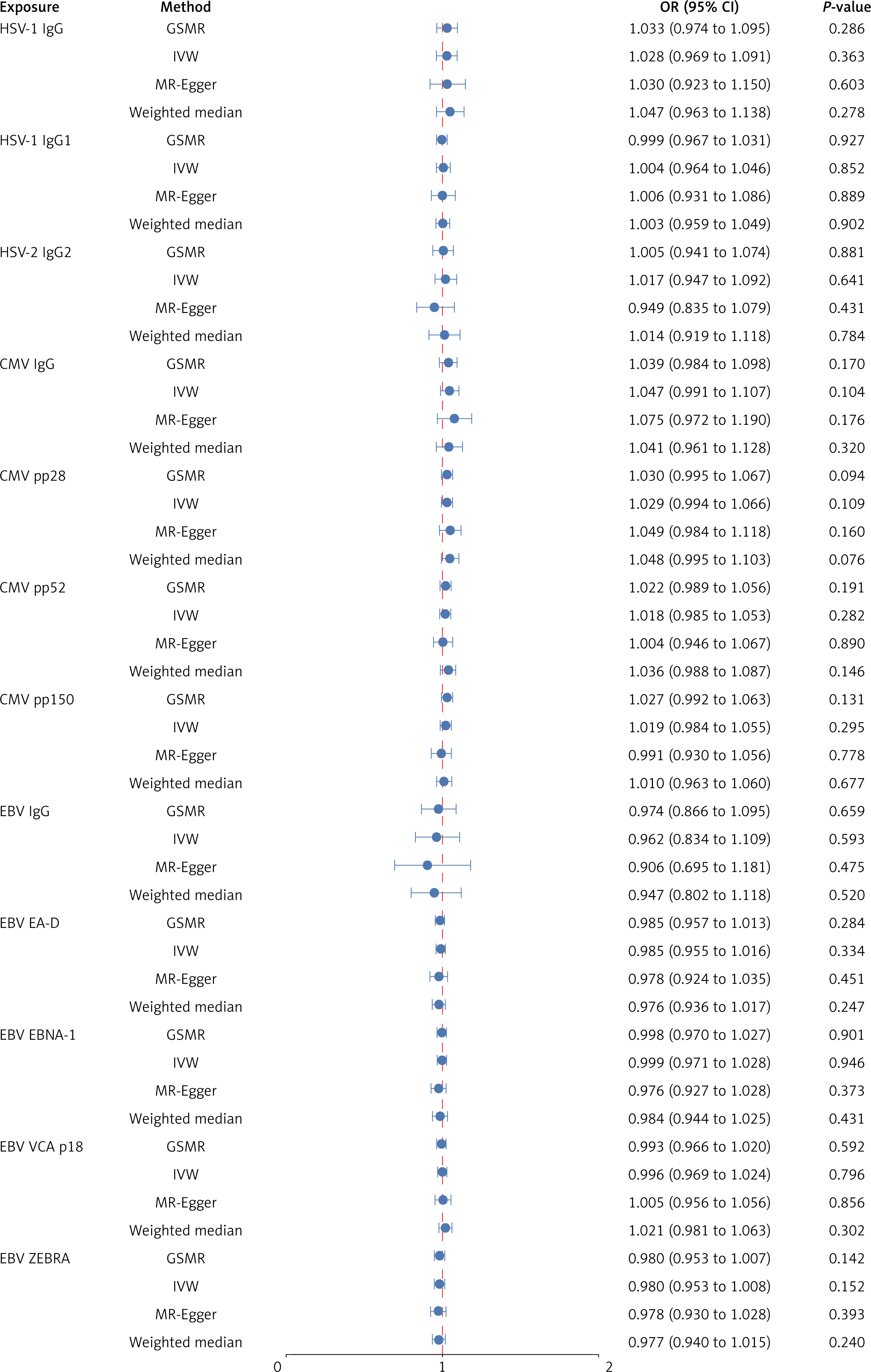
In GSMR analyses, no significant causal relationship was observed between IPF and genetically predicted herpesvirus-related antibody traits (Figure 4). Likewise, in IVW, MR-Egger, and weighted median analyses, the results did not support a causal relationship between IPF and HSV, CMV, or EBV.
Figure 4
Forest plot of the causal association between idiopathic pulmonary fibrosis and herpesvirus-related antibody traits
HSV – herpes simplex virus, CMV – cytomegalovirus, EBV – Epstein-Barr virus, OR – odds ratio, CI – confidence interval, GSMR –
generalized summary data based Mendelian randomization, IVW – inverse variance weighted.
Discussion
The relationship between herpesvirus-related immune responses and IPF has been unsettled for years. Clarifying these associations could provide a theoretical basis for future large-scale antiviral therapeutic trials in IPF. In the MR analyses, we found that genetically predicted anti-EBV VCA p18 antibodies and anti-EBV IgG levels were associated with IPF risk, consistent with a potential causal relationship under MR assumptions. Early detection and appropriate treatment of EBV infection may help alleviate the progression or worsening of IPF symptoms.
Viruses have long been thought to play an important role in the development and progression of IPF. In a systematic review and meta-analysis by Mostafaei et al. [27], the overall prevalence of viruses in IPF patients was found to be 53.72%, with HSV (77.7%) and EBV (72.0%) being the most common. The herpesvirus family has received the most attention as a causative and exacerbating factor in IPF. Significant associations with IPF risk have been found for HSV-1, EBV, CMV, HHV-7, and HHV-8 [9, 28]. Furthermore, Tang et al. [29] detected herpesvirus DNA in 97% of IPF patients’ lung tissue samples, with EBV being the predominant virus. Their study also found that sporadic IPF patients often harbor two or more herpesviruses, suggesting that genetic factors may render familial IPF patients more susceptible to fewer viral infections, while sporadic IPF may be linked to multiple viral infections. Genetic predispositions could potentially increase susceptibility to herpesvirus infections, thereby elevating the risk of developing IPF. Seibold et al. [30] reported that a specific polymorphism in the MUC5B gene, strongly associated with IPF risk, might enhance susceptibility to viral infections, including herpesviruses, by modulating lung immune responses.
Most existing studies are retrospective case-control studies or meta-analyses, or conducted on relatively small cohorts. Due to the low incidence of IPF, conducting large-scale prospective studies to investigate actual causal relationships is challenging. MR is a method that leverages genetic variants associated with specific exposures as IVs to infer causal effects while minimizing confounding [31]. MR methods use SNPs as IVs to infer causal relationships between exposures and outcomes. Variants are assigned randomly to offspring by their parents at conception. Therefore, the MR method is generally less susceptible to confounding and reverse causality, making it conceptually analogous to the random assignment method used in randomized controlled trials [32, 33]. Therefore, we used MR analysis to explore the potential association between genetically predicted herpesvirus-related antibody traits and IPF risk. We found that genetically predicted elevated anti-EBV VCA p18 levels were positively associated with IPF, whereas genetically predicted anti-EBV IgG levels were inversely associated with IPF. These findings suggest heterogeneous associations between EBV-related immune response traits and IPF risk under Mendelian randomization assumptions. However, further prospective and mechanistic studies are required to clarify these relationships. EBV is known to establish latent infections in host epithelial and B cells, leading to chronic antigen stimulation and persistent inflammatory responses. This chronic stimulation can activate various signaling pathways, including NF-κB and PI3K/AKT, which are crucial for cell survival, proliferation, and cytokine production [34]. Such sustained activation can contribute to tissue fibrosis and immune dysregulation, aligning with our observations of EBV’s strong effect. Furthermore, EBV’s ability to evade host immune responses through mechanisms such as downregulation of MHC class I molecules and expression of viral proteins that inhibit apoptosis can exacerbate epithelial injury and immune dysregulation [35]. The involvement of EBV in autoimmune diseases, as suggested by its association with systemic lupus erythematosus and rheumatoid arthritis, further underscores its role in immune dysregulation [35].
It is crucial to note that EBV infection triggers a multifaceted immune response, leading to the production of distinct antibodies targeting various viral antigens. These antibodies can reflect different stages and characteristics of the infection, ranging from acute to latent phases [36, 37]. For example, antibodies against EBV VCA and EA-D are often associated with active or recent infection, while antibodies against nuclear antigens are indicative of a more chronic or latent infection state [13]. EBV proteins, such as EBNA-1, ZEBRA, EA-D, and VCA-p18, are key targets in serology assays. Different serological profiles may be associated with the incubation and clearance phases of EBV infection [38, 39]. These phases could contribute to IPF progression through distinct mechanisms. During latency, chronic antigen stimulation may trigger persistent inflammatory responses, leading to fibrosis, whereas acute inflammation and tissue damage during viral clearance could further accelerate pulmonary fibrosis [29]. One of the key factors contributing to progressive pulmonary fibrosis is the excessive accumulation of extracellular matrix (ECM), primarily derived from myofibroblasts [1]. Prolonged EBV infection may lead to excessive deposition of ECM [40]. Additionally, Sides et al. [41] found that EBV can promote epithelial-mesenchymal transition (EMT), which subsequently generates myofibroblasts. All of these mechanisms of chronic immune stimulation and the initiation of fibrosis further support our findings.
The differential association of EBV antibodies with disease risk can be explained by statistical explanation and their distinct biological roles. In this study, we adopted a relatively conservative statistical approach to enhance the reliability of the results. Specifically, we used a comprehensive set of genetic instruments for each antibody, which allowed us to discern these subtle differences in associations. The statistical power to detect associations varies across antibody traits and is influenced by differences in sample size and the proportion of variance explained by the genetic instruments. Consequently, some antibody phenotypes may have limited power to detect modest causal effects. The use of stringent instrument selection criteria and multiple sensitivity analyses helps to improve the robustness of the findings and reduce the likelihood of spurious associations, although it does not eliminate the possibility of bias. From a biological perspective, antibodies such as IgA against VCA, which signify active viral replication, may correlate with ongoing tissue damage and inflammation in the lungs, processes that are known to contribute to the pathogenesis of IPF [40]. This is supported by studies showing a higher prevalence of EBV DNA and active replication markers in the lung tissues of IPF patients compared to controls [36, 37]. Antibodies like IgG against EBNA, which reflect a latent infection, may not directly contribute to the acute inflammatory processes but could indicate a chronic state of viral persistence. This persistent infection might contribute to a low-grade, chronic inflammatory milieu that, over time, could facilitate fibrotic changes in the lung tissue [13]. Furthermore, the presence of EBV latent membrane protein 1 (LMP1) antibodies, specifically associated with more severe disease progression in IPF, suggests a role for this viral protein in modulating host cellular responses. LMP1 is known to mimic a constitutively active TNF receptor, activating signaling pathways that can lead to EMT, a process implicated in the development of pulmonary fibrosis [41]. These biological findings provide a potential mechanistic context for observed associations between EBV-related immune response traits and idiopathic pulmonary fibrosis [36, 41].
For CMV and HSV, we found no evidence of a causal relationship between HSV and CMV antibodies and the risk of IPF. This finding is consistent with the results reported by Yan et al. [42], who also observed no causal association between herpesvirus infections – including HSV and CMV – as well as herpesvirus-related IgG levels, and IPF. This suggests that HSV and CMV infections may not play a direct causal role in the development and progression of IPF, and also reflects the complexity of interactions between CMV and the host, which may vary depending on factors such as age, immune status, and genetic predisposition [43]. However, Yonemaru et al. [13] found elevated CMV and HSV IgG titers in IPF patients compared to controls. A meta-analysis showed that chronic infections with herpesviruses such as EBV, CMV, HHV7, and HHV8 were significantly associated with increased risk of IPF [9]. Lasithiotaki et al. [44] attempted to infect primary macrophages from IPF patients and healthy controls with wild-type HSV-1, and found that latent HSV-1 infection may have regulatory effects on inflammation, fibrosis, angiogenesis, and wound healing. Zhang et al. [45] used HSV infection-related GWAS data from the FinnGen study and conducted an analysis via the IVW method. They reported a potential causal effect of HSV infection on the risk of developing IPF. Our study findings differ from those of the abovementioned research, and this discrepancy may arise from differences in study design, sample size, or the specific IVs used. To enhance the robustness of our results, our study analyzed antibody-related data for HSV, CMV, and EBV using the GSMR method. The biological mechanisms underlying the potential relationship between HSV-1-related immune response traits and fibrosis remains unclear, and further research is needed to determine whether the findings reflect causal effects, shared immune pathways, or non-specific host–virus interactions. Additionally, genetic and environmental factors may play important roles in determining the impact of the virus on IPF. Costa et al. [46] provide valuable insights into how iNK cell receptors can modulate the immune response during chronic viral infections such as HIV. In the context of our study, it is plausible that similar mechanisms involving iNK cells could contribute to the chronic inflammation observed in IPF. While our research focused primarily on the potential causal relationship between herpesvirus antibodies and IPF, existing evidence suggests that the persistence of viral antigens, including those from EBV, may influence immune regulation, including the expression and function of iNK cells. This, in turn, might affect the balance between pro-inflammatory and anti-inflammatory responses, contributing to the fibrotic process. Murdaca et al. [47] provide a comprehensive overview of the involvement of Th17 cells in the pathogenesis of various autoimmune and inflammatory diseases. Th17 cells are known to produce interleukin-17 (IL-17), a pro-inflammatory cytokine that can drive tissue inflammation and damage. Given the chronic inflammatory nature of IPF, it is conceivable that Th17 cells and IL-17 may play a role in the progression of the disease.
There are some limitations to our study. First, our sample was composed of individuals of European ancestry, which may limit the generalizability of our findings to other ethnic groups. Second, we found a significant association between genetically predicted Epstein–Barr virus (EBV)-related antibody traits and IPF only in the GSMR and IVW analyses, and further research is needed to confirm and expand on these findings, especially in larger clinical cohorts. Third, the exposure variables were derived from GWAS of serologically measured antibody titers, which reflect host immune response to viral antigens rather than direct measures of active or persistent infection. Therefore, these traits may not fully capture ongoing viral activity, and interpretation in terms of infection status should be made with caution. Finally, we lacked additional details related to IPF, such as family history, genetic factors, age, sex, lifestyle, and comorbidities, preventing us from performing further stratified analyses. Future studies should focus on collecting data from independent populations and obtaining more SNPs or increasing the sample size.
Some studies have shown that antiviral therapy may have a positive impact on delaying disease progression in IPF patients. Tang et al. [29] observed improvement in two EBV-positive IPF patients after short-term valganciclovir treatment and found that EBV was no longer detectable in their sputum. Egan et al. [48] treated 14 EBV-positive patients with advanced IPF using ganciclovir, and after 8 weeks of treatment, 64.3% (9/14) of the patients showed symptom improvement. However, these studies have limitations such as small sample size, short treatment time, and lack of a control group. At this stage, antiviral therapy cannot yet be considered an effective treatment for IPF, and further research is still needed. Our study aimed to contribute to future therapeutic strategies and elucidate the underlying mechanisms of IPF. Nevertheless, the role of EBV antibody levels in IPF progression and the influence of individual genetic susceptibility remain unclear. Future research using longitudinal multi-omics analyses, integrating various types of data, may help clarify these uncertainties.
In conclusion, our integrative MR study, combining bidirectional and GSMR approaches, provides novel evidence suggesting an association between EBV-related antibody response traits and the risk of IPF. Specifically, elevated antibody levels against EBV VCA p18 were significantly associated with risk of developing IPF, whereas higher anti-EBV IgG levels showed an inverse association. These findings suggest heterogenous effects of EBV-specific immune response traits on IPF susceptibility and highlight the need for further studies to clarify underlying mechanisms. By suggesting a potential causal relationship between EBV-related antibody response traits and IPF, our study offers new insights into the potential involvement of host–virus immune interactions in disease pathogenesis and may inform future mechanistic and translational research.

