

Introduction
As a kind of antioxidant enzyme, peroxiredoxin 3 (PRX3) is mainly located in mitochondria and reduces hydrogen peroxide into water with its two cysteines. By controlling mitochondrial reactive oxygen species (ROS), PRX3 is involved in diverse cellular processes such as growth, differentiation, apoptosis, and malignancy [1–3]. According to previous reports, the expression of PRX3 was upregulated in cervical cancer [4, 5]. Strong cytoplasmic staining of PRX3 was observed in the surrounded cells of cervical cancer nests, which was consistent with the pattern of Ki67 as a marker of active cell proliferation [5]. However, the precise mechanism for the upregulation of PRX3 is not well known.
The development of cervical cancer is causally linked to high-risk human papillomavirus (HR-HPV), and the integration of HPV oncogenes (e.g. E6 and E7) into the genome of host cells is considered to be a key step towards carcinogenesis. Previous studies indicated that oxidative stress was involved in the integration process. The oncogenes of HR-HPV induced ROS production and subsequent DNA damage of host cells, promoting genomic instability of the cells and the integration of HPV DNA [6, 7]. In addition, an increase in oxidative DNA damage facilitated HPV genome amplification in cancer cells [8]. Since alteration of PRX3 was involved in the persistent HPV infection and cervical carcinogenesis [9], the present study was conducted to investigate the link between PRX3 and HR-HPV in cervical cancer.
Material and methods
Patients and samples
This study was approved by the Ethics Committee of Yantai Affiliated Hospital of Binzhou Medical University (No. 2017-007R) and was conducted between October 2017 and December 2019. After informed consent from all patients, we collected blood samples from patients with squamous cervical cancer 3 days before and after the operation. Samples from patients with hysteromyoma were collected at the same time and were used as controls. General information including age, body mass index (BMI), and blood pressure (BP) was recorded before blood sampling. Fasting blood sugar (FBS), fasting insulin (FINS), total cholesterol (TC), and triglyceride (TG) were routinely examined in the blood.
Examination of HPV infection in cervices
The status of HPV infection in cervical tissues was examined using a HPV genoarray test kit (Hybribio Company, Hong Kong, China). Briefly, DNA was extracted from cervical tissues and amplified by PCR. The PCR products were then hybridized on a HPV genoarray membrane that contained probes corresponding to 23 HPV subtypes (HR-HPVs: 16, 18, 3l, 33, 35, 39, 45, 51, 52, 53, 56, 58, 59, 66, 68, 73, 82; low-risk HPVs: 6, 11, 42, 43, 81, 83). Biotin was used as a positive control and distilled water as a negative control. The membrane was visualized through NBT/BCIP to determine the status of HPV infection.
Detection of oxidative markers in cervices
Oxidative markers including oxidative DNA adduct 8-hydroxy-2-deoxyguanosine (8-OHdG) and glycoxidation product carboxymethyl lysine (CML) were detected in cervical cancer tissues and control samples by enzyme-linked immunosorbent assay (ELISA). The Highly Sensitive 8OHdG ELISA kit was purchased from Japan Institute for the Control of Aging (Fukuroi 437-0122, Japan), and the Human CML ELISA kit was purchased from Kamiya Biomedical Company (Seattle, WA 98168 USA). For 8OHdG detection, DNA was extracted from tissues and digested in distilled water. 50 µl of the sample or standard and 50 µl of the primary antibody were added to each well. After fully mixing, the plates were covered and incubated at 4ºC overnight. The contents of the plates were poured off and the plates were washed thoroughly. 100 µl of secondary antibody was added to each well and incubated at room temperature for 1 h. The contents of the plates were poured off and the plates were washed thoroughly. 100 µl of the reconstituted enzyme substrate was added to each well and incubated at room temperature for 15 min. Finally, 100 µl of the reaction terminating solution was added and the absorbance was read at 450 nm. The amount of 8OHdG in each sample was determined using a standard curve by plotting absorbance vs. concentration of standards. For CML detection, tissues were rinsed in ice-cold PBS to remove excess blood and weighed before homogenization. The homogenates were centrifuged for 15 min at 1500 × g and the supernatants were used for the assay. 100 µl of calibrators or samples were added to each well of the antibody pre-coated plate, and 100 µl of PBS was added to the blank control well. 10 µl of balance solution was dispensed into 100 µl specimens and mixed thoroughly. 50 µl of conjugate was added to each well (except the blank control well) and incubated at 37°C for 1 h. After removal of incubation mixture and washing five times, 50 µl Substrate A and 50 µl of Substrate B were added to each well. After incubation at 37°C for 15 min, 50 µl of stop solution was added to each well and the absorbance was read at 450 nm. A calibration curve was constructed by plotting the average absorbance for each calibrator on the horizontal (X) axis against the concentration on the vertical (Y) axis, and the concentration of samples was calculated from the calibration curve.
Detection of serum PRX3
Blood samples from the subjects were centrifuged at 1500 × g and 4°C for 10 min for serum separation. We performed ELISA to detect serum PRX3 by following the manufacturer’s instructions (NeoScientific Company, Cambridge, MA, USA). Except for the pre-treatment of samples, the detection procedure was basically the same as described in the above section. Each sample was assayed in triplicate and measured at the absorbance of 450 nm on a spectrophotometer.
Immunohistochemistry
The cervical tissues were processed to be formalin-fixed and paraffin-embedded blocks. Slides were incubated with mouse monoclonal antibodies against human PRX3 and E6 of HPV16 or HPV18 respectively (1 : 1000 dilution according to the suggestion of the manufacturer Abcam, Cambridge, UK). Immunostaining scores were evaluated by two pathologists according to the product of positive cell percentage and staining intensity: < 5% – 0, 5–20% – 1, 21–50% – 2, 51–75% – 3, > 75% – 4; no color – 0, light yellow – 1, yellow – 2, deep yellow – 3.
Detection of PRX3 and E6 expression in cervical cancer cell lines
Cervical cancer cell lines including CaSki, HeLa, and C33A were purchased from the Cell Bank of Chinese Academy of Sciences (Shanghai, China). These cells were cultured at 37°C in a humidified incubator with 5% CO2 and were passaged at a 1 : 4 split ratio. Cells at passage 3 were re-cultured in six-well plates with a density of 1 × 10⁶ cells/ml. Seventy-two hours later, total protein was extracted from the cells to detect the expression of PRX3 and E6 of HPV16 or HPV18 respectively by Western blot (WB). The primary antibodies against PRX3 and E6 were purchased from Abcam, Cambridge, UK. GAPDH was used as the internal control. Cells from each well of the culture plate were considered as one sample and we detected twelve samples for each cell line. The intensities of the signals were analyzed with a Gel-Pro Analyzer and presented as integrated optical density (IOD).
Statistical analysis
Serum PRX3 was compared by t test. Analysis of variance was used to compare the expression of PRX3 and E6 in cervical cancer cell lines, while Pearson’s correlation coefficient was applied to analyze the association between the expression of PRX3 and E6. P < 0.05 was considered to be statistically significant.
Results
In the present study, we included fifty-six patients with invasive squamous cervical cancer and sixty control subjects with hysteromyoma. The FIGO stages of the cancer patients were 39 for stage I and 17 for stage II, among which 10 cases showed cancer positivity in pelvic lymph nodes. The patients did not receive chemotherapy or radiotherapy before the operation. As indicated in Table I, there was no significant difference between cancer patients and controls in relation to age, BMI, BP, FBS, FINS, TC, and TG.
Table I
Comparison of clinical and biochemical data between cervical cancer patients and controls
Status of HPV infection and oxidative stress in cervical cancer patients
As determined by HPV genoarray test kit, all the tumor samples were infected with HR-HPVs, among which forty-eight samples were positive for HPV16, four samples were positive for HPV18, and four samples were positive for HPV33. No relationship was found between HPV infection and BMI. All of the control patients were negative for HPVs. The levels of 8-OHdG were 7.5 ±1.2 ng/ml in cancer tissues and 2.9 ±0.3 ng/ml in controls (cancer vs. control: t = 8.20, p < 0.001), while the levels of CML were 116.9 ±5.7 µg/ml in cancer tissues and 52.5 ±4.3 µg/ml in controls (cancer vs. control: t = 10.89, p < 0.001).
Serum levels of PRX3 in cervical cancer patients
We performed ELISA to detect the levels of serum PRX3 in cervical cancer patients and controls with hysteromyoma. Pre-operative serum PRX3 was 30.2 ±4.6 ng/ml in cervical cancer patients and 14.6 ±2.1 ng/ml in controls (cancer patients vs. controls: t = 2.320, p = 0.026). Further analysis showed higher serum PRX3 in node positive patients than in negative cases (t = 2.110, p = 0.035). Interestingly, serum levels of PRX3 in cancer patients decreased to 17.0 ±1.3 ng/ml 3 days after the operation, which was comparable to that of control patients (15.3 ±1.1 ng/ml). In addition, we did not note a significant association between serum PRX3 and clinical or biochemical parameters including BMI, BP, FBS, FINS, TC, and TG (data not shown).
Expression of PRX3 and E6 of HPV16 or HPV18 in cervical cancer tissues
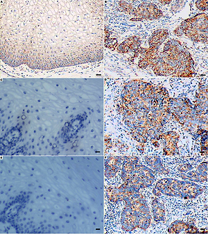
As shown in Figures 1 A, B, PRX3 was extensively expressed in the cytoplasm of normal cervical epithelia, but the staining intensity was much weaker as compared to that of cervical cancer cells. The immunostaining score was 3.6 ±0.9 in normal cervical epithelia and 10.6 ±1.4 in cancer cells (F = 2.819, p = 0.000). In addition, the E6 of HPV16 or HPV18 was expressed in nearly all cervical cancer cells as illustrated in Figures 1 D, F respectively. The staining intensity for HPV18 E6 was slightly weaker than that of HPV16 E6, but the immunostaining scores did not reach a significant difference between the two subtypes. Pearson’s correlation analysis showed a positive association between the expression of PRX3 and HPV16 E6 (r = 0.532, p = 0.031) or HPV18 E6 (r = 0.515, p = 0.041). The staining scores of either PRX3 or HPV16 E6 were not associated with clinicopathologic parameters including stage, cell grade, and lymph nodes (Table II).
Figure 1
Representative results of immunohistochemistry for PRX3 and E6 of HPV16 or HPV18. A – PRX3 was extensively expressed in normal cervical epithelia. B – Intensive staining was observed in cervical cancer cells. C – Negative control of HPV16 E6 staining in normal cervical epithelia. D – The E6 of HPV16 was expressed in nearly all cervical cancer cells. E – Negative control of HPV18 E6 staining in normal cervical epithelia. F – The E6 of HPV18 was expressed in nearly all cervical cancer cells but the intensity was weaker than that of HPV16 E6. Scale bar = 50 μm; original magnification × 200
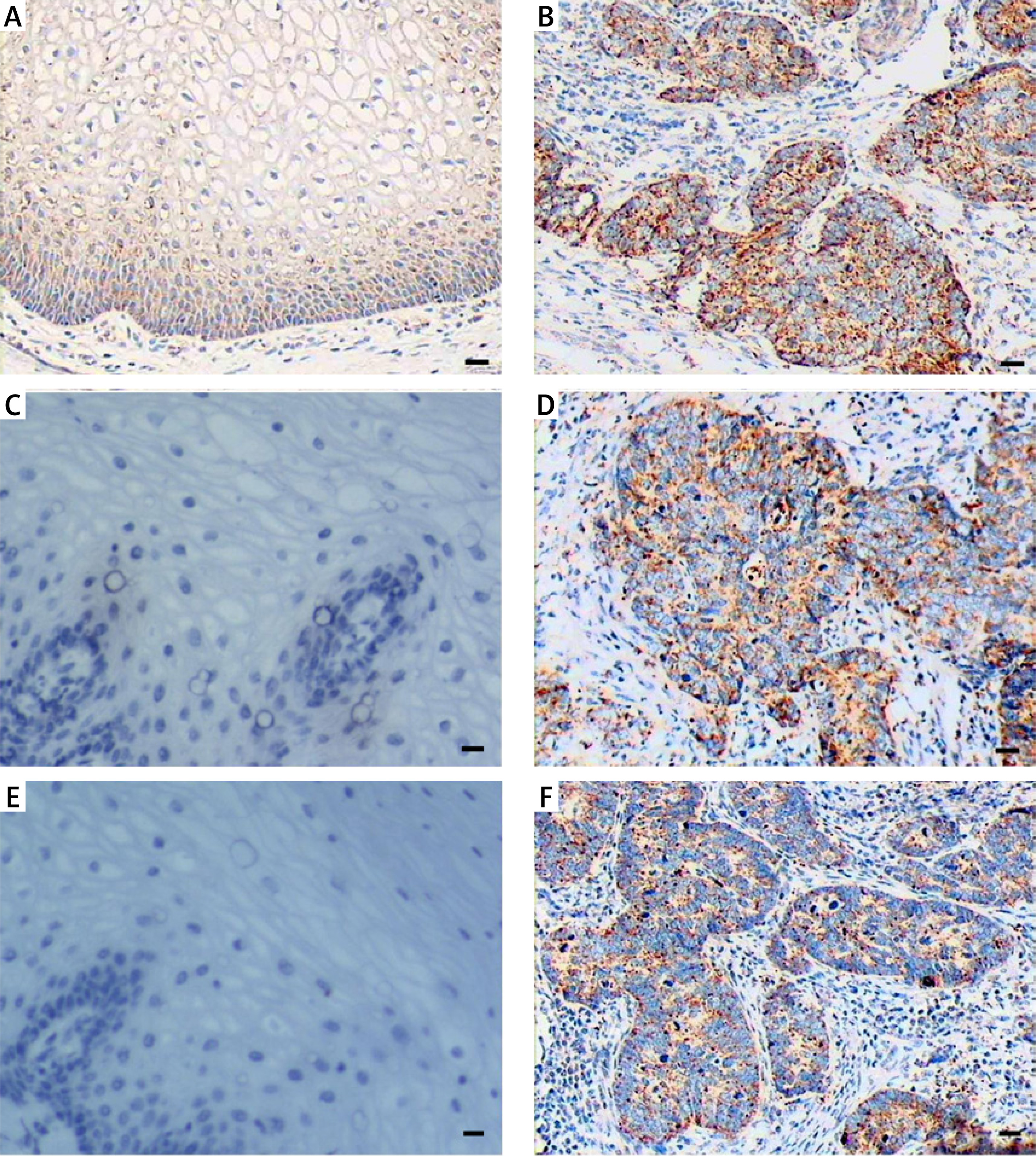
Table II
Association of staining scores of PRX3 or HPV16 E6 with clinicopathologic parameters
Expression of PRX3 and E6 of HPV16/HPV18 in cervical cancer cell lines
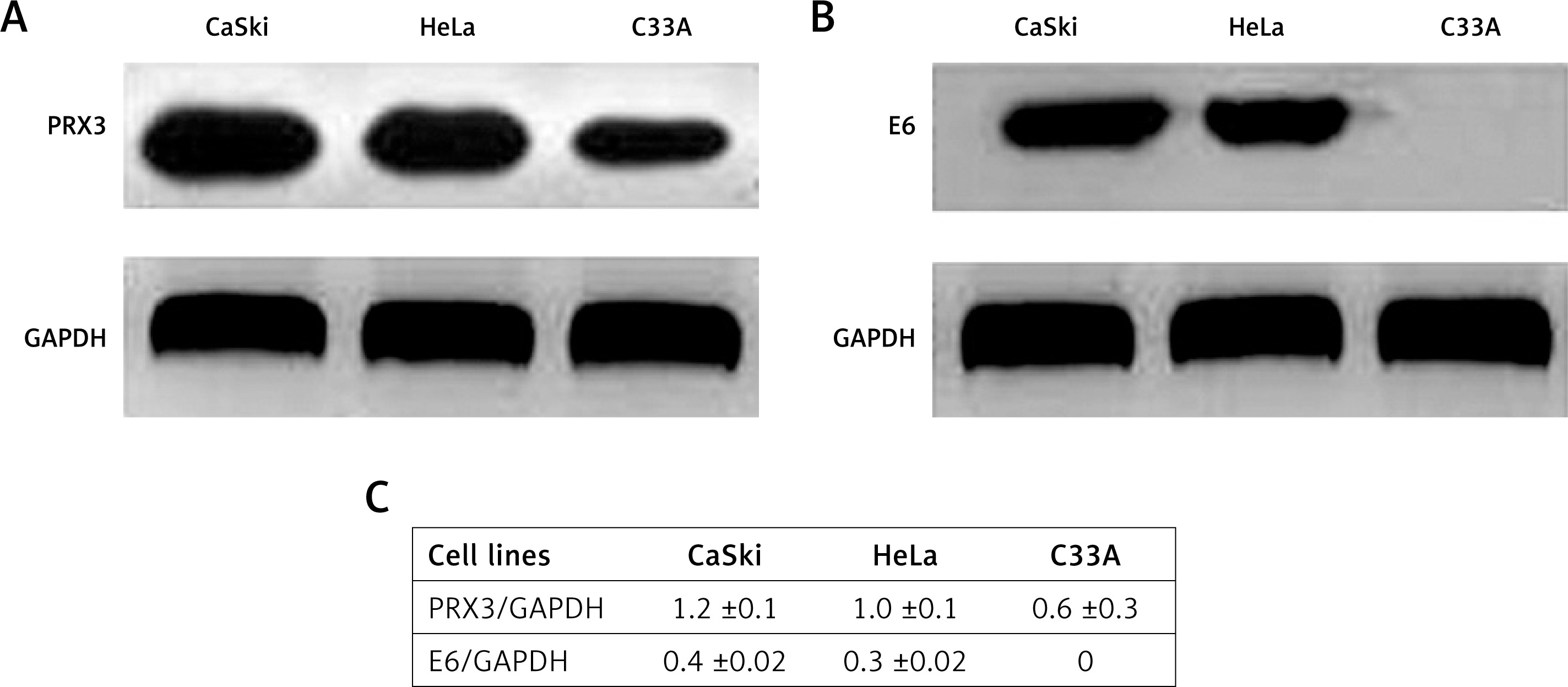
To confirm the association between HPV amplification and PRX3 expression, we detected the expression of PRX3 and E6 of HPV16/HPV18 in cervical cancer cell lines, including CaSki, HeLa, and C33A. As indicated in Figures 2 A, B, the expression of PRX3 was significantly increased in HR-HPV-containing cells (CaSki and HeLa) as compared to that in HPV-negative C33A cells. Figure 2 C shows the mean IOD values of PRX3 and HPV16/HPV18 E6 in each cancer cell line. In addition, the expression of PRX3 was positively associated with that of E6 of HPV16 (r = 0.651, p = 0.027) or HPV18 (r = 0.560, p = 0.031).
Figure 2
Expression of PRX3 and E6 as determined in cervical cancer cell lines. A – WB analysis for PRX3 indicated that the signals of CaSki and HeLa cells were stronger as compared to that of C33A cells. B – WB analysis for E6 of HPV16 and HPV18 in CaSki and HeLa cell lines, respectively. C – Analysis of variance showed higher expression of PRX3 in CaSki and HeLa cells than in C33A cells. CaSki vs. C33A: F = 3.480, p = 0.002; HeLa vs. C33A: F = 2.901, p = 0.006
Discussion
In the present study, we demonstrated upregulation of PRX3 by HR-HPV in cervical cancer cells, suggesting an important role of PRX3 in the response to HR-HPV induced oxidative stress. Since the serum PRX3 was increased in cervical cancer patients, we suppose that serum PRX3 might be a potential indicator of HR-HPV amplification.
The production of ROS is increased because of active proliferation of cervical cancer cells [10]. Another factor that contributes to the ROS production might be the amplification of HR-HPV [11], which induces oxidative stress, as indicated in the present study. Increased ROS production and development of oxidative stress lead to oxidation of proteins and peroxidation of membrane lipids [12]. Based on a review of previous literature, we suppose that the upregulation of PRX3 might be transcriptional regulation by genes including c-Myc, nuclear factor E2-related factor 2 (Nrf2), forkhead box transcription factor FOXO3A or FOXM1, and small non-coding microRNAs (miRNAs). According to previous reports, infection with HPV16 is closely associated with c-Myc amplification [13, 14], which plays an important role in cervical cancer development and progression [15]. Since PRX3 is a downstream target of c-Myc [3], the interaction of c-Myc and PRX3 might be a candidate pathway for the upregulation of PRX3. In lung squamous cell carcinoma, PRX3 is overexpressed in an Nrf2 dependent manner, which indicates a potentially important role of the Nrf2-PRX3 pathway in the tumor [16]. Chiribau et al. reported that FOXO3A mediated PRX3 expression, resulting in resistance to oxidative stress in human cardiac fibroblasts [17], which was confirmed by other researchers in pheochromocytoma cells, cardiac cells, and endothelial cells [18–20]. In addition, a study conducted by Cunniff et al. reported the co-existence of cytoplasmic FOXM1 and mitochondrial PRX3 in mesothelioma cells [21]. In endometrial cancer stem cells, increased expression of PRX3 was induced by FOXM1 for the maintenance of mitochondrial function and cell survival [22]. MicroRNAs (miRNAs) are a class of small non-protein-coding molecules. Up to now, miR-23b and miR-383 have been reported to control cell growth and cancer progression by negatively regulating PRX3 expression in several types of solid tumors [23, 24].
The serum levels of PRX3 in cancer patients have been investigated in recent years. For example, serum PRX3 is significantly higher in patients with hepatocellular carcinoma and could be used as a biomarker for the diagnosis and prognosis of the disease [25, 26]. However, the alteration of serum PRX3 in cervical cancer patients has not been reported up to now. Here we presented an increase of serum PRX3 in cervical cancer patients, especially in patients with cancer-positive lymph nodes. The serum PRX3 had no significant association with either the staining scores of PRX3 in cancer tissues or clinicopathologic features of patients, unlike in patients with hepatocellular carcinoma [25, 26]. The biological characteristics of cervical cancer are quite different from those of hepatocellular carcinoma. Cancer cells in the cervix grow locally and invade mainly the surrounding tissues or organs. Patients in the present study were at stages I and II, in which distant metastasis was absent. In contrast, the liver is an organ with abundant blood supply and blood-based metastasis is a common occurrence in hepatocellular carcinoma. Therefore, the oxidative stress in liver cancer can be reflected in circulating blood. In the present study, we did not find a significant association between serum PRX3 and biochemical parameters such as FBS and FINS. The present results were consistent with our previous reports that PRX3 responded mainly to oxidative stress induced by a rapid increase of glucose and insulin [27, 28].
In conclusion, we demonstrated the link between PRX3 and HR-HPV in cervical cancer cells. Upregulation of PRX3 reflects a positive response of PRX3 to the oxidative stress in cervical cancer cells induced by HR-HPV amplification and cell growth. Further studies are needed to elucidate the underlying mechanism for the interaction between PRX3 and HR-HPV.

