

Introduction
The brain is a sensitive and adapting organ with high metabolic activity and oxygen consumption [1–3]. Following brain ischaemia, subsequent reperfusion may lead to neurological deterioration with impaired redox balance, neuro-inflammation, and altered blood-brain barrier (BBB) permeability [4, 5]. Thus, it is important to understand the mitochondrial responses after impaired oxygen delivery in cerebral I/R injury.
PGC-1α (PPAR (peroxisome proliferator-activated receptor)-γ coactivator-1α) along with nuclear respiratory factor-1, nuclear respiratory factor-2, and sirtuin 1 (SIRT1) regulate mitochondrial function [6, 7]. PGC-1α regulates cell survival by targeting p53 activation under metabolic stress [8]. Prolonged oxidative stress leads to mitochondrial dysfunction. Potential regulators of mitochondrial dysfunction are PGC-1α, PINK1, Nrf-2, and antioxidant genes [9–11]. Nrf-2, an oxidative stress-sensitive transcription factor, binds to the antioxidant response element (ARE) and increases expression of antioxidant enzymes [12, 13]. Previous investigations with acute kidney injury and liver ischaemia reperfusion injury demonstrated the importance of PGC-1α expression [14–16]. In this study we aimed to explore the functional role of PGC-1α after cerebral I/R injury and its relationship with Nrf-2-dependent cellular protection. In the current setting, we compared the cerebral aftereffects of wild-type mice and PGC-1α-overexpressed mice following cerebral ischaemia/reperfusion injury.
Material and methods
Animals and the focal cerebral I/R mouse model
The experiments were performed as per the regulations of American Animal Protection Legislation. The Institutional Animal Care and Use Committee of Gannan Medical University approved the use of the animals and the complete protocol used for the development of middle cerebral artery occlusion (MCAO). Wild-type (000664 C57BL/6J) and transgenic PGC-1α C57BL/6-Tg(Ckm-Ppargc1a)31Brsp/J mice at the age of 8-10 weeks were obtained from the Jackson Laboratory, ME 04609, United States. PGC-1α complementary DNA was placed downstream of a 6.5-kilobase (kb) muscle creatine kinase promoter sequence. PGC-1α transgenic mice were generated by standard DNA microinjection and identified by PCR-based genotyping. For gene expression analyses, muscles were dissected from 3-month-old wild-type or transgenic C57BL/6 mice. All experiments were performed in transgenic line 31 except where indicated. All the animals were maintained in the controlled conditions of 12/12-hr light-dark cycle, temperature 23±2°C, and food and water ad libitum. The MCAO model was developed as described previously [17, 18]. The mice were anaesthetised by 1.5–2% isoflurane. A filament coated with silicon resin was initiated into the left carotid artery to induce ischaemia. Reperfusion was attained by removing the filament after 1 h of occlusion. Successful I/R surgery was demonstrated and recorded by the measurement of cerebral blood flow by using laser-Doppler flowmetry.
Infarct volumes and neurological scoring
To analyse the infarct volume the brains were sectioned after I/R injury and stained with 2% TTC (2,3,5-triphenyltetrazolium chloride) for 15 min. Followed by which the sections were fixed with 10% formalin neutral buffer solution (pH 7.4), and the infract lesions were measured. The results were calculated as compared to total cerebral volume. The neurological score of WT and PGC-1α overexpression mice was performed after I/R injury using behavioural tests. The deficit scores were set as follows: 0 – no deficits; 1 – difficulty in fully extending the contralateral forelimb; 2 – unable to extend the contralateral forelimb; 3 – mild circling to the contralateral side; 4 – severe circling; 5 – falling to the contralateral side. The higher scores represent more severe motion impairment.
ROS
The ROS in the tissues were measured using Reactive Oxygen Species (ROS) Fluorometric Assay Kit, E-BC-K138-F, Elabscience. The tissues (without the fibre, fat, and blood vessels) were washed with reagent 3 (buffer solution) to remove the blood and other contaminants. The minced tissues in pre-cooled reagent 3 working solution were enzymatically digested at 37˚C in a water bath for 30 min, then the reaction was stopped and the mixture was filtered using nylon mesh. The collected cells were centrifuged at 600 g for 15 min. The pellets containing the cells were washed with reagent 3 working solution. The cell suspension was used for ROS determination using 10 µM DCFH-DA working solution (reagent 1). After incubation at 37˚C for 30 min, the cells were centrifuged at 1200 g for 15 min. The cells were further washed using reagent 3 working solution three times and read at excitation wavelength 500 nm and emission wavelength 525 nm. The results were expressed as ROS percentage compared to controls.
Western blot analysis
The total protein from the cerebral ischaemic hemispheres and neuro-2A cells were extracted after the treatment schedule. Equal protein loading (30 µg) of the samples on 10% SDS-PAGE gels were performed. The polyvinylidene difluoride (PVDF) membrane transferred proteins were blocked with blocking buffer. The blots were washed with TBST and incubated with primary antibodies (overnight, 4˚C). The blots were washed with TBST and incubated with secondary antibodies (1 h RT). The blots were detected using an ECL detection kit, and densitometric analysis was performed and normalised with β-actin.
Inflammatory cytokine expressions: ELISA
Inflammatory cytokine expressions were determined using the ELISA kit (Mouse IL-1β ELISA Kit [ab100704], Abcam; Mouse IL-6 ELISA Kit [ab100712], Mouse TNF-α ELISA Kit [ab100747]). The relative interleukin levels were expressed in comparison with controls.
OGD/R
Neuro-2A cells were transfected with control scramble or si-Nrf-2 for 72 h and used for OGD/R treatment (1% O2) and allowed for 1 hr reoxygenation in glucose/FBS medium. After treatment, the cells were lysed (tris, EDTA, β-mercaptoethanol, protease inhibitor, and phosphatase inhibitor) for total protein isolation and used for protein expression analysis using western blot.
Statistical analysis
The results comprise three independent experiments in triplicate; the data are represented as mean ± standard deviation. The statistical analysis utilised one-way ANOVA with Dunnett’s or Tukey’s multiple comparison test and GraphPad software. ***p < 0.001 compared to sham mice. +++p < 0.001 compared to ischaemia reperfusion mice.
Results
Cerebral I/R injury down-regulated PGC-1α expression and induced apoptosis
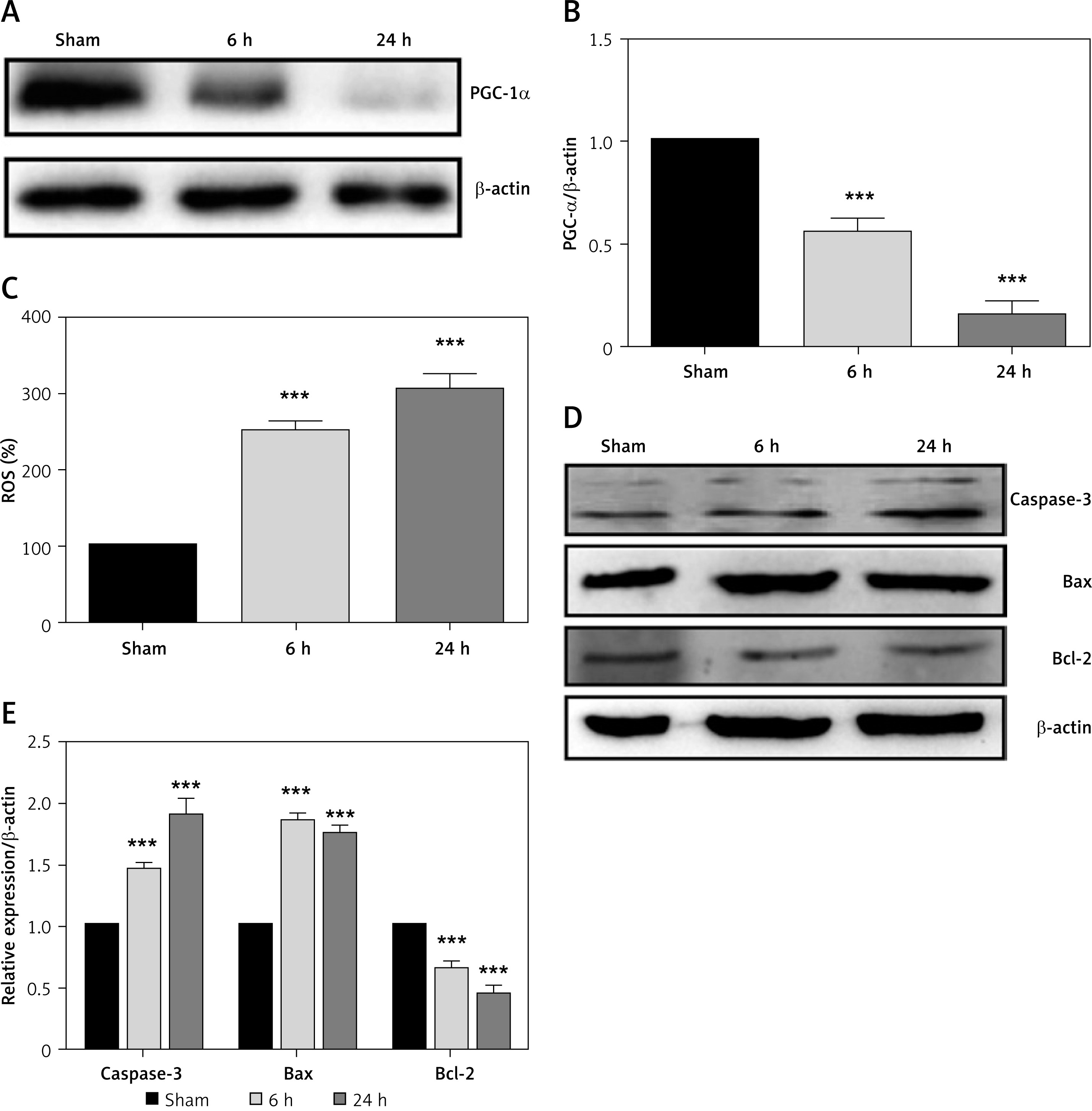
Compared to sham mice, PGC-1α expression is down-regulated in cerebral I/R injury (6 h and 24 h) mice (Figures 1 A, B). Figure 1 C shows elevated ROS content (250% at 6 h and 290% at 24 h) in cerebral I/R injury mice as compared to control mice (100%). Furthermore, apoptosis markers (caspase-3, Bax) are up-regulated and anti-apoptosis marker (Bcl-2) down-regulated at 6 h and 24 h cerebral I/R injury (Figures 1 D, E).
Figure 1
Cerebral I/R injury down-regulates PGC- 1α expression and induces apoptosis. A – Western blot of PGC-1α expression during cerebral I/R injury (6 and 24 h) compared to sham mice. B – Densitometry of PGC-1α expression/β-actin (ImageJ). C – Reactive oxygen species levels in mice 6 and 24 h after cerebral I/R injury. D, E – Western blot and densitometry of caspase -3, Bax, Bcl-2 expression 6 and 24 h after cerebral I/R injury
PGC-1α overexpression in mice reduces brain damage during cerebral I/R injury
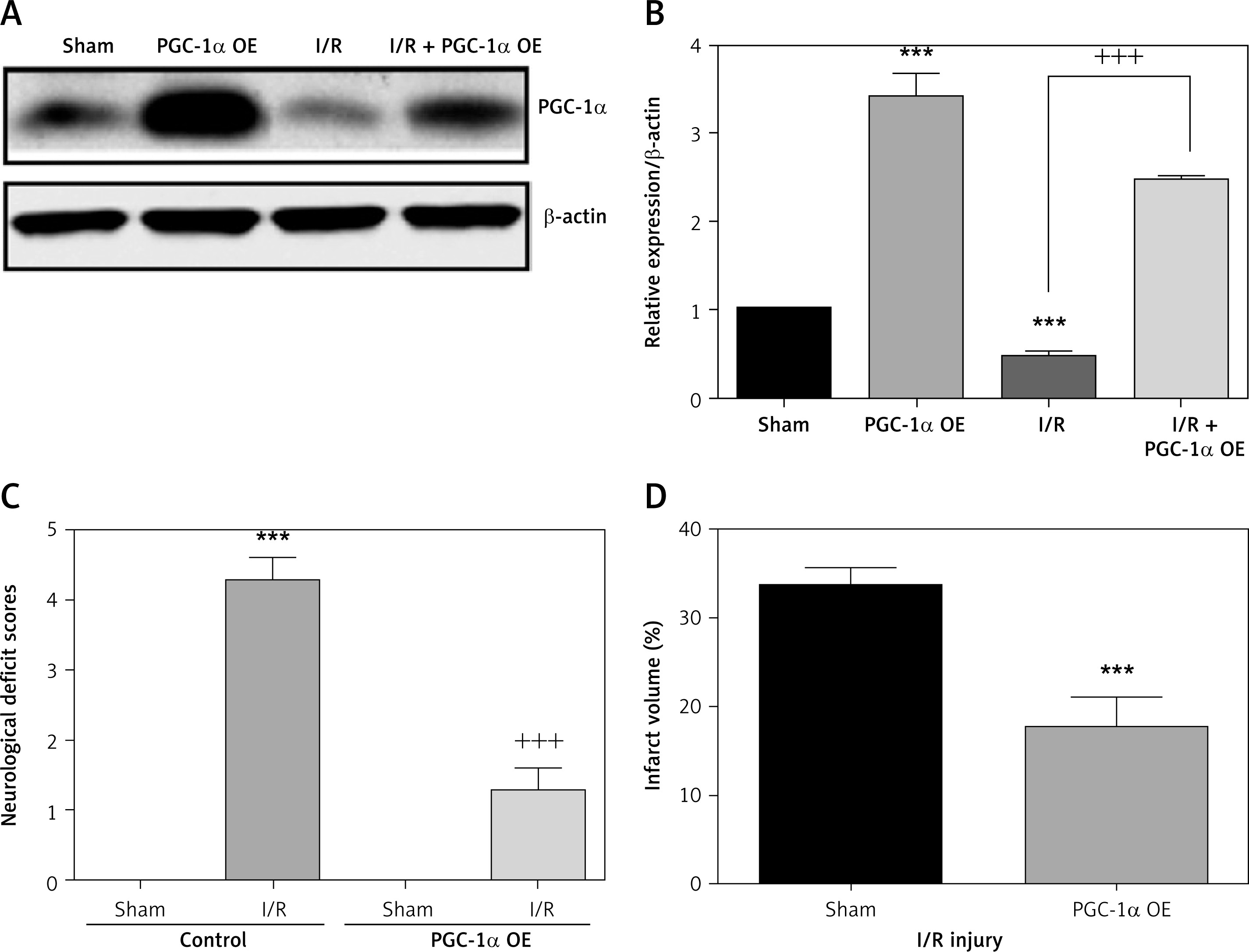
The expression of PGC-1α was up-regulated in PGC-1α overexpression mice compared to wild-type mice. Figure 2 C shows that I/R injury mice had neurological deficit scores of 4 compared to sham mice (deficit score- 0). However, PGC-1α-overexpressed mice showed a reduced neurological deficit score of 1, compared to that of wild-type I/R mice. Figure 2 D demonstrates that PGC-1α-overexpressed mice (15%) had reduced cerebral infarct volume compared to control mice (33%) after I/R injury (Figure 2 D).
PGC-1α overexpression in mice reduces inflammation and cytokine expression
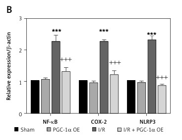
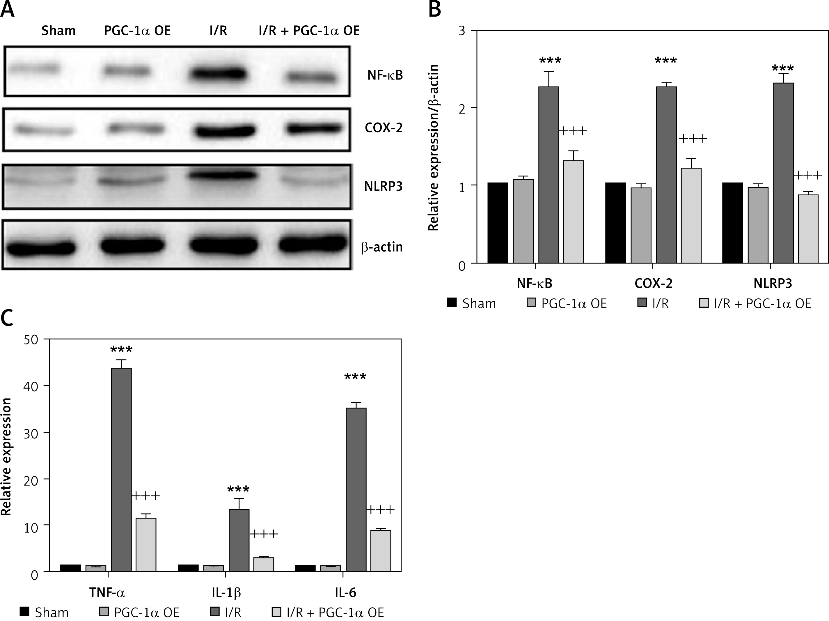
Cerebral I/R injury up-regulated inflammation through NF-κB, COX-2, NLRP3, and inflammatory cytokine expressions compared to sham mice. However, inflammatory protein (NF-κB, COX-2, NLRP3) and inflammatory cytokine (TNF-α, IL-1β, IL-6) expressions were down-regulated in PGC-1α-overexpressed mice compared to wild-type mice after I/R injury (Figures 3 A–C).
Figure 3
PGC-1α-overexpression mice suppress inflammation. A, B – Effect of PGC-1α overexpression on inflammatory protein NF-kB, COX-2, and NLRP3 expressions compared to sham rats during I/R injury. C – Relative expression of cytokines (TNF-α, IL-1β, IL-6) in WT and PGC-1α-overexpression mice after I/R injury
PGC-1α induced Nrf-2 pathway in cerebral I/R injury mice and in an in vitro OGD/R model
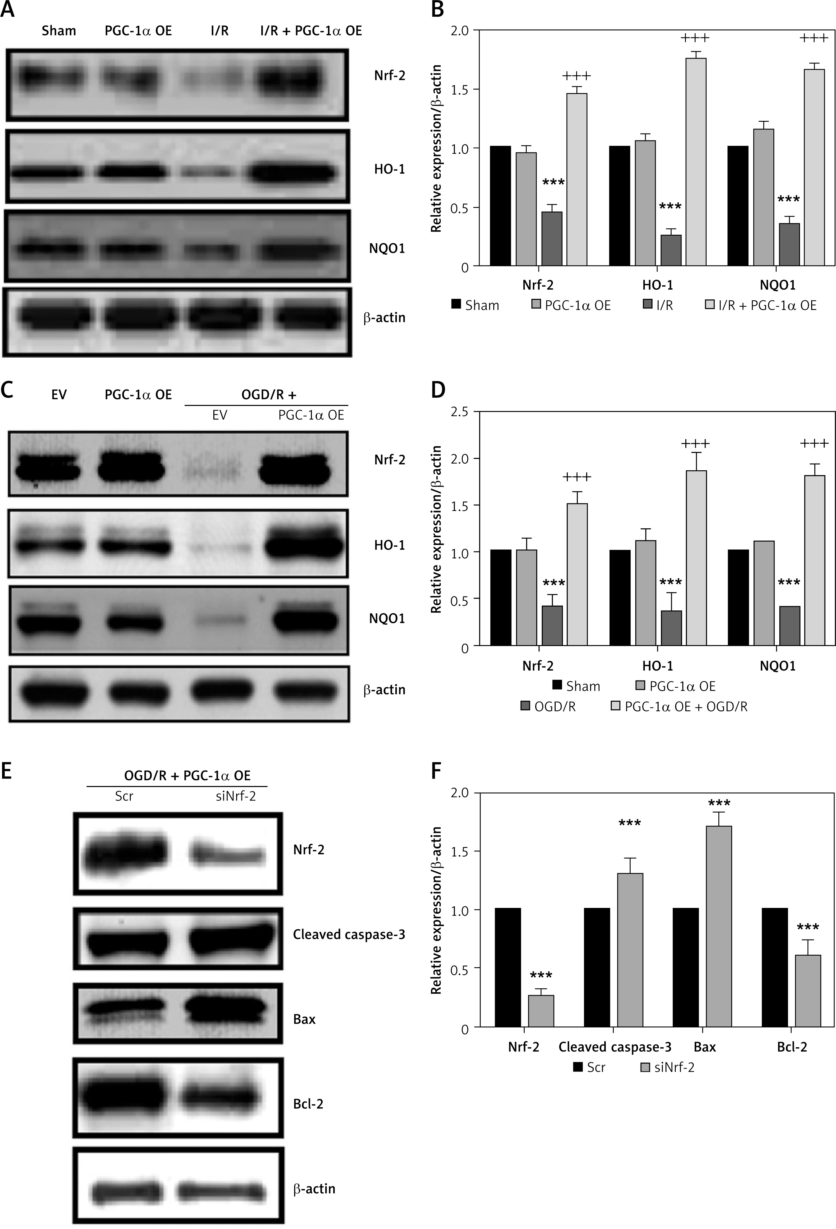
We determined Nrf-2 and its related protein expressions during I/R injury in PGC-1α overexpression mice. The results showed up-regulated Nrf-2, HO-1 and NQO1 protein expressions in PGC-1α-overexpressed mice compared to WT mice after I/R injury (Figures 4 A, B). The results are consistent with the in vitro OGD/R model, showing up-regulated expressions of Nrf-2, HO-1, and NQO1 proteins in PGC-1α overexpression cells compared to controls (Figures 4 C, D). Furthermore, the direct association of PGC-1α and Nrf-2-mediated protection was confirmed through si-Nrf-2 studies. The Nrf-2 knockdown during OGD/R up-regulated apoptosis through increased caspase-3 and Bax expressions with suppression of Bcl-2 expression compared to WT mice (Figures 4 E, F).
Figure 4
PGC-1α regulates Nrf-2 expression and apoptosis. A, B – Western blot and densitometry of Nrf-2, HO-1, NQO1 expressions in WT and PGC-1α-overexpression mice after I/R injury. C, D – Western blot and densitometry of Nrf-2, HO-1, NQO1 expressions in WT and PGC-1α overexpression cells in the OGD/R model. E – Western blot results of apoptosis markers regulated by PGC-1α/Nrf-2. F – Densitometry analysis of apoptosis proteins regulated by PGC-1α/Nrf-2
Discussion
The study demonstrates that PGC-1α reduces cerebral damage after I/R injury through suppression of inflammation and apoptosis.
Cerebral ischaemia leads to oxidative stress with inflammatory responses and cellular apoptosis [19]. Cerebral injury induced a redox imbalance followed at 6 and 24 h with increased apoptosis. Importantly, cerebral I/R injury down-regulated the PGC-1α expression. PGC-1α, a key mitochondrial biogenesis protein, regulates cellular energy needs and promotes cytoprotection [20]. Regulation of mitochondrial biogenesis by PGC-1α in neuronal cells has been previously demonstrated [21]. Other studies reveal their role in maintaining the redox balance and inflammatory response [22, 23]. Moreover, AMPK mediates PGC-1α expression [24]. We developed PGC-1α-overexpression mice to understand the protective role of PGC-1α after cerebral I/R injury. Improved brain infarct volume and fewer neurological deficits were observed after I/R injury.
Progression of sustained oxidative stress leads to neuro-inflammation. Free radicals released accordingly activate NF-κB and its target genes, thus magnifying the overall disease status [25, 26]. Cerebral I/R injury showed the up-regulation of inflammatory cytokines, NF-κB, COX-2, and NLRP3 expressions. The PGC-1α-overexpressed mice down-regulated the inflammatory protein expressions. Importantly, PGC-1α-overexpressed mice regulated the key transcription factor Nrf-2 and its downstream proteins, HO-1 and NQO1 expressions. Increased ROS and stress conditions regulate Nrf-2 activation through SIRT1-PGC-1α signalling [27]. Nrf-2 is mainly associated with Keap1 (Kelch-like ECH associated protein 1) protein in the cytoplasmic compartment [28, 29]. Under oxidative stress, Nrf-2 translocates into the nucleus and binds to antioxidant response elements (ARE) to activate cytoprotective enzymes (GSH, heme oxygenase 1, NQO-1, GPX, GST) [30–32]. The present results showed that PGC-1α regulates apoptosis through the Nrf-2 pathway. Consistent with our findings, antagonism between PGC-1α and inflammation have been reported. For instance, in multiple sclerosis, PGC-1α suppresses IL-6 and chemokine (C-C motif) ligand and thereby enhances mitochondrial antioxidant status [33]. Similarly, PGC-1α suppresses muscle inflammation in Duchenne muscular dystrophy and denervation-induced muscle atrophy [34, 35].
In conclusion, in mice PGC-1α suppresses ROS, inflammatory responses, and apoptosis through Nrf-2 activation during cerebral I/R injury. Thus, PGC-1α could be a therapeutic target for protection against I/R injury and its associated inflammation.

