Introduction
Oxidative stress generated by insecticides causes the formation of reactive oxygen species (ROS) in living organisms and accordingly induces changes in antioxidant systems [1, 2]. Oxidative stress, which may cause an imbalance in the oxidant-antioxidant status, regulates inflammatory mediators and inhibits neutrophil infiltration [3], is effective in insecticide-induced tissue damage.
Neonicotinoids are known to have an important role as synthetic neuroactive insecticides and many of them are used in crops and livestock for pest control [4]. Imidacloprid (IMC), one of the neonicotinoid insecticides, is widely used to eliminate pests in agriculture [5]. Residues of the IMC can be encountered in the food chain and therefore it is important to investigate the side effects of this insecticide. Previous studies have reported adverse effects of IMC in mammals [6–8]. Furthermore, side effects of IMC on reproductive ability have been reported in parenteral and offspring rats [9].
Vitamin D (VD) is a steroid hormone that is frequently incepted from dietary sources. In addition, it is also synthesized in the skin following exposure to UVB light [10, 11]. The balance between oxidants and antioxidants causes oxidative stress when disrupted in favor of oxidants. Vitamin D has both a pro- and anti-oxidative effect potential. Vitamin D deficiency is quite common worldwide and affects people of all ages [12]. The association of vitamin D deficiency with coronary artery disease has been demonstrated [13–15]. and obese people are at greater risk [16]. The male reproductive system includes widely distributed vitamin D receptors (VDR) [17]. The expression of VDR and VD metabolizing enzymes in the male reproductive system indicates the function of the testis in VD production and regulation [18]. Vitamin D has important functions in the male reproductive system and VD deficiency has been reported to have significant effects on the male reproductive system [19]. Vitamin D deficiency (VD) and intake of high-fat diets may cause morphological and physiological changes related to infertility in the testis [20].
Irisin is a recently discovered peptide hormone and is synthesized in rats during exercise [21]. Moreover, it was determined that the level of irisin increased during oxidative stress status [22]. The peptide is found in skeletal and cardiac muscle tissue as well as brain, skin, spleen, testis, pancreas and stomach tissues [23]. Irisin regulates glucose and lipid homeostasis. Anti-apoptotic, anti-inflammatory and anti-oxidative properties of irisin have attracted the attention of scientists, since these properties are associated with the stages of many diseases such as cancer, myocardial infarction, lung injury, kidney diseases, atherosclerosis, obesity, liver diseases and type 2 diabetes [24]. Hence, it is important to find substances that could reverse the degenerative effects of IMC in mammalian tissues.
The aim of the present study is to examine the protective effects of VD in IMC-induced testis injury and to determine its possible effect on irisin immunoreactivity.
Material and methods
Animals, diets and experimental protocols
A total of 32 male Wistar albino rats aged 8–10 weeks (200–220 g), supplied by Adiyaman University Experimental Animal Production and Research Center, were divided into five groups: the control group (n = 6), corn oil (CO) group (n = 6), VD group (n = 6), IMC group (n = 7), and IMC + VD group (n = 7). The animals were maintained and fed ad libitum with standard rodent pellet diet and water in an ambient environment with 1/2 daylight/1/2 day dark cycle at 25 ±3°C for 15 days for adaptation. The animals in the control group were maintained on a standard rodent pellet diet and water ad libitum without any treatment during the 8-week experimental period. The animals in the CO group were administered a daily dose of 2 ml of CO by oral gavage for 8 weeks. The VD group was given a dose of VD 200 IU/day via an oral dropper for 8 weeks. The IMC group was administered orally with 20 mg/kg IMC dissolved in CO (a total of 2 ml volume) per day for 8 weeks, while the IMC + VD group was administered orally 20 mg/kg IMC dissolved in CO (a total of 2 ml volume) and 200 IU/day VD via an oral dropper for 8 weeks. Testis tissues obtained from rats, which were anesthetized with ketamine and xylazine (75 and 10 mg/kg, respectively), were fixed in 10% buffered formalin. Some fresh tissue samples were stored at –20°C for total antioxidant status (TAS) and total oxidant status (TOS) measurements. This animal study was approved by the Ethical Committee of Experimental Animals of Firat University (Protocol number: 2017/67).
Biochemical methods
The testis tissues were washed with cold PBS buffer, homogenized in ice with 0.1 M phosphate buffer (1/10 ratio at pH 7.4) and centrifuged at 1800 rpm for 5 min for evaluating TAS and TOS levels. Tissue TAS [25] and TOS [26] levels were measured by using TAS and TOS Assay Test Kits (Rel Assay Diagnostic, Gaziantep, Turkey) with an autoanalyzer (Olympus AU2700) to assess the degree of oxidant damage.
Histopathological method
Formalin fixed tissue samples were processed and blocked with paraffin, and then sectioned into 5-μm sections and stained with hematoxylin and eosin (HE). These slides were blindly examined under a light microscope (Leica DM500 microscope –Leica DFC295).
Immunohistochemical method
Briefly, 4–6 µm thick sections taken from paraffin blocks were transferred on poly-L-lysine coated slides. Deparaffinized and re-hydrated sections were boiled in a microwave oven and placed at room temperature for about 20 min and transferred into hydrogen peroxide for blocking endogenous peroxides. Subsequently the sections were washed with PBS for 3 × 5 min and nonspecific immunoglobulins were blocked with Ultra V Block solution for 5 min. In the next step, irisin primary antibody (Irisin Rabbit Polyclonal H-067-17, Phoenix Pharmaceuticals, Inc., California, USA) (at a ratio of 1/200) was applied on the sections for 60 min. Subsequently, an anti-mouse/rabbit IgG secondary antibody was applied for 30 min at room temperature. These sections were next washed with PBS and were incubated with streptavidin peroxidase. After these processes the sections were visualized with 3-amino-9-ethylcarbazole (AEC) and the background was colorized with Mayer’s Hematoxylin. The sections were covered with aqua medium. Manufacturers’ proposed methods were applied for positive and negative control stains. The stained sections were stored at room temperature in the dark until examination. Histopathological scores for the immunoreactivity were counted using the prevalence interval (0.1: < 25%, 0.4: 26–50%, 0.6: 51–75%, 0.9: 76–100%) and intensity (0: absent, +0.5: very low, +1: low, +2: moderate, +3: severe). The histopathological score was determined as prevalence x intensity.
Statistical analysis
Statistical evaluations were performed by SPSS software, version 15.0. The one-sample Kolmogorov-Smirnov test was used to determine whether the data were distributed normally. One-way analysis of variance was used to compare TAS, TOS and immunoreactivity variables. The differences between the variables of groups were determined using the Tukey HSD multiple comparison test. The obtained results were exhibited as mean ± standard deviation (SD). The significance level was considered as p < 0.05.
Results
Biochemical results
In biochemical analysis, similar TAS (Table I) and TOS (Table II) levels were seen in the control, VD, and CO groups. A statistically significantly lower TAS level and a statistically significantly higher TOS level were detected in the IMC group (p < 0.05) compared to the control group. Compared to the IMC group, the higher TAS level and lower TOS level in the IMC + VD group were insignificant (p < 0.05).
Histopathological results
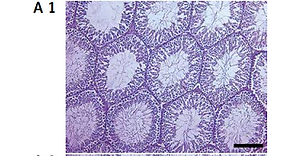
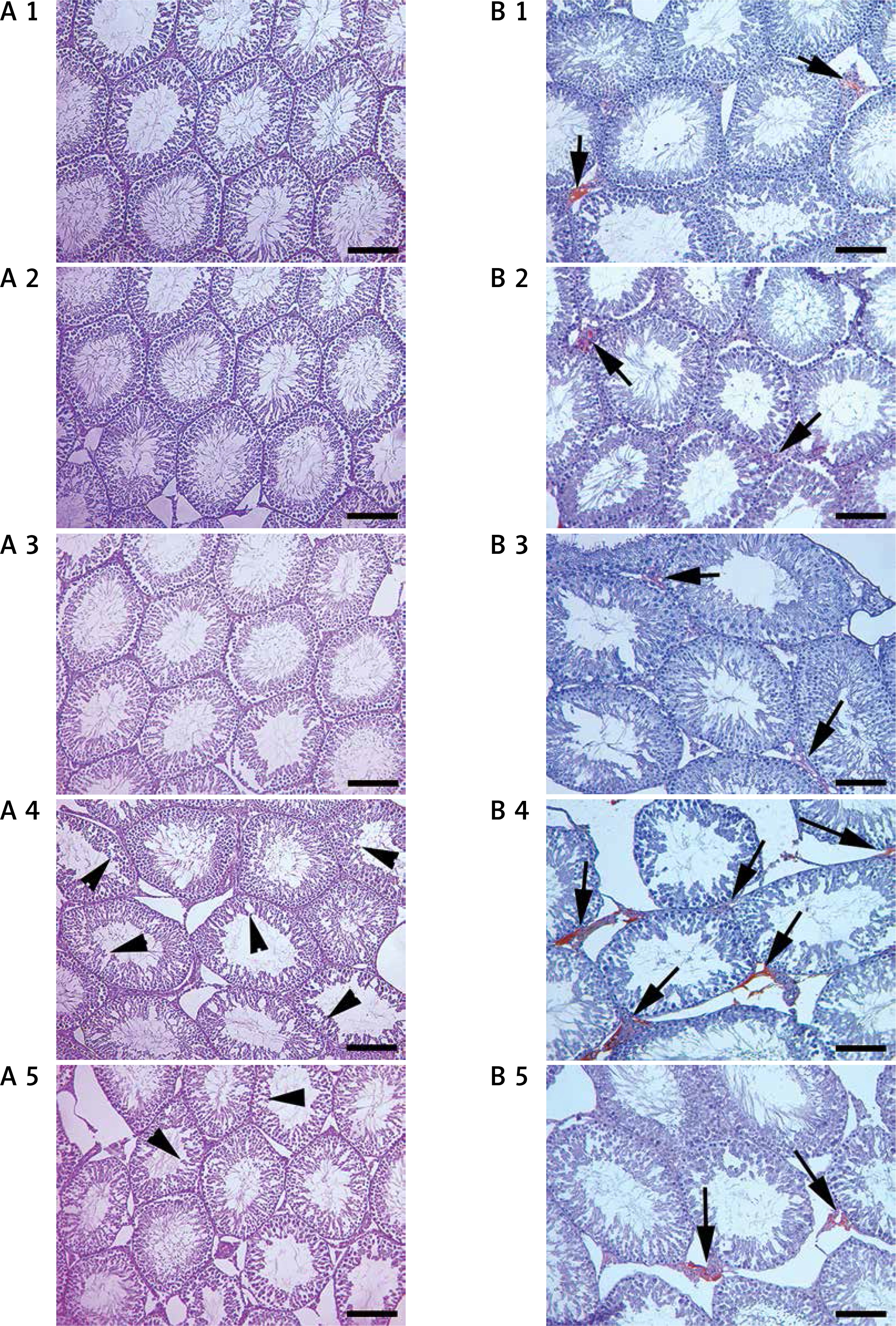
The testes of the control group (Figure 1. A 1), VD group (Figure 1. A 2) and CO (Figure 1. A 3) group had normal histological structures. The testes of the IMC group (Figure 1. A 4) had degenerative changes in the seminiferous tubule epithelium and interstitial edema compared to the control group. In comparison to the IMC group, degenerative changes in the seminiferous tubule epithelium and interstitial edema were at a lower level in the IMC + VD group (Figure 1. A 5).
Figure 1
A – Testis histopathology of the control, VD, CO, IMC, and IMC + VD groups. Scale bar is 100 µm in the HE stained figures. Arrowheads indicate degeneration. A 1 – Control group: normal histological view of testis. A 2 – VD group: normal histological view of testis. A 3 – CO group: normal histological view of testis. A 4 – IMC group: severe degeneration, edema, and desquamation in testis. A 5 – IMC + VD group: light degeneration and edema in testis. B – Irisin immunohistochemistry of the control, VD, CO, IMC, and IMC + VD groups, respectively. Scale bar is 200 µm in the irisin stained figures. Arrows indicate immunopositivity. B 1 – Control group: slight interstitial positivity. B 2 – VD group: slight interstitial positivity. B 3 – CO group: slight interstitial positivity. B 4 – IMC group: severe interstitial positivity. B 5 – IMC + VD group: slight interstitial positivity
Immunohistochemical results
Immunohistochemically similar irisin immunoreactivity (Table III) was observed in the interstitial space of testis tissue in the control group (Figure 1. B 1), VD group (Figure 1. B 2) and CO group (Figure 1. B 3). Irisin immunoreactivity was significantly higher in the IMC group (Figure 1. B 4) compared to the control group (p < 0.05). Conversely, irisin immunoreactivity was insignificantly lower in the IMC + VD group (Figure 1. B 5) compared to the IMC group (p > 0.05).
Discussion
Organophosphate compounds are commonly known as pesticides and/or insecticides. Insecticides and pesticides induce toxic effects causing atrophy in the testis of rats [27]. ROS are effective in the pathogenesis of testicular damage induced by organophosphate compounds in rat models [28] and ROS is associated with the defective morphology of sperm cells [29].
IMC-like insecticides are reproductive toxic substances in male animals, causing disruption of testicular tissues, spermatogenic cells and decreased sperm motility [30] and may induce necrosis in the seminiferous tubules along with histopathological alterations [27]. In this study, significantly increased TOS and decreased TAS levels, as well as degeneration in the germinal epithelium of seminiferous tubules and interstitial edema, were observed only in the IMC group. Therefore, the negative effect of IMC on testicular tissue of male rats is thought to be related to oxidative stress.
In previous studies, decreased spermatocytes, reduced or lack of spermatogenesis, weak or arrested spermatogenesis in some seminiferous tubules and mild edema in the interstitial space, as well as increased apoptosis of germ cells and fragmentation of seminal DNA, have been reported in IMC-treated rats [31, 32]. In IMC-treated rabbits, it was observed that the seminiferous tubule lumen was empty and interstitial tissue widened probably as a result of the elimination of Leydig cells and in some tubules this space was compressed. Furthermore, vacuolization in the seminiferous tubules and pyknosis in the spermatocytes have been reported [33]. In the present study, histopathological observations confirmed that IMC caused testicular damage including degeneration in the germinal epithelium of seminiferous tubules and edema in the interstitial space in the testis tissue, similar to previous studies [31–34].
It has been reported that VD deficiency disrupted both testicular development and spermatogenesis in mice and that mature seminiferous tubules remain at a lower percentage [34]. VD-deficient rats showed degenerative changes in the germinal epithelium and significantly reduced Leydig cell and sperm numbers, while in the normal VD diet group, only body weight decreased [35]. In the current study, depending on the IMC administration, we observed histopathological damage, decreased TAS and increased TOS levels. In the histopathological examination of the IMC + VD-treated group, it was observed that VD ameliorated testis injuries by decreasing degenerative tubules and interstitial edema. However, VD showed an insignificant effect on the oxidative state. Taking into consideration these positive effects of VD on testis morphology, VD is thought to be useful in decreasing IMC-induced testicular tissue damage.
Depleted antioxidant levels were reported on IMC-treated male rats [31]. Decreased TAS and increased TOS levels were reported in the testis tissue of diabetic rats and VD administration was observed to improve these biochemical parameters by increasing TAS and decreasing TOS levels [36]. In the current study, while a significant decrease in TAS and increase in TOS levels were observed in the IMC group, an insignificant increase in TAS levels and decrease in TOS levels were detected in the IMC + VD group.
Irisin is a peptide that has been studied with increasing interest recently. However, its importance is still not fully understood. The localization of this peptide has been determined in many tissues [37–39]. Gur et al. [40] determined that irisin is located in the cytoplasm of the cells of all examined tissues including testis. Furthermore, irisin immunoreactivity in seminiferous tubules, fetal spermatogenic cells, ductus epididymis in fetal human epididymis, Leydig cells in fetal and adult testis has been observed [23]. In our study, however, irisin immunoreactivity was observed only in the interstitial area. Additionally, irisin is thought to be associated with the antioxidant mechanism. It has been reported that irisin might protect cells from ROS-induced severe cellular oxidative damage via the activation of antioxidative mechanisms [41]. In another study, it was reported that hyper- and hypothyroidism are associated with up-regulation of serum irisin in male rats, probably as a reaction to oxidative damage or myopathy observed under both conditions [42]. Irisin levels have been observed to be increased in type 1 diabetes [39] and in hypothyroid patients [37]. In the current study, similar to previous studies [35, 39, 42], irisin immunoreactivity increased in the IMC-treated group compared to the control group. Increased immunoreactivity was insignificantly reduced in the IMC + VD group compared to the IMC group. This increase in irisin immunoreactivity may be a response to oxidative stress caused by IMC in testis tissue.
In conclusion, in this study, it was found that Vitamin D administered at a dose of VD 200 IU/day prevents testicular damage in rats given 20 mg/kg IMC daily for 8 weeks. ROS is thought to be a cause of testicular damage induced by IMC. Furthermore, VD was determined to have an effect in improving oxidative stress and especially testicular damage induced by IMC. Therefore, VD is thought to contribute to the improvement of testicular injury caused by IMC in male rats. The annual use of IMC and similar insecticides worldwide reaches thousands of tonnes. Both field observations and experimental studies have shown that these compounds have significant negative effects on male fertility as well as many different side effects. In addition to their secondary effects on fertility, primary damage to the testis and spermatogenic cells can range from insufficient motility to atrophy. In this context, further studies on vitamin D, which demonstrated protective properties in terms of the testis and hence fertility, should be conducted and the specific dosage and administration method should be well studied. This is important because high doses of vitamin D may be harmful even for individuals with vitamin D deficiency.