
Introduction
Ferroptosis is a programmed cell death driven by iron-dependent lipid peroxidation [1, 2]. The hallmarks of ferroptosis are reduced glutathione peroxidase 4 (GPX4) activity and an inadequate glutathione (GSH)-dependent antioxidant response, ultimately leading to the excessive accumulation of membrane lipid peroxides. The buildup of polyunsaturated fatty acids (PUFAs), along with excessive iron and reactive oxygen species (ROS) generation, drives ferroptosis, which is accompanied by specific morphological changes not seen in apoptotic cells. For example, apoptotic cells are characterized by chromatin condensation, membrane blebbing, and mitochondrial outer membrane permeabilization, whereas ferroptotic cells display condensed mitochondria with reduced cristae [3].
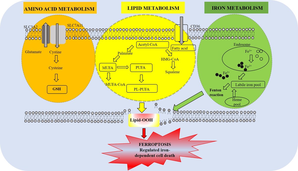
The proportion of PUFAs in the cell membrane determines how sensitive the cell is to ferroptosis. Incorporation of PUFAs into membrane phospholipids depends on the activities of acetyl-CoA synthetase long-chain family member 4 (ACSL4) and lysophosphatidylcholine acyltransferase 3 (LPCAT3). Acetyl-CoA synthetase long-chain family member 3 (ACSL3), however, directs the synthesis of monounsaturated fatty acids (MUFAs) that inhibit ferroptosis by reducing membrane PUFAs. Mechanistically, ferroptosis occurs at the intersection of three processes: metabolic reprogramming of lipid and amino acid pathways, ROS generation via the Fenton reaction, and dysregulation of iron homeostasis via transferrin receptor-mediated uptake [4, 5].
The selenium (Se)-dependent enzyme GPX4 is required to maintain cell membrane integrity and prevent ferroptosis. By converting membrane phospholipid hydroperoxides into non-toxic lipid alcohols, GPX4 acts as the main defense against lipid peroxidation. When GPX4 is absent, lipid peroxides accumulate excessively and can trigger a chain reaction of membrane lipid oxidation leading to ferroptotic cell death [6, 7]. GPX4 becomes inactive when GSH is depleted or upon inhibition of the cystine/glutamate antiporter system xc−, which consists of a light chain, also known as SLC7A11, and a heavy chain, SLC3A2 [8, 9]. Cells treated with GPX4 inhibitors become highly sensitive to ferroptosis, which has important therapeutic implications, especially for cancer, where GPX4 is crucial for tumor cell survival. GPX4 also plays a role in the homeostasis of immune cells. In the absence of GPX4, T regulatory (Treg) cells accumulate lipid peroxides and undergo ferroptosis. This reduction of Treg cell suppression may enhance antitumor immunity [10].
Here, we analyze the interplay between iron, lipid, and amino acid metabolism in ferroptosis and describe how ferroptosis-associated metabolic alterations affect cancer cells. We discuss the context-dependent effects of ferroptosis, which can contribute to tissue damage and tumor initiation, and present the major strategies that induce ferroptosis. Finally, we describe the role of ferroptosis-inducing nutrients (FINs) in cancer and outline potential future directions for translating ferroptosis-targeting strategies into effective and safe clinical therapies.
Ferroptosis-associated metabolic reprogramming
Lipid metabolism
Membrane vulnerability to oxidative damage depends on the presence of the reactive double bonds in PUFAs. Sensitivity of cells to ferroptosis is largely determined by ACSL4, an enzyme whose primary function is to mediate conversion of long-chain PUFAs into active acyl-CoA derivatives [11, 12] responsible for incorporating PUFAs such as arachidonic acid, adrenic acid, eicosapentaenoic acid, docosahexaenoic acid, epoxyeicosatrienoic acids, and hydroxyeicosatetraenoic acid into membrane phospholipids, thereby increasing the membrane’s susceptibility to lipid peroxidation [13]. Apart from remodeling the cell membrane composition by enriching its PUFA-based phospholipid content, ACSL4 has been implicated in regulating the biosynthesis of eicosanoids and steroid hormones, mediating metabolic reprogramming, and promoting antitumor immunity.
ACSL4 may facilitate the cross-talk between fatty acid metabolism and ferroptosis. Fatty acyl-CoAs generated by ACSL4-catalyzed reactions can either provide energy to cells via β-fatty acid oxidation or enter the lipid biosynthesis pathway [14]. Once PUFAs are integrated into phospholipids, they can undergo oxidation enzymatically via lipoxygenases (LOXs) or through iron-dependent free radicals to produce lipid hydroperoxides. Excessive peroxidation of PUFA-containing phospholipids in the cellular membrane is the major signal that triggers ferroptosis in both normal and cancer cells [15, 16], since the oxidative attack of the carbon–carbon double bonds of PUFA-containing phospholipids leads to the rupture of cell membranes [6].
Lipid peroxides can be generated in the cell by Fe2+-dependent non-enzymatic Fenton reaction or by ACSL4/LPCAT3/acid-15-lipoxygenase (ALOX15)-mediated enzymatic reactions, where, upon conversion of free intracellular PUFAs into their fatty acyl-CoA form by the enzyme ACSL4, they undergo LPCAT3-mediated esterification into phospholipids. ALOX15 then converts free and esterified PUFAs into their hydroperoxide forms [6, 15–19]. Thus, ACSL4-mediated lipid peroxidation represents a promising therapeutic target that can be exploited to either protect healthy cells from oxidative injury or to induce ferroptosis in tumor cells.
Redox homeostasis and iron metabolism
The cell maintains its redox homeostasis through a balanced action of both enzymatic and non-enzymatic antioxidant systems. The GSH/GPX4 axis, the NADPH-dependent coenzyme Q10 (CoQ10)/ferroptosis suppressor protein 1 (FSP1) system, and the tetrahydrobiopterin pathway [5–7] mediate protective responses against phospholipid peroxidation. The functionality of this response is determined by effective cystine absorption, mitochondrial reducing equivalents, and transcriptional regulation via stress-response pathways, such as the Nrf2-Keap1-p62 pathway.
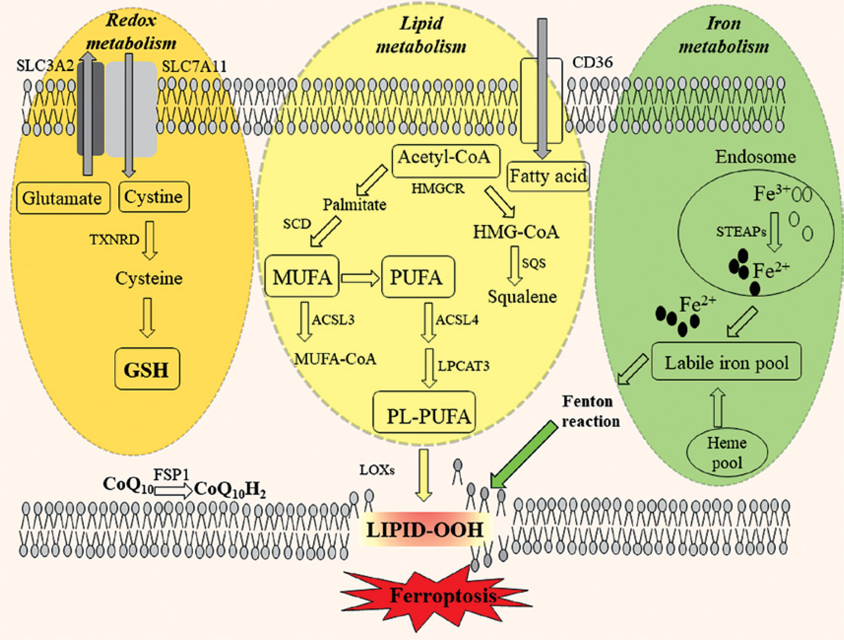
When cystine import via system xc– is restricted or when pharmaceuticals inhibit GPX4 activity, cells are unable to degrade lipid hydroperoxides, leading to ROS accumulation [10, 11]. Mechanistically, ferroptosis is based on iron-driven redox catalysis where, in the Fenton reaction, ferrous iron (Fe2+) is converted into Fe3+, which leads to the formation of highly reactive •OH that targets PUFAs in membrane phospholipids [12]. The formed lipid peroxides are highly reactive and can further propagate excessive ROS accumulation in various cellular compartments, leading to oxidative damage, disruption of membrane integrity, and cell death [20, 21] (Figure 1; [22]). The exact mechanism underlying iron’s involvement in ferroptosis remains unclear. However, it is established that increased cellular free iron content is usually observed during the induction of ferroptosis [23] and that excessive heme and non-heme iron can directly induce ferroptosis [24]. In addition, iron chelators were shown to disrupt ferroptotic cell death in vitro and in vivo, whereas exogenous iron supplementation sensitizes cells to ferroptosis inducers [1].
Figure 1
The interplay between lipid and iron metabolism in ferroptosis
ACSL4 – acyl-CoA synthetase long-chain family member 4, CD36 – cluster differentiation 36, CoQ10 – coenzyme Q10, FSP1 – ferroptosis suppressor protein 1, GSH – glutathione, HMGCR – 3-hydroxy-3-methylglutaryl-CoA reductase, LOX – lipoxygenase, LPCAT3 – lysophosphatidylcholine acyltransferase 3, MUFA – monounsaturated fatty acid, PL-PUFA – phospholipid-containing polyunsaturated fatty acid, PUFA – polyunsaturated fatty acid, SCD1 – stearoyl-coenzyme A desaturase 1, SLC – solute carrier family, SQS – squalene synthase, STEAP – six-transmembrane epithelial antigen of the prostate metalloreductase family, TXNRD – thioredoxin reductase 1
Iron transport proteins
Transferrin (TF) is a glycoprotein that reversibly binds iron in the circulation and redirects it to various organs. TF can bind to two ferric ions (Fe3+), and its iron-binding properties depend on pH changes. Low plasma TF levels are observed in patients with chronic inflammation and/or liver and kidney disease [25]. Amino acid starvation experiments showed that TF is required for the induction of ferroptotic cell death, most likely due to cysteine (Cys) starvation that leads to cellular GSH depletion [26]. Gao et al. suggested that TF serves as a positive regulator of ferroptosis because iron-free apotransferrin or TF-free serum failed to induce ferroptosis in mouse embryonic fibroblasts (MEF). In contrast, the addition of recombinant iron-containing holo-TF induced cell death in MEFs [26]. Iron-bound TF interacts with the dimeric glycoprotein transferrin receptor 1 (TfR1) on the plasma membrane, which is encoded by the TFRC gene, and the TF-TfR1 complex is internalized via receptor-mediated endocytosis. In the acidic endosomal environment, iron is reduced from Fe3+ to Fe2+ by the enzyme six-transmembrane epithelial antigen of prostate 3 (STEAP3), and released into the cytosol as TfR1 is recycled back to the cell surface. In malignant cells, increased TFRC expression reflects the high iron demand for cell growth and proliferation and is positively correlated with ferroptosis induced by erastin [27, 28] or artemisinin derivatives [29]. Yang and Stockwell reported that oncogenic Ras sensitizes cells to erastin-induced ferroptosis by increasing TFRC expression, whereas TFRC knockdown reduces sensitivity to erastin-induced ferroptosis in cells bearing a Ras mutation [30]. Additionally, TFRC knockdown inhibits Cys-depletion-induced ferroptosis [26]. TfR1 may serve as a biomarker of ferroptosis sensitivity, as the recently identified anti-TfR1 antibody, generated by immunizing mice with membrane fractions from lymphoma cells treated with the ferroptosis inducer piperazine-erastin, recognizes only ferroptotic (and not apoptotic) cells [5].
Other metal ion transporters that mediate the uptake of iron include SLC39 family members, solute carrier family 39 member 14 (SLC39A14/ZIP14) and solute carrier family 39 member 8 (SLC39A8/ZIP8), which directly transport non-transferrin-bound iron (NTBI) across the cell membrane. SLC39A8 knockout in mice causes severe anemia, hematopoiesis dysregulation, and neonatal lethality, whereas SLC39A8–/– newborn mice have significantly reduced levels of iron [31]. SLC39A14 knockout in mice impairs gluconeogenesis and reduces hepatic NTBI absorption [32]. In contrast, conditional hepatic SLC39A14 knockout blocks iron accumulation in the liver and ferroptosis-mediated liver fibrosis, suggesting that SLC39A14-mediated alterations in iron content promote ferroptosis [25].
Iron storage proteins
Ferritin, a cytosolic protein complex involved in various metabolic and inflammatory processes, consists of ferritin heavy chain 1 (FTH1) and ferritin light chain (FTL), which are responsible for iron oxidization and storage. It typically consists of 24 homologous subunits that spontaneously assemble into a spherical protein shell, with each subunit carrying an active center that converts Fe2+ to Fe3+, enabling its storage as ferrihydrite. FTH1 confers ferroxidase activity by converting Fe2+ to Fe3+, which is important for the incorporation of iron into the ferritin core. Ferritin exerts protective effects against ferroptosis in various animal models, as evidenced by findings that FTH1 depletion in Drosophila larval wing disks leads to severe growth defects due to mitochondrial dysfunction and ferroptosis [33]. Also, overexpression of mitochondrial ferritin in neuroblastoma SH-SY5Y cells was shown to inhibit erastin-induced ferroptosis [34].
The degradation of ferritin is achieved by ferritinophagy, which represents a form of nuclear receptor coactivator 4 (NCOA4)-mediated selective autophagy, as the knockdown of NCOA4 was shown to block ferritin degradation and suppress erastin-induced ferroptosis in fibroblasts and pancreatic cancer cells, while NCOA4 overexpression promotes ferroptosis by degrading ferritin [23]. Increased ferritinophagy is required for ferroptosis induced by Cys starvation [35] and implicated in promoting ferroptotic cancer cell death [36, 37]. It should be noted that lysosomal degradation of ferritin, which is independent of autophagy, also promotes dihydroartemisinin-induced ferroptotic cancer cell death, suggesting the existence of an alternative mechanism of ferritin degradation during ferroptosis [38].
Heme, present in hemoglobin and myoglobin, is a primary dietary iron source in mammals. The enzyme heme oxygenase 1 (HMOX1) facilitates the degradation of heme into the gasotransmitter carbon monoxide (CO), free iron, and biliverdin, which is further converted into bilirubin by biliverdin reductase. Although heme itself induces HMOX1 expression, a wide range of stress-related factors, including inflammatory cytokines, heat shock, endotoxins, and heavy metal exposure, also upregulate the enzyme, pointing to a potential role for HMOX1 in maintaining redox balance. HMOX1 can either activate or suppress ferroptosis depending on the cellular context. In HT1080 fibrosarcoma cells, the ferroptosis-inducing agent (FIN) erastin increases HMOX1 expression, which can be further enhanced by hemin, an HMOX1 activator, or attenuated by the HMOX1 inhibitor zinc protoporphyrin IX (Table I) [39]. Moreover, HMOX1 knockout is associated with reduced ferroptosis, whereas HMOX1 overexpression increases susceptibility to erastin-induced ferroptosis [39, 40]. However, HMOX1 can also play an anti-ferroptotic role, as illustrated by findings that decreased HMOX1 levels in liver cancer cells or renal proximal tubular cells are associated with increased sensitivity to ferroptosis [41, 42].
Table I
Ferroptosis-inducing agents, their targets, and mechanisms of action
| FIN | Target | Mechanism of action | Compound | Cancer type | Reference |
|---|---|---|---|---|---|
| Class I | SLC7A11 | GSH depletion via inhibition of SLC7A11 activity | Erastin | Melanoma; non-small-cell lung cancer; lung adenocarcinoma; ovarian cancer; anaplastic thyroid cancer; hepatocellular carcinoma | [120–126] |
| Sorafenib | Hepatocellular carcinoma; gastric cancer; clear cell renal cell carcinoma; differentiated thyroid cancer | [42, 127–129] | |||
| IKE | Diffuse large B-cell lymphoma; sarcoma; glioblastoma; non-small-cell lung cancer | [117, 127, 130–132] | |||
| Cyst(e)inase | Prostate cancer; breast cancer; lung cancer; pancreatic tumors; ovarian cancer | [117, 133–135] | |||
| Gemcitabine | Pancreatic cancer; biliary tract cancer; solid tumor | [85, 136] | |||
| Class II | GPX4 | GPX4 inactivation | RSL3 | Fibrosarcoma; triple-negative breast cancer; hepatocellular carcinoma; lung cancer; anaplastic thyroid cancer | [17, 117, 126, 137–139] |
| ML-162 | Anaplastic thyroid cancer; triple-negative breast cancer | [126, 137] | |||
| ML-210 | Glioblastoma; lung cancer; non-small-cell lung cancer;head and neck cancer | [126, 140–142] | |||
| Cisplatin | Gastric cancer; head and neck cancer; non-small-cell lung cancer | [143–146] | |||
| Class III | CoQ10 | CoQ10 depletion; GPX4 degradation | FIN56 | Lung cancer; glioblastoma | [111, 147, 148] |
| Class IV | Redox-active iron | Iron oxidation; GPX4 inactivation | Hemin | Lung cancer | [149] |
| FINO2 | Fibrosarcoma | [150] |
Propagation of lipid oxidation can be halted by GPX4-mediated enzymatic reduction or by antioxidants such as CoQ10, vitamin E, or lipophilic radical traps [14]. It should be noted that the regulation of redox processes is closely linked to cellular energy utilization, as NADPH produced in the pentose-phosphate pathway enables the regeneration of GSH from glutathione disulfide (GSSG) and the reduction of oxidized CoQ10 (ubiquinone) to reduced CoQ10 (ubiquinol). Maintenance of stable NADPH levels within the mitochondrial matrix is mediated by mitochondrial nicotinamide-nucleotide transhydrogenase (NNT), which helps limit ROS leakage from the electron transport chain [17, 18]. When the supply of NADPH is exhausted due to metabolic inhibition, oncogenic reprogramming, or oxidative overload, antioxidant regeneration is impaired, allowing lipid peroxidation to proceed more readily [43].
Adaptive transcriptional programs driven by ATF4, Nrf2, and HIF-1α are initiated under moderate oxidative stress and increase cellular antioxidant capacity, maintaining iron homeostasis by upregulating genes such as GPX4, SLC7A11, FTH1, and HMOX1 [20, 23, 24].
Although the hormetic response delays ferroptosis-induced cellular collapse, it also may contribute to treatment resistance in tumors exposed to ongoing oxidative stress [25]. Cancer cells have been shown to frequently exploit these redox pathways to maintain an equilibrium that supports their growth. However, such a “redox-addicted” state could create a vulnerability to therapy, in which deliberate interference, such as blocking cystine import, inhibiting GPX4, or disrupting NADPH recycling, can trigger ferroptosis preferentially in cancerous cells [27, 28, 44, 45].
Recent lipidomic and metabolomic analyses revealed that ferroptotic redox imbalance has important signaling consequences [46]. For instance, highly reactive and toxic secondary lipid peroxidation products, such as 4-hydroxy-2-nonenal (4-HNE) and malondialdehyde (MDA), may covalently modify proteins and trigger kinase cascades linked to autophagy, inflammation, and immune modulation [30, 47]. These findings indicate that, by interfering with immunogenic cell death, metabolic adaptation, and mitochondrial quality control, ferroptosis is closely associated with redox-dependent signaling processes.
Amino acid metabolism
Excessive availability of certain amino acids (AAs) can promote ROS generation via the electron transport chain, leading to both oxidative and metabolic stress. An approach based on modulating AA metabolism to regulate ferroptosis and treat various pathophysiological conditions has been explored in preclinical and clinical studies [48, 49], and AAs were classified as either ferroptosis-promoting or ferroptosis-inhibiting factors. It has been observed that the impact of AAs on ferroptosis varies between different cell types and requires the involvement of multiple regulatory factors, such as mammalian target of rapamycin complex 1 (mTOR), activating transcription factor 4, nuclear factor E2-related factor 2 (Nrf2), and hypoxia-inducible factor 1α (HIF-1α) [50–55]. When AAs are present in sufficient quantity in cells, mTORC1 localizes to the lysosome for activation by Rag GTPases [54]. In addition, some transcription factors and stress-responsive proteins, such as methyltransferase-like protein 16 (SAMTOR), an S-adenosylmethionine (SAM) sensor, cytosolic arginine sensor for mTORC1 subunit 1 (CASTOR1), and sestrin 2, can sense the cellular contents of methionine (Met), Arg, and leucine (Leu), respectively [56–58]. Growing evidence supports the important role of sestrin 2 in mediating resistance to ferroptosis through mTORC1 and Nrf2 activation in several disease models [59–62]. It should be noted that the functions of amino acid-sensing proteins are general and cannot be correlated to the effect of specific AAs on ferroptosis.
Ferroptosis-promoting amino acids
The differences in cellular AA profiles primarily reflect differential expression of metabolic enzymes or specific AA transporters across cell types [63, 64]. For instance, most cancer cells depend on glutamine (Gln) to replenish the TCA cycle during aerobic glycolysis, which is paradoxical, as Gln is an abundant, nonessential AA that can be synthesized from glucose [65, 66]. Excessive Gln catabolism may result in disruption of the TCA cycle and the electron transport chain, hyperpolarization of the mitochondrial membrane potential, and accumulation of lipid peroxides. In contrast, the inhibition of Gln catabolism confers resistance to ferroptosis [67]. Thus, the high Gln uptake rate observed in Gln-dependent cancer cells cannot be solely explained by its role as a nitrogen donor in AA and nucleotide biosynthesis and reflects additional, important Gln roles in cancer cells, such as maintaining mTOR kinase activation, mitochondrial membrane potential, and nicotinamide adenine dinucleotide phosphate (NADPH) production for redox homeostasis and synthesis of macromolecules [68]. Suzuki et al. reported that glutaminase 2 (GLS2), a central regulator of glutaminolysis, suppresses HCC progression in vivo by promoting ferroptosis through α-ketoglutarate (α-KG)-dependent lipid ROS production, suggesting that GLS2 acts as a tumor suppressor [69]. In oral squamous cell carcinoma, the Gln transporter promotes ferroptosis by augmenting the Gln flux to the TCA cycle [70]. Furthermore, Prange et al. demonstrated that Gln restriction enhances the cytotoxicity of immune cells against tumor cells, thereby inhibiting tumor cell proliferation [71]. However, unlike cancer cells, adult cardiomyocytes predominantly rely on fatty acid metabolism [72]. In pathophysiological states, such as pulmonary arterial hypertension, activation of the MYC oncogene upregulates the expression of the Gln transporters SLC1A5 and SLC7A5, driving right ventricular hypertrophy through Gln catabolism. Interestingly, GLS inhibition in cardiomyocytes does not alleviate oxidative stress in these cells [65, 73].
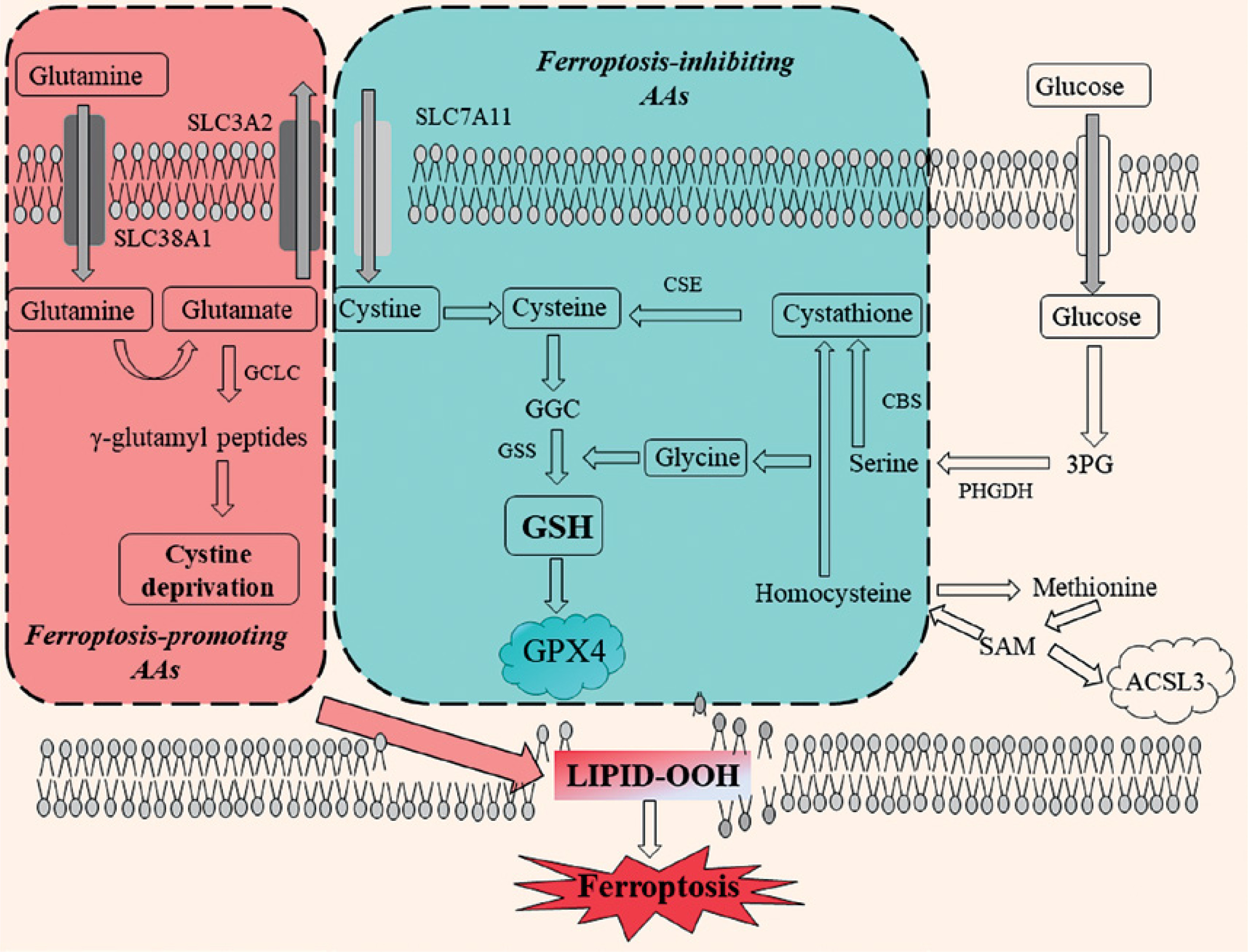
Solute carrier family 38 member 1 (SLC38A1) and SLC1A5 can increase the intracellular availability of glutamine, which contributes to production of glutamate (Glu), which can be exchanged with cystine through SLC7A11. Inhibition of SLC7A11 is associated with ferroptosis because it reduces cystine uptake, leading to depletion of intracellular cysteine (Cys) and accumulation of glutamate [65] (Figure 2 [22]). Glu accumulation induced by a system xc– inhibitor in lung adenocarcinoma sensitizes cells to ferroptosis via suppression of adenylate cyclase 10 (ADCY10)-dependent Yes-associated protein 1 (YAP1) signaling [74].
Figure 2
Amino acid metabolism pathways that promote or inhibit ferroptosis
AAs – amino acids, ACSL3 – acyl-CoA synthetase long-chain family member 3, CBS – cystathionine beta-synthase, CSE – cystathionine gamma-lyase, GCLC – glutamate-cysteine ligase catalytic subunit, GSH – reduced glutathione, GSS – glutathione synthetase, GPX4 – glutathione peroxidase 4, 3PG – 3-phosphoglycerate, PHGDH – phosphoglycerate dehydrogenase, SAM – S-adenosylmethionine, SLC38A1 – solute carrier family 38 member 1, SLC1A5 – solute carrier family 1 member 5, SLC7A11 – solute carrier family 7 member 11, SLC3A2 – solute carrier family 3 member 2.
Tumor cells also often have high metabolic demands for branched-chain AAs (BCAAs) and Met. BCAAs are metabolized by transaminases BCAT1 (mainly expressed in the mitochondria) and BCAT2 (predominantly expressed in the cytosol) [73] upon being transported into cells through L-type AA transporters. BCAT2 has been reported as a novel inhibitor of ferroptosis. A recent study, using a high-throughput CRISPR/Cas9-based genetic screen in HCC cells to identify ferroptosis-inhibiting proteins, identified BCAT2 as a ferroptosis suppressor. BCAT2 activation by ectopic expression protected liver and pancreatic cancer cells from ferroptosis, both in vitro and in vivo, by specifically antagonizing system xc– inhibition, whereas RNA interference-mediated inhibition triggered ferroptosis [75].
Increased Met metabolism is indicated by upregulation of methionine adenosyltransferase 2A (MAT2A) activity and solute carrier family 43 member 2 expression in tumor cells, resulting in inhibition of the antitumor activity of CD8+ T cells [76]. In lung and pancreatic cancer, KRAS (Kirsten rat sarcoma viral oncogene homolog) mutations promote BCAA metabolism [77].
Ferroptosis-inhibiting amino acids
As substrates of GSH synthesis, glycine (Gly) and Cys can regulate GPX4-mediated antioxidant defense. Gly uptake, synthesis, and catabolism are regulated by Nrf2 and are associated with tumorigenesis and malignancy [78]. Gly can inhibit ferroptosis in various disease models, and its transporter, GLYT1, is induced by Nrf2 and may contribute to reduced ROS production. The GLYT1 inhibitor bitopertin has been reported to increase Nrf2 stability by inhibiting Kelch-like ECH-associated protein 1 (Keap1)-mediated Nrf2 ubiquitination [79]. However, prolonged and excessive exposure of cells to Gly leads to GSH depletion and accumulation of lipid peroxidation products, which may be due to Gly-induced methylation, as Gly has been shown to promote SAM-mediated methylation of the GPX4 promoter [80].
Cys is required for the production of α-KG, pyruvate, and taurine. In vivo studies of conversion of Cys to taurine by Cys dioxygenase type 1 (CDO1) indicated that CDO1 overexpression renders cells susceptible to ferroptosis due to reduced intracellular GSH levels [81, 82]. Cys extracellular uptake is mediated by the Cys/Glu antiporter SLC7A11, which imports one cystine molecule in exchange for an intracellular Glu molecule [83] (Figure 2). If extracellular Cys is depleted, homocysteine is synthesized de novo through the activities of cystathionine β-synthase and cystathionine γ-lyase in the Met-mediated sulfur transfer pathway, which also induces Cys release in lysosomes through the ATF4/AhR-mediated amino acid starvation response [84].
Glutathione and GPX4 axis
The GSH/GPX4 axis integrates amino acid, redox, and Se metabolism into a unified defense against lipid peroxidation. GSH, a tripeptide consisting of Glu, Cys, and Gly, is the primary antioxidant within cells and acts as a cofactor for GPX4. System xc– mediates the exchange of intracellular Glu for extracellular cystine, which is converted into Cys upon entering the cell through NADPH-dependent thiol-disulfide exchange reactions, accelerated by glutathione reductase (GSR) and thioredoxin (Trx) systems [37, 85] (Figure 2). GSH is formed in two consecutive ATP-dependent reactions in which γ-glutamylcysteine ligase (GCL) condenses Cys with Glu, whereas glutathione synthetase (GSS) ligates it with Gly [4]. GSH maintains thiol homeostasis and redox signaling by detoxifying hydrogen peroxide and lipid hydroperoxides under physiological conditions [5]. GPX4 facilitates the two-electron reduction of phospholipid hydroperoxides (PLOOHs) to their respective alcohols (PLOHs), using two molecules of GSH as electron donors. GSH is converted into GSSG during this process [86]. GSR utilizes NADPH to convert GSSG back into GSH [5, 6]. This redox cycle inhibits the propagation of lipid radical chain reactions, preventing potential membrane damage. As GSH levels decrease or GPX4 activity is inhibited, PLOOHs accumulate and subsequently decompose into reactive aldehydes such as 4-HNE and MDA. Decreased GPX4 levels may result from perturbed activity of enzymes involved in selenoprotein synthesis, Se deficiency, or defects in the mevalonate-isopentenyl pathway [23, 43]. Additionally, post-translational modifications affecting protein stability or activity may alter ferroptotic sensitivity. For instance, the deubiquitinase OTUB1 inhibits SLC7A11 degradation, while the interaction between Beclin-1 and SLC7A11 prevents cystine uptake under stress conditions [87]. MDA and 4-HNE represent important markers of lipid peroxidation associated with ferroptosis, as their accumulation is associated with increased membrane permeability, membrane disruption, and, ultimately, ferroptotic cell death [6, 10].
Another emerging ferroptosis suppressor that has demonstrated a protective effect against iron-induced cell death in conditions of GPX4 deficiency is FSP1, which primarily functions as a membrane lipid radical-scavenging system [88]. A recent study by Doll et al. showed that lipid radicals can be captured independently of GPX4 through the FSP1-CoQ10-NADPH pathway that restores reduced CoQ10 at the plasma membrane [24, 89], thus providing an alternative defense mechanism against GPX4 deficiency (Figure 2). The inhibitory effect of FSP1 on ferroptosis is mediated via the reduced form of CoQ10, ubiquinol, which scavenges lipid-derived radicals [89]. FSP1 plays a role in resistance to ferroptosis in various cancer cell lines [90, 91] and developing specific inhibitors that target FSP1 and/or disrupt oncogene-mediated regulation of FSP1 expression may enhance tumor cell sensitivity to chemotherapy [92]. In addition, significantly reduced FSP1 levels were observed in several pathophysiological conditions, such as preeclampsia and cerebral ischemia/reperfusion (I/R) injury [93, 94]. FSP1 overexpression significantly stimulated the PI3K/AKT/GSK3β pathway and enhanced resistance to ferroptosis, suggesting that FSP1 may represent a potential target for the treatment of I/R injury [94].
Nrf2-Keap1-p62 signaling
Nrf2 signaling is a key cellular defense mechanism that regulates iron metabolism, redox balance, and the oxidative stress response. The transcription factor Nrf2 regulates the expression of more than 200 genes by binding to antioxidant response elements in their promoter regions [1–3]. Under basal conditions, Nrf2 is confined in the cytoplasm by its repressor, Keap1, which acts as a substrate adaptor for the Cullin-3 (Cul3)-Rbx1 E3 ubiquitin ligase complex, facilitating Nrf2 ubiquitination and subsequent proteasomal degradation [4, 5]. Keap1 functions as a redox sensor due to its reactive Cys residues (Cys151, Cys273, and Cys288). ROS, lipid peroxides, or xenobiotics can alter Keap1’s conformation, preventing its interaction with Nrf2 and inhibiting Nrf2 ubiquitination [6]. The stabilized Nrf2 translocates to the nucleus and forms heterodimers with small Maf proteins, which bind to antioxidant response element (ARE) sequences in the promoters of genes that provide cellular protection against oxidative stress [7, 10].
The consequence of such transcriptional reprogramming is enhanced efficacy of anti-ferroptotic pathways involved in the regulation of iron levels and antioxidant defense. Increased expression of SLC7A11, the catalytic subunit of glutamate-cysteine ligase (GCLC), GPX4, and GSS promotes cystine import into the cell via system xc–, and enhance glutathione production, thus facilitating the degradation of lipid peroxides [11, 12]. In addition, Nrf2 regulates the expression of FTH1, FTL, and ferroportin (SLC40A1), which sequester and export labile iron, limiting the availability of iron that can generate ROS in the Fenton reaction [95]. Nrf2 also increases the transcription of heme oxygenase-1 (HMOX1), whose byproducts, biliverdin and CO, possess radical-scavenging capabilities. Resistance to ferroptosis is augmented by the selective autophagy receptor p62/SQSTM1, which interacts with Keap1 by specifically binding to its Kelch domain. Under prolonged oxidative stress, a positive feedback loop is established in which Nrf2 increases p62 transcription, while p62 accumulation stabilizes Nrf2 [47]. Persistent Nrf2 activation caused by KEAP1 and/or CUL3 alterations is often associated with head-and-neck squamous carcinoma, non-small-cell lung carcinoma, and HCC [29, 30], and leads to increased expression of GPX4 and SLC7A11, which inhibit lipid peroxidation, thereby establishing a milieu that promotes tumor growth [96, 97].
The Nrf2-Keap1-p62 pathway interacts with several metabolic redox circuits. For instance, Nrf2 is involved in the transcriptional upregulation of glucose-6-phosphate dehydrogenase (G6PD), 6-phosphogluconate dehydrogenase (PGD), and isocitrate dehydrogenase 1 (IDH1). The activity of these enzymes is mandatory for the production of NADPH, which serves as the reducing agent for the regeneration of GSH and CoQ10 from their oxidized states [23]. Dysregulation of Nrf2 signaling is associated with iron accumulation, as well as reduced GSH and decreased GPX4 expression. In murine models, hepatocytes lacking Nrf2 show enhanced vulnerability to ferroptosis induced by erastin and Ras-selective lethal 3 (RSL3), whereas deletion of Keap1 confers a protective effect [26]. Similarly, pharmacological inhibition of Nrf2 with agents such as brusatol or trigonelline has shown potential to re-sensitize tumors to ferroptosis inducers [27, 28].
In addition, recent findings report the role of Nrf2 in regulating lipid metabolism. Nrf2 was shown to suppress the expression of the enzymes ACSL4 and ALOX15, which are involved in the incorporation and oxidation of PUFAs into membrane phospholipids, thus indirectly reducing the pool of lipids available for peroxidation [32, 33, 47].
Mitochondrial ROS and NADPH systems
Mitochondria are a major intracellular source of ROS. During normal respiration, the formation of superoxide anion radical (O2•–) principally occurs at complex I (NADH dehydrogenase) and complex III (cytochrome bc complex) and is caused by a limited quantity of electrons that escape the electron-transport chain (ETC) and interact with molecular oxygen [1, 2]. The enzymes superoxide dismutase 1 (SOD1) and superoxide dismutase 2 (SOD2), located in the intermembrane space/cytosol and mitochondrial matrix, respectively, convert O2•– into hydrogen peroxide [3, 4]. Perturbed mitochondrial homeostasis induced by oxygen deprivation and ETC inhibition increases electron leakage, which results in excessive ROS accumulation and mitochondrial lipid peroxidation [5–7]. Thus, mitochondrial dysfunction sensitizes cells to ferroptosis, and inhibition of oxidative phosphorylation promotes evasion of ferroptosis triggered by cystine deficiency [10, 11].
Mitochondria play a key role in heme synthesis and the assembly of iron-sulfur (Fe-S) clusters [12]. An imbalance in the mitoferrin transporter system that carries Fe2+ into mitochondria leads to an accumulation of Fe2+ in the mitochondrial labile iron pool and promotes the Fenton reaction that generates •OH, which initiates peroxidation of polyunsaturated cardiolipins in the inner mitochondrial membrane [14]. Lipid peroxidation results in a loss of polarization of the mitochondrial inner membrane, disorganization of the cristae, and the release of pro-oxidant species into the cytosol [95].
Mitochondrial ferritin (FtMt) sequesters excess free iron, and its increased expression is associated with amelioration of oxidative damage in cardiomyocytes, neurons, and osteoblasts [98]. Also, efficient iron utilization is mediated by the iron–sulfur cluster scaffold protein IscU and iron chaperone frataxin, whose perturbations lead to excessive iron accumulation and hypersensitivity to ferroptosis [99]. These data suggest that mitochondrial iron-buffering capacity influences the outcome of adaptive cellular oxidative signaling.
The role of NADPH in protection against ferroptosis
NADPH provides essential reducing power for the function of GPX4 and FSP1. Enzymes responsible for mitochondrial NADPH production include IDH2 and NNT. IDH2 mediates the oxidative decarboxylation of isocitrate to α-KG, whereas NNT links proton motive force to the transfer of hydride equivalents from NADH to NADP+ [25]. Decreased activity of either enzyme is associated with reduced mitochondrial NADPH levels, diminished GSH levels, and increased cellular susceptibility to ferroptosis [26].
Maintenance of cytoplasmic NADPH levels is mediated by the pentose phosphate pathway, which is driven by G6PD and PGD, along with ME1 and IDH1 [27]. By transcriptionally coordinating the expression of numerous antioxidant genes, Nrf2 plays a key role in linking cellular NADPH-generating pathways to antioxidant responses [28]. In conditions of cystine starvation, cells respond to the reduction of GSH by boosting the regeneration of CoQ10 through the FSP1-CoQ10-NADPH pathway. As expected, simultaneous inhibition of NNT or G6PD leads to a collapse of proferroptotic antioxidant defenses [43].
The mitochondrial TCA cycle and glutaminolysis considerably influence ferroptotic responses. Gln catabolism, mediated by enzymes GLS2 and glutamate dehydrogenase, results in the formation of α-KG and can contribute to NADH/NADPH production. However, ROS production in mitochondria is stimulated by excessive α-KG oxidation, leading to lipid peroxidation [32]. Inhibition of α-KG dehydrogenase activity or glutaminolysis reduces mitochondrial ROS production and protects cells against ferroptosis, an effect that is especially pronounced under cystine limitation [33]. Furthermore, by accelerating Gln degradation, MYC and KRAS-driven oncogenic pathways may connect metabolic perturbations to ferroptotic hypersensitivity in specific cancer cells [47].
Taken together, mitochondria play a key role in the interplay between metabolic disruption, compromised antioxidant defenses, and redox homeostasis, functioning as both generators and amplifiers of pro-ferroptotic signals. The interactions between the GSH/GPX4 axis, the Nrf2-Keap1-p62 signaling network, and mitochondrial ROS-NADPH mediate iron and cystine availability, maintain reducing equivalents through NADPH regeneration, and detoxify lipid peroxides. Further studies of the interactions between mitochondrial and metabolic circuits may reveal the bioenergetic foundations of ferroptosis, paving the way for novel therapeutic strategies targeting mitochondrial antioxidants.
Therapeutic implications of ferroptosis
The existence of strong associations between ferroptosis and metabolic and oxidative perturbations suggests that highly metabolically active cancer cells may be more predisposed to FINs due to higher iron demand and ROS accumulation [84, 100]. Some cancer cells that exhibit resistance to apoptosis and traditional therapies, such as mesenchymal and dedifferentiated cells, are highly susceptible to ferroptosis [101–103]. Interestingly, because ferroptosis can modulate tumor immunity and immune responses, targeting this pathway may represent an effective strategy to inhibit tumor initiation and progression. Interventions that modulate ferroptosis have shown high efficacy in cancer treatment [104–106]. Associations between ferroptosis and the tumor-suppressive functions of immuno- and radiotherapy [107–109] enable ferroptosis-based combination therapy to augment the effectiveness of conventional treatment, target resistant tumors, and prevent tumor recurrence. However, growing evidence suggests that ferroptosis has context-dependent effects at different stages of tumorigenesis. For example, while induction of ferroptosis may enable chronic liver disease progression to HCC, it is also implicated in limiting the development of already established HCC [110, 111]. Kim et al. reported that pharmacological induction of ferroptosis suppresses tumor growth more effectively when FINs are administered during early tumor development [112].
Mechanisms of ferroptosis induction by radio- and immunotherapy
Generally, ferroptosis induction for cancer therapy can be achieved through radiotherapy, immunotherapy, and/or pharmacological agents. Radiotherapy is a widely used treatment modality that delivers ionizing radiation to induce tumor cell death [113]. Radiotherapy induces ferroptosis to suppress tumor growth through multiple mechanisms [108]: i) direct DNA damage by forming DNA double-strand breaks, which reduces SLC7A11 expression in an ataxia-telangiectasia mutated protein-dependent manner, leading to reduced cystine uptake and increased ferroptosis [114]. Also, DNA damage can inhibit SLC7A11 by upregulating the cyclic GMP-AMP synthase-stimulator of interferon genes (cGAS–STING) pathway and the activating transcription factor 3 (ATF3)/SLC7A11/GPX4 regulatory axis [115]; ii) increased ACSL4 expression that promotes excessive accumulation of PUFA-containing phospholipids [108]; iii) through the bystander effect, irradiated tumor cell-derived microparticles may induce ROS accumulation and ferroptosis in unirradiated neighboring cells [116]. In immunotherapy, activated CD8+ T cells release interferon gamma (IFNγ) and inhibit SLC7A11, thereby promoting PUFA accumulation via ACSL4 [117–119].
Pharmacological inducers of ferroptosis
In addition to radio- and immunotherapy, four classes of compounds have been identified as FINs with therapeutic potential in cancer, based on their mechanism of action (summarized in Table I) [120–150].
Several other systemic pharmacological agents approved for inflammatory, infectious, cardiometabolic diseases, and psychiatric disorders have been shown to induce ferroptosis in various cancer cells (Table I). Sulfasalazine (SAS), an anti-inflammatory drug used to treat rheumatoid arthritis, induces ferroptosis by inhibiting SLC7A11 [151, 152]. SAS enhances the therapeutic effectiveness of other chemotherapeutic agents, such as daunorubicin in acute myeloid leukemia [153] and paclitaxel in ovarian clear cell carcinoma [151, 154]. It also improves the efficacy of radiotherapy and anti-PD-1 immunotherapy by promoting ferroptosis [155, 156].
Statins reduce blood cholesterol levels but also induce ferroptosis by inhibiting the GSH/GPX4 and FSP1/CoQ10/NAD(P)H antioxidant pathways [102, 157–160]. For instance, simvastatin induces ferroptosis in cancer cells by inhibiting 3-hydroxy-3-methylglutaryl-coenzyme A reductase expression, thereby downregulating the mevalonate pathway and reducing GPX4 expression [160]. In contrast, lovastatin reduces programmed death-ligand 1 (PD-L1) expression in lung cancer cells, thereby sensitizing the tumor cells to immunotherapy [161].
Dopamine receptor D2 (DRD2) antagonist haloperidol is used for the treatment of psychiatric disorders and inhibits tumor growth by inducing ferroptosis [162, 163]. In HCC, haloperidol can increase sorafenib- and erastin-induced ferroptosis by inhibiting sigma-1 receptor signaling, which is involved in oxidative stress metabolism [164]. In glioblastoma multiforme, it enhances temozolomide-induced ferroptosis by inhibiting DRD2 [162]. Zalcitabine (2ʹ,3ʹ-dideoxycytidine) is a nucleoside analog reverse transcriptase inhibitor that binds to mitochondrial DNA polymerase gamma and is used for the treatment of human immunodeficiency virus. In pancreatic cancer cells, zalcitabine induces ferroptosis by modulating the STING1/TMEM173-dependent DNA damage response and promoting mitochondrial DNA stress [165]. Brequinar is a dihydroorotate dehydrogenase inhibitor that affects pyrimidine synthesis. It was used to selectively block GPX4low tumor growth by triggering ferroptosis. In combination with sulfasalazine, brequinar suppresses the growth of GPX4-high tumors [166]. Brequinar also acts synergistically with AMPK activators to suppress tumor growth through ferroptosis [167].
Several bioactive natural compounds, such as artemisinin, withaferin A (WA), β-elemene (β-ELE), and curcumenol, also show promise as FINs. Artemisinin, derived from the Chinese herb Artemisia annua, can promote ferritinophagy and increase intracellular free iron levels [44]. Its derivatives, artesunate and dihydroartemisinin, have been shown to synergize with sorafenib to induce ferroptosis in HCC [168, 169]. Dihydroartemisinin also enhances cisplatin-induced toxicity in pancreatic ductal adenocarcinoma (PDAC) [170]. WA, isolated from Withania somnifera, induces ferroptosis in high-risk neuroblastoma tumors via targeting and inhibiting GPX4 activity [171, 172]. WA enhanced ferroptotic responses in sorafenib-resistant HCC cells [173] and, in combination with anti-PD-1 immunotherapy and the C-X-C chemokine receptor 2 inhibitor, significantly reduced liver metastasis of colorectal cancer and improved survival in wild-type mice with liver tumors by inducing GPX4-associated ferroptosis in hepatocytes [174]. β-ELE from Curcuma wenyujin activates transcription factor EB-mediated lysosome degradation of GPX4 and has been shown to induce ferroptosis in NSCLC cells [175, 176]. Curcumenol exploits epigenetic mechanisms to suppress lung cancer cell growth by inducing ferroptosis via the lncRNA H19/miR-19b-3p/FTH1 axis [177].
Novel drug delivery systems, such as liposomes, hydrogels, and nanoparticles, may allow precise control of drug release while reducing toxicity to healthy tissues [178]. For example, BQR@MLipo, brequinar-loaded liposomes that target mitochondrial ferroptosis, were shown to effectively inhibit bladder cancer growth [178]. Additionally, targeted RSL3 delivery can be achieved using injectable alginate hydrogel RTFG@SA [179] or BNP@R nanoparticles that enable acid-activatable photodynamic therapy [139]. Novel nanodrug delivery systems offer numerous advantages, including improved water solubility and cell uptake, prolonged drug circulation time, increased drug accumulation in tumors, and reduced drug toxicity [180]. At present, several nano-drugs are used in clinical applications, including doxorubicin liposomes and paclitaxel albumin nanoparticles [180, 181].
Combination therapeutic strategies
Growing evidence supports the observation that FINs can overcome resistance to certain conventional chemotherapies. For instance, RSL3 enhances the antitumor efficacy of cisplatin by increasing ROS levels and lipid peroxidation [182, 183], whereas combined treatment with sulfasalazine and erastin may overcome cisplatin resistance [143]. Several reports showed that erastin also increased the efficacy of cisplatin [183, 184], doxorubicin [185], docetaxel [186], vemurafenib [103], and temozolomide [187] in specific cancer models.
Programmed cell death 1 (PD-1)/PD-L1 immune checkpoint pathway targeting [188] represents a promising therapeutic approach. Immune checkpoint inhibitor (ICI)-based immunotherapy, such as anti-CTLA4 and anti-PD-1/PD-L1 antibodies, specifically targets dysfunctional immune cells and activates CD8+ T cells to eradicate tumor cells [189, 190]. A growing body of evidence demonstrates that combining ICIs with FINs can synergistically inhibit tumor growth in preclinical models [117]. Two important findings are associated with combination therapy, including FINs and ICIs: i) ferroptosis induction potentiates antitumor effects of CD8+ T cells; and ii) immunotherapy can promote cancer cell ferroptosis.
IFNγ derived from CD8+ T cells sensitizes tumor cells to ferroptosis by activating the JAK/STAT1 pathway to decrease SLC7A11 and SLC3A2 levels [117]. However, IFNγ can also induce ferroptosis by increasing ACSL4 transcription, which stimulates STAT1/IRF1-mediated arachidonic acid incorporation into C16 and C18-acyl chain-containing phospholipids [191]. Ferroptosis contributes to the therapeutic response to PD-L1 inhibition, as illustrated by findings that the ferroptosis inhibitor lipoxstatin-1 significantly diminishes the efficacy of PD-L1 inhibitors [117]. Also, restoring tumor cells’ sensitivity to ferroptosis could enhance immunotherapy efficacy, as ferroptosis resistance correlates with tumor cells’ unresponsiveness to ICIs. For instance, ICI-resistant TYRO3high tumors can be resensitized to anti-PD1 therapy by inhibiting TYRO3-mediated Akt/Nrf2 signaling, thereby restoring ferroptosis [192]. Also, a combination of GPX4 inhibitors and anti-PD-1 therapy induced an immune response and suppressed tumor growth in immunocompetent mice bearing triple-negative breast cancer [186]. However, it was observed that increased CD8+ T cell infiltration due to GPX4 inhibition-induced ferroptosis in HCC cells was counteracted by PD-L1 upregulation on tumor cells [174]. This can be explained by findings that ferroptosis triggers the infiltration of immunosuppressive myeloid-derived suppressor cells (MDSCs) through increased release of high-mobility group box-1 (HMGB1), a signaling molecule involved in the pathogenesis of chronic liver disease, thereby accelerating liver tumorigenesis [193]. In line with this finding, the triple combination of checkpoint blockade, pharmacological FINs, and MDSC suppression improves survival in wild-type mice with liver tumors and effectively inhibits liver metastasis [174]. The components of multidrug combination therapy based on FINs and immunotherapy can be tailored to enhance antitumor efficacy by eliciting a robust antitumor immune response while preventing ferroptosis-triggered immunosuppression.
Resistance to ferroptosis can contribute to radioresistance in some cancer cells, which is associated with cancer recurrence and an unfavorable prognosis [194]. Ionizing radiation can induce SLC7A11 and GPX4 to safeguard cells against ferroptosis, and growing evidence suggests that inhibiting SLC7A11 or GPX4 can sensitize radioresistant cancers to radiotherapy [111, 195]. The combination of radiotherapy with class I FINs that inhibit SLC7A11 or class II or III FINs that inhibit or deplete GPX4 (Table I) acted synergistically to induce ferroptosis [111]. Suppressors of cytokine signaling 2, which promote ubiquitination-mediated degradation of SLC7A11, have been proposed as a potential biomarker for predicting HCC radiosensitivity [196]. Multiple mechanisms operate to regulate the interplay between ferroptosis and radiosensitivity. For instance, in Keap1 mutant lung cancers refractory to radiotherapy, resistance to ferroptosis and radiotherapy is caused by upregulation of the Nrf2/FSP1 anti-ferroptosis axis [197]. In radioresistant esophageal squamous cell carcinoma, stanniocalcin 2 (STC2), which is involved in various processes of malignant tumors, activates protein methyltransferase 5 (PRMT5) and increases SLC7A11 levels, thereby enhancing resistance to ferroptosis [194].
Role of ferroptosis in non-cancer diseases
Dysregulation of ferroptosis has been linked to various neurodegenerative and cardiovascular diseases (CVDs) [198, 199]. For instance, ferroptosis has been implicated as a potential factor contributing to the progression of neurodegenerative diseases such as Alzheimer’s disease (AD) and Parkinson’s disease (PD). In AD, ferroptosis is associated with the presence of amyloid-beta (Aβ) peptides, which promote the accumulation of redox-active iron leading to GSH depletion [198, 200]. In PD, which is characterized by loss of dopaminergic neurons in the iron-rich substantia nigra, dopamine metabolism can generate oxidative stress and ROS accumulation that facilitates ferroptosis [201]. Interestingly, PD pathology is associated with significantly reduced GSH levels in the substantia nigra, which makes these neurons vulnerable to iron-catalyzed lipid peroxidation and ferroptotic death [202].
Ferroptosis is implicated in the development and progression of atherosclerosis [203], heart failure [204], myocardial I/R injury [205], and myocardial infarction [206]. Atherosclerotic processes are associated with ferroptotic death of endothelial and foam cells, which contributes to plaque instability and rupture [203]. Also, CVDs are frequently associated with perturbed iron homeostasis. The high metabolic rate of the myocardium is accompanied by an increased rate of oxygen consumption and ROS accumulation [207]. In I/R injury, extensive lipid peroxidation is caused by the severe depletion of GSH during ischemia and the release of iron from damaged mitochondria, contributing to myocardial injury following infarction [198, 207].
Tissue function in neurodegenerative diseases and acute injuries may be preserved by inhibiting ferroptosis through sequestering iron required for lipid peroxidation [208]. Several potential therapeutic candidates that chelate the catalytically active iron have been identified, such as deferoxamine (DFO), deferasirox (DFX), dexrazoxane (DZR), and deferiprone (DFP). DFO showed promising results in mitigating secondary brain injury [209] and protecting kidneys against I/R injury by inhibiting ferroptosis in renal tubular cells [210].
Conclusions and future perspectives
Ferroptosis was first described in 2012 [1] and, although it has been extensively investigated in several pathophysiological conditions, its exact molecular mechanisms remain incompletely elucidated [197]. Several studies have suggested that cellular susceptibility to ferroptosis is determined by metabolic state and cellular context [46, 197, 211–214]. Results of studies investigating the connections between ferroptosis and metabolism suggest that certain metabolic enzymes play important roles in multiple ferroptosis-associated metabolic pathways. For instance, ACSL3 can promote the integration of different fatty acids into triacylglycerols and phospholipids, and also direct their mitochondrial β-oxidation [197]. Another issue that should be resolved is whether lipid peroxidation occurs only in the plasma membrane [88, 215] since a recent study by von Krusenstiern et al. suggests that the endoplasmic reticulum membrane represents a major site of lipid peroxidation [216].
Ferroptosis-inducing pharmacological agents represent a promising therapeutic strategy for treating chemoresistant cells and/or specifically targeting drug-tolerant persister cancer cells [217–223]. However, some literature findings report that both induction and inhibition of ferroptosis contribute to the effectiveness of cancer immunotherapy, depending on the cellular context [112, 212, 218, 219]. Such a paradoxical finding requires further clarification, possibly by studying the metabolic perturbations that modulate ferroptosis in a specific cellular context. Additional challenges that should be addressed in future research include the development of standardized animal models to facilitate comparison of study results. Current models primarily rely on FINs to treat xenograft tumors and do not account for potential side effects associated with long-term FIN administration. In addition, the mechanisms of resistance to FINs are yet not well understood [46]. Since the expression of key molecules in ferroptosis, such as GSH and CoQ10, is regulated by multiple metabolic pathways, integrating transcriptomic, proteomic, metabolomic, and lipidomic studies of ferroptosis-associated pathways is necessary for future research on the role of ferroptosis in specific pathophysiological states [220]. Addressing these challenges will facilitate the development of personalized ferroptosis-based strategies for cancer treatment.