

Introduction
Breast cancer is the most common cancer diagnosed worldwide in women, accounting for approximately 25% of all new cancer cases in women globally, according to 2022 data [1]. In Poland, among women in 2022, breast cancer represented 23.6% of all cancer cases registered in the National Cancer Registry. It also accounted for 15% of cancer-related deaths among women [2]. Over the years, survival outcomes for patients with early-stage breast cancer have significantly improved, primarily due to the use of adjuvant therapies such as chemotherapy, radiotherapy, and hormone therapy, which reduce the risk of both local recurrence and distant metastases [3]. Improved survival outcomes in breast cancer have led to a heightened focus on the long-term impacts of treatment. As the number of women cured of breast cancer increases significantly, the likelihood of developing subsequent cancers and dying from them also rises. Compared with the general population, breast cancer survivors have a significantly higher risk for all second primary cancers (SPC) and non-breast cancers [4]. Following a breast cancer diagnosis, the risk of developing a second primary malignancy is estimated to be approximately 30% higher compared to the risk observed in the cancer-free population. The risk of developing a second malignancy was higher with young age at initial diagnosis, elevated body mass index, and smoking. In contrast, it was lower among women with higher educational attainment, those who were postmenopausal, and those with a history of full-term pregnancy [5]. The development of subsequent malignancies following primary breast cancer may result from shared risk factors that predispose individuals to both the first and second cancers, such as genetic susceptibility or other established risk factors, or from adverse effects related to the treatment of the primary tumour. Numerous scientific studies focus on analysing the risk of developing a second malignancy in relation to the type of breast cancer treatment received – such as radiotherapy, chemotherapy, surgical intervention, or hormone therapy – to assess the impact of treatment on subsequent cancer risk [6–10]. The most documented second primary cancers following breast cancer include endometrial cancer, ovarian cancer, thyroid malignancies, lung cancer, soft tissue sarcomas, and leukaemia [10–13].
In this study, we use both terms “second malignancy” and “secondary cancer” interchangeably. While “second malignancy” more precisely refers to histologically distinct, independent tumours, the term “secondary cancer” is widely used in clinical literature to describe cancers occurring after treatment of a primary tumour. For clarity, we define second primary cancers as new, non-metastatic malignancies arising in a different organ or tissue, distinct from recurrence or metastasis of the original breast cancer.
Several factors need to be assessed. The detected cancer could be a recurrence of previously treated breast cancer or a new primary cancer. A subsequent disease may result from past treatment, genetic mutations, or common risk factors like smoking and others. This research presents a two-centre, hospital-based analysis conducted in Poznan and Gorzow Wielkopolski, focusing on women previously treated for primary breast cancer. The study examines the development of secondary malignancies in this group, with particular focus on the link between treatment type and the appearance of additional cancers. By analysing treatment-specific patterns and their impact on the timing of second cancer development, the study aims to shed light on potential long-term risks tied to different treatment options.
Material and methods
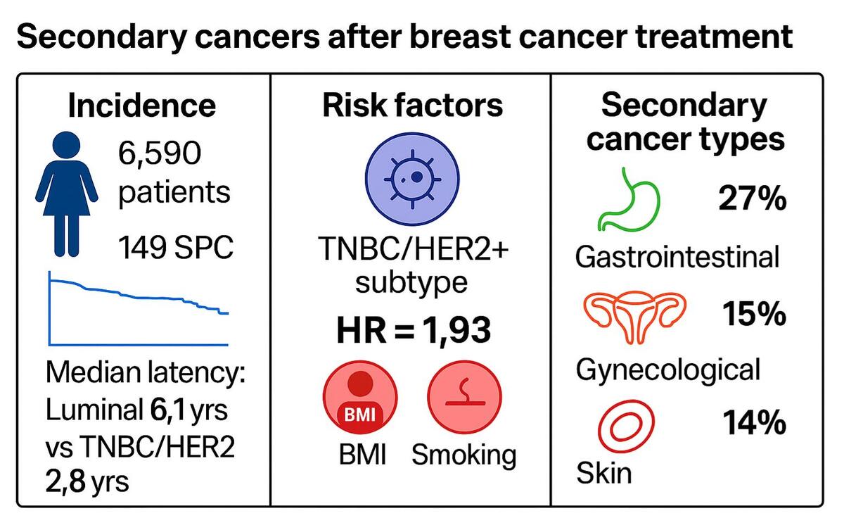
This retrospective, hospital-based cohort study included 6590 female patients from 2 centres – in Poznan (the majority of participants) and Gorzow Wielkopolski. From this group, 149 cases were identified who were diagnosed between 2017 and 2022 with a second, secondary malignancy after previous breast cancer treatment. It is important to note that the initial diagnosis and treatment of primary breast cancer in these patients occurred over a much broader time span, in some cases more than 40 years earlier. Patient data were collected through a systematic review of hospital electronic medical records and institutional oncology databases at both participating centres. All women treated for primary breast cancer between 2017 and 2022 were screened, and cases of second primary malignancies were identified based on pathology reports and clinical documentation. To ensure data completeness, only patients with full diagnostic and treatment records available within the participating institutions were included. Because some patients may have sought treatment for subsequent cancers at other hospitals, this approach may have introduced selection bias. To minimise this risk, cross-checks were performed against regional cancer registries and pathology archives whenever feasible, and cases with incomplete documentation were excluded from the analysis.
Exclusion criteria: patients with prior malignancies, synchronous cancers, or incomplete clinical records. Second primary malignancies were defined as histologically distinct cancers diagnosed at least 6 months after the initial breast cancer diagnosis. Verification of second cancers was performed through pathology reports and clinical documentation. Censoring was applied at the date of last contact or death.
The scope of the analysis included descriptive statistics on the time to detection of the second malignancy, analysis of the relationship between treatment categories and the occurrence of the second malignancy, and assessment of the impact of therapy on the time to its development.
Patient characteristics were recorded under the following headings:
Incidence of breast carcinoma among admitted patients.
Patient characteristics (sex, age, weight, height, number of surgeries, smoking status).
Cancer type.
Treatment used (chemotherapy, hormone therapy, and radiotherapy, either as monotherapy or combined therapies).
Post-treatment characteristics (time to detection of the second malignancy, mortality).
Data were collected in a spreadsheet. Sensitive personal data such as surname, first name, postal code, town, and patients’ phone numbers were removed to ensure anonymity and compliance with data protection regulations. Each patient was assigned a unique patient identifier (ID) to allow for unambiguous record identification during analysis. The patients’ age at the time of the first surgery was calculated based on the difference between the date of the first surgery and date of birth. A new variable “Treatment” was created based on the variables “Chemotherapy”, “Hormone Therapy”, and “Radiotherapy”, indicating whether the patient received monotherapy or combined treatment.
Adjuvant treatment decisions were made based on contemporary national and international guidelines, tailored to the individual patient’s disease stage, biological subtype, and overall health. Common chemotherapeutic regimens included anthracycline-based and/or taxane-based protocols. Patients with HER2-positive disease received anti-HER2 targeted therapy (trastuzumab with or without pertuzumab) concurrently with chemotherapy. Endocrine therapy for hormone receptor-positive disease consisted of tamoxifen or aromatase inhibitors (letrozole, anastrozole). Radiotherapy was administered following breast-conserving therapy and in selected high-risk cases post-mastectomy, with standard fractionation delivering 45–50 Gy to the whole breast/chest wall, often followed by a tumour bed boost. To ensure high quality of statistical analysis, a data cleaning and standardisation process was carried out. These actions aimed to eliminate errors, unify data formats, and improve the consistency of variables in the dataset of patients who underwent mastectomy or breast-conserving therapy.
Statistical analysis
All statistical analyses were conducted using complete case data to maximise analytical reliability and avoid the bias associated with data imputation. Statistical significance was evaluated at the conventional threshold of α = 0.05 (Type I error), and all reported p-values are two-sided. All computations and data visualisations were performed using R version 4.4.3 (R Core Team, 2024). Descriptive statistics were computed for demographic (e.g. age, BMI) and clinical variables (e.g. cancer subtype, treatment type) using means with standard deviations and medians with interquartile ranges for continuous variables, and counts with percentages for categorical data. Group comparisons for continuous variables were conducted using Welch’s permutation t-test with 10,000 iterations to ensure robustness against violations of the normality and homoscedasticity assumptions. The nonparametric nature of the test enhances inferential reliability, particularly in scenarios involving small or imbalanced samples. Pearson’s chi-square test of independence was used to evaluate associations between categorical variables, such as cancer subtypes and treatment types. Monte Carlo simulation with 10,000 replicates was employed to estimate p-values, thereby increasing the accuracy of inference in contingency tables with small expected cell counts. The magnitude of these associations was subsequently quantified by computing Cramér’s V and interpreting the result via the Fei scale. This scale delineates effect sizes as negligible (≤ 0.1), weak (0.1–0.3), moderate (0.3–0.5), and strong (≥ 0.5). To assess the multivariate relationships between cancer treatment modalities and the occurrence of second primary malignancies, multinomial logistic regression models were fitted. To assess the main effects and interactions, the marginal means and Type II ANOVA were applied. To complement the regression analysis, correspondence analysis was conducted to visually explore the complex interdependencies between the categorical variables in a low-dimensional space. The time-to-event data, specifically the time interval between primary breast cancer surgery and the diagnosis of a subsequent malignancy, were subjected to analysis using Kaplan-Meier survival curves and log-rank tests to facilitate group comparisons. Furthermore, the employment of Cox proportional hazards regression models was undertaken to quantify the effect of clinical covariates (e.g. treatment type, cancer subtype, age, BMI, smoking status, number of surgeries) on the risk of developing a second malignancy. Proportional hazards assumptions were verified using Schoenfeld residuals; no violations were detected.
Results
The study focused on a cohort of 149 patients who were diagnosed with a second primary cancer following their treatment for breast cancer. Comprehensive demographic and clinical data pertaining to these patients are meticulously detailed in Table I, which includes variables such as age at diagnosis and treatment history.
Table I
Demographic and clinical characteristics of patients (n = 149) undergoing mastectomy/BCT – overall
In the analysed group of patients who underwent mastectomy or breast-conserving therapy, the demographic and clinical characteristics were also compared based on the subtype of the initial breast cancer (Table II).
Table II
Demographic and clinical characteristics of patients undergoing mastectomy/BCT – relative to breast cancer subtype one
Most patients in both groups underwent surgery, with 95% in the luminal group and 96% in the triple-negative breast cancer and HER2-positive breast cancer (TNBC and HER2+) group. The primary type of intervention performed was mastectomy, accounting for 59% in the luminal group and 61% in the TNBC + HER2+ group. There was no significant difference in the frequency of various surgical procedures between the groups (p > 0.9). On average, patients in the TNBC + HER2+ group were younger at the time of surgery, with an average age of 55 years (SD = 13), compared to an average age of 60 years (SD = 10) for patients with luminal cancer. However, this age difference did not reach statistical significance (p = 0.3). The most common breast cancer subtype was luminal cancer, which was found in 94 (80%) patients. The combined subtypes of TNBC and HER2+ were identified in 23 (20%) patients. Among the study group of patients diagnosed with a second cancer, malignant tumours of the digestive system were the most prevalent, constituting 27% of cases. Gynaecological cancers were the second largest category at 15%, followed by non-melanoma skin tumours at 14%. Respiratory and thoracic cancers accounted for 9.4% of patients, while haematological and urological cancers each represented 8.7% of cases. Breast cancer was noted as a second cancer in 5.4% of instances. Other less common cancers included thyroid, eye, brain, central nervous system tumours, and tumours of uncertain or unknown nature, as well as in situ tumours, making up a total of 6% of cases (Figure 1 and Table III).
Figure 1
Distribution of second cancer occurrence depending on the subtype of the first breast cancer among 117 patients. The left bar represents luminal subtype; the right bar represents TNBC + HER2+ subtypes
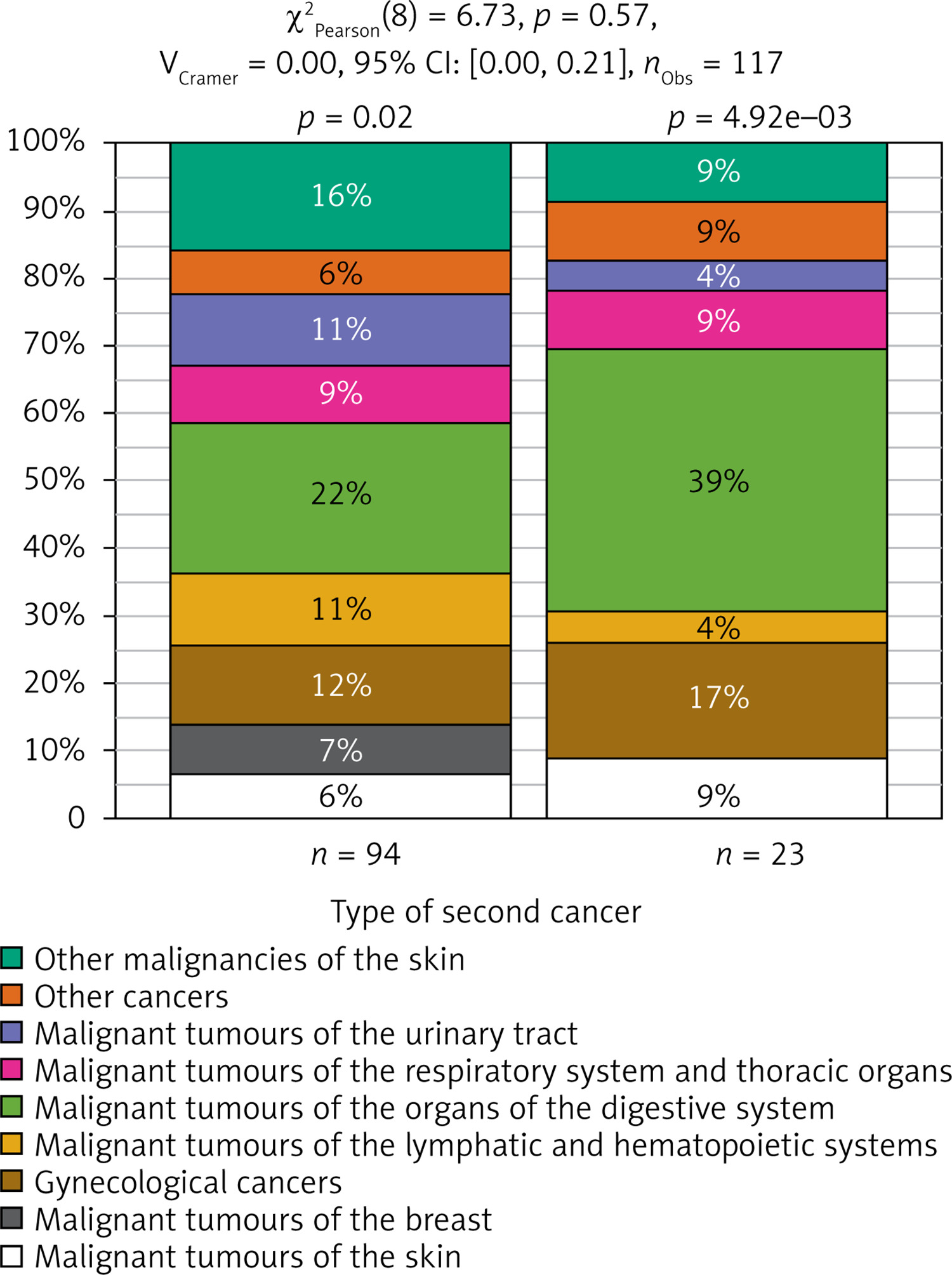
Table III
Most frequent secondary cancer sites in the study cohort (N = 149)
In the analysis of the patient cohort, chemotherapy emerged as the predominant treatment modality, with 97 (70%) patients receiving this intervention, while 42 (30%) patients did not. Data were missing for 10 patients in this category. A comparable distribution was noted for hormone therapy, with 97 (71%) patients engaged in this treatment, 40 (29%) patients not receiving it, and incomplete data recorded for 12 cases. Radiotherapy was employed in 76 (54%) patients, whereas 65 (46%) patients did not undergo this treatment, with data missing for 8 cases. Upon reviewing the combinations of treatments, the most prevalent regimen was the combination of chemotherapy, radiotherapy, and hormone therapy, which constituted 25% of the cases. This was followed by the combination of chemotherapy and hormone therapy, at 19%, and the combination of chemotherapy and radiotherapy, at 15%. Moreover, the analysis indicated several monotherapy options: hormone therapy (15%), chemotherapy (9.7%), and radiotherapy (3%). Notably, 2.2% of patients did not receive any form of treatment.
Analysis of the relationship between subtypes of the first and second cancer types
Among the 94 patients classified with the luminal subtype, the most frequently observed malignancies were gastrointestinal cancers (18%) and non-melanoma skin cancers (13%). Additionally, there were instances of gynaecological cancers (9.4%), haematological malignancies (8.5%), urinary cancers (8.5%), and respiratory and thoracic cancers (6.8%). In the subgroup consisting of 23 patients with TNBC and HER2-positive subtypes, gastrointestinal malignancies were notably prevalent, comprising 7.7% of the cases. The p-value obtained from the χ2 test of independence was 0.5, indicating that there is no statistically significant relationship between the subtype of the primary breast cancer and the classification of the subsequent malignancy.
Analysis of the impact of treatment on the risk of second cancer
Table IV illustrates the distribution of categories of second malignancies by treatment type in patients with luminal subtype cancers. The analysis included 8 cancer categories and 8 treatment regimens, which encompassed no treatment, monotherapies, and combination therapies (chemotherapy, hormone therapy, and radiotherapy). The predominant categories of second malignancies observed were gastrointestinal malignancies (23% of patients), non-melanoma skin tumours (15% of patients), and gynaecological cancers (12% of patients). The most frequently utilised treatment regimen was the combination of chemotherapy with radiotherapy and hormone therapy, accounting for 29% of patients; conversely, no treatment was reported in a single patient (1.1%). Statistical analysis performed using the χ2 test with a simulated p-value yielded a value of p = 0.83, suggesting no significant relationship between treatment regimen and malignancy category. Table V presents the distribution of second malignancy categories among 23 patients with triple-negative breast cancer and HER2-positive subtypes, organised by treatment received. This analysis included 7 cancer categories and 4 treatment regimens, which featured chemotherapy alone and various combinations of therapies (chemotherapy alongside hormone therapy and/or radiotherapy). The most prevalent categories of second malignancies identified were gastrointestinal malignancies (39% of patients) and gynaecological cancers (17% of patients). The most common treatment regimen was chemotherapy combined with radiotherapy, utilised by 43% of patients. The χ2 test with a simulated p-value produced a value of p = 0.41, indicating no statistically significant relationship between treatment and the category of second malignancy.
Table IV
Distribution of second cancers depending on the treatment used among 91 patients with the luminal subtype
| Treatment | None | Chemo | Chemo + Hormo | Chemo + Radio | Chemo + Radio + Hormo | Hormo | Radio | Radio + Hormo | Total | P-value1 |
|---|---|---|---|---|---|---|---|---|---|---|
| Category of second cancer | 0.92 | |||||||||
| Malignant melanoma of the skin | 0 (0%) | 1 (1.1%) | 3 (3.3%) | 0 (0%) | 1 (1.1%) | 0 (0%) | 0 (0%) | 1 (1.1%) | 6 (6.6%) | |
| Malignant tumour of the breast | 0 (0%) | 0 (0%) | 2 (2.2%) | 0 (0%) | 2 (2.2%) | 1 (1.1%) | 0 (0%) | 2 (2.2%) | 7 (7.7%) | |
| Malignant tumours of the female genital organs | 1 (1.1%) | 0 (0%) | 3 (3.3%) | 0 (0%) | 2 (2.2%) | 3 (3.3%) | 0 (0%) | 2 (2.2%) | 11 (12%) | |
| Malignant tumours of the lymphatic and hematopoietic systems | 0 (0%) | 1 (1.1%) | 2 (2.2%) | 0 (0%) | 4 (4.4%) | 1 (1.1%) | 1 (1.1%) | 1 (1.1%) | 10 (11%) | |
| Malignant tumours of the organs of the digestive system | 0 (0%) | 0 (0%) | 4 (4.4%) | 1 (1.1%) | 4 (4.4%) | 8 (8.8%) | 0 (0%) | 4 (4.4%) | 21 (23%) | |
| Malignant tumours of the respiratory system and thoracic organs | 0 (0%) | 1 (1.1%) | 0 (0%) | 0 (0%) | 2 (2.2%) | 2 (2.2%) | 0 (0%) | 3 (3.3%) | 8 (8.8%) | |
| Malignant tumours of the urinary tract | 0 (0%) | 0 (0%) | 2 (2.2%) | 1 (1.1%) | 4 (4.4%) | 0 (0%) | 0 (0%) | 1 (1.1%) | 8 (8.8%) | |
| Other cancers | 0 (0%) | 0 (0%) | 1 (1.1%) | 0 (0%) | 3 (3.3%) | 1 (1.1%) | 0 (0%) | 1 (1.1%) | 6 (6.6%) | |
| Other malignancies of the skin | 0 (0%) | 1 (1.1%) | 4 (4.4%) | 0 (0%) | 4 (4.4%) | 2 (2.2%) | 1 (1.1%) | 2 (2.2%) | 14 (15%) | |
| Total | 1 (1.1%) | 4 (4.4%) | 21 (23%) | 2 (2.2%) | 26 (29%) | 18 (20%) | 2 (2.2%) | 17 (19%) | 91 (100%) | |
Table V
The distribution of second cancers depending on the treatment used among 23 patients with TNBC and HER2+ subtypes
| Treatment | None | Chemo | Chemo + Hormo | Chemo + Radio | Chemo + Radio + Hormo | Hormo | Radio | Radio + Hormo | Total | P-value1 |
|---|---|---|---|---|---|---|---|---|---|---|
| Category of second cancer | 0.40 | |||||||||
| Malignant melanoma of the skin | 0 (0%) | 1 (4.3%) | 1 (4.3%) | 0 (0%) | 0 (0%) | 0 (0%) | 0 (0%) | 0 (0%) | 2 (8.7%) | |
| Malignant tumours of the female genital organs | 0 (0%) | 2 (8.7%) | 0 (0%) | 1 (4.3%) | 1 (4.3%) | 0 (0%) | 0 (0%) | 0 (0%) | 4 (17%) | |
| Malignant tumours of the lymphatic and hematopoietic systems | 0 (0%) | 0 (0%) | 0 (0%) | 1 (4.3%) | 0 (0%) | 0 (0%) | 0 (0%) | 0 (0%) | 1 (4.3%) | |
| Malignant tumours of the organs of the digestive system | 0 (0%) | 0 (0%) | 2 (8.7%) | 5 (22%) | 2 (8.7%) | 0 (0%) | 0 (0%) | 0 (0%) | 9 (39%) | |
| Malignant tumours of the respiratory system and thoracic organs | 0 (0%) | 0 (0%) | 0 (0%) | 2 (8.7%) | 0 (0%) | 0 (0%) | 0 (0%) | 0 (0%) | 2 (8.7%) | |
| Malignant tumours of the urinary tract | 0 (0%) | 0 (0%) | 1 (4.3%) | 0 (0%) | 0 (0%) | 0 (0%) | 0 (0%) | 0 (0%) | 1 (4.3%) | |
| Other cancers | 0 (0%) | 0 (0%) | 0 (0%) | 1 (4.3%) | 1 (4.3%) | 0 (0%) | 0 (0%) | 0 (0%) | 2 (8.7%) | |
| Other malignancies of the skin | 0 (0%) | 0 (0%) | 1 (4.3%) | 0 (0%) | 1 (4.3%) | 0 (0%) | 0 (0%) | 0 (0%) | 2 (8.7%) | |
| Total | 0 (0%) | 3 (13%) | 5 (22%) | 10 (43%) | 5 (22%) | 0 (0%) | 0 (0%) | 0 (0%) | 23 (100%) | |
Analysis of time to detection of second cancer
In the cohort of patients characterised by luminal subtype (N = 86), the mean time until detection of a second malignancy was 8.4 years (SD = 7.1). The median duration was recorded at 6.1 years (IQR: 3.0–11.0), with extreme values spanning from 0.4 to 41.0 years. This wide range of follow-up times reflects the retrospective nature of the dataset. The time to detection of a second malignancy was calculated as the interval between the date of the initial breast cancer surgery and the date of diagnosis of the second cancer. Conversely, patients diagnosed with the TNBC or HER2+ subtypes (N = 22) exhibited a reduced mean time to the detection of a second malignancy, averaging 5.2 years (SD = 7.2). The median time within this subgroup was 2.8 years (IQR: 1.0–7.0), with a range of 0.3 to 33.3 years. Figure 2 delineates the Kaplan-Meier survival curves illustrating the time to detection of a second cancer across the 2 patient groups: those with the luminal subtype and those with HER2+ or TNBC. The curves highlight the distinctions in the timing of second cancer diagnoses between the investigated cohorts.
Figure 2
Kaplan-Meier survival curve for time to detection of a second cancer (in years) depending on the subtype of the first breast cancer
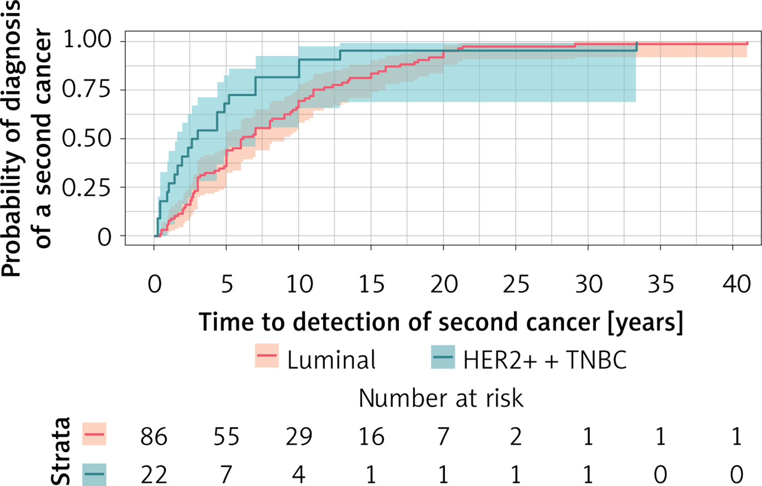
It is evident that within the TNBC + HER2+ subgroup, there is an accelerated detection of second cancers, thereby affirming the statistical significance of the observed differences between the groups (p = 0.024). The confidence intervals presented indicate a higher level of uncertainty regarding estimates for the TNBC + HER2+ group, which can be attributed to the smaller sample size of this cohort. Additionally, the table positioned below the graph details the number of patients remaining under observation in the subsequent years.
A faster “dropout” of patients from the TNBC + HER2+ group is clearly visible, which additionally emphasises the shorter time to occurrence of the second cancer. These results indicate significant differences in the risk of developing a second cancer depending on the subtype of the first breast cancer.
Analysis of the effect of treatment on time to second malignancy detection
Table VI presents the descriptive statistics for the time until the detection of a second malignancy (measured in years) categorised by treatment type. The median time to the detection of a second malignancy exhibited significant variation among different treatment groups. The longest median duration was observed in patients who received chemotherapy, with a median of 9.8 years (standard deviation = 9.5), as well as in those who underwent combined chemotherapy and hormone therapy, showing a median of 8.5 years (standard deviation = 6.3). Conversely, the shortest median time was noted in patients who did not receive any postoperative treatment, with a median of 1.8 years (standard deviation not available due to a single observation), and in the group that received radiotherapy alone, which had a median of 5.9 years (standard deviation = 4.5). Additionally, the median timing for the detection of a second malignancy varied considerably among groups. The highest median was recorded for patients who underwent chemotherapy, at 8.5 years (interquartile range: 1.0–19.5), while the lowest was observed in patients who received no treatment, with a median of 1.8 years (no interquartile range available). The range of results (minimum–maximum) indicated substantial variability among groups, particularly in patients receiving combined chemotherapy and radiotherapy, with a range spanning from 0.3 to 33.3 years. It is important to note that some treatment groups, such as those receiving no treatment or radiotherapy alone, were relatively small, which may have implications for the stability and generalisability of these findings.
Table VI
Descriptive statistics of time to detection of a second cancer (in years) depending on the treatment used after mastectomy/BCT surgery
Modelling the impact of first breast subtype, treatment, and specialised covariates on time to subsequent cancer detection
In the initial analysis presented in Table VII, we evaluated the impact of tumour subtype as well as specific treatment modalities – including chemotherapy, hormone therapy, and radiotherapy – on the time to the onset of a second cancer. None of the treatments examined showed a statistically significant effect on risk. Notably, the TNBC + HER2+ subtype displayed a tendency towards an increased risk (hazard ratio = 1.51, p = 0.2); however, this finding did not achieve statistical significance.
Table VII
Results of Cox proportional hazards model analysis for assessing the impact of factors related to the subtype of the first breast cancer and the treatment used after mastectomy/BCT on the time to diagnosis of the second cancer (in years) – impact of individual therapies
In the second analysis (Table VIII), which evaluated combinations of therapies (e.g. chemotherapy combined with hormone therapy), the triple-negative breast cancer subtype with HER2 positivity was found to be significantly associated with an elevated risk (hazard ratio [HR] = 1.93, p = 0.048). Additionally, certain combinations of therapies, such as chemotherapy plus hormone therapy (HR = 0.53, p = 0.061) and chemotherapy combined with radiotherapy and hormone therapy (HR = 0.58, p = 0.087), demonstrated a tendency to lower the risk, although these findings did not achieve statistical significance.
Table VIII
Results of Cox proportional hazards model analysis for assessing the impact of factors related to the subtype of the first breast cancer and the treatment used after mastectomy/BCT on the time to diagnosis of the second cancer (in years) – impact of monotherapy and combination therapy
Key findings
Patients diagnosed as TNBC or HER2 positive exhibited a considerably shorter median duration until the detection of a second malignancy, recorded at just 2.8 years. In stark contrast, individuals belonging to the luminal subtype experienced a much longer median interval of 6.1 years before a subsequent cancer was identified (p = 0.024). Furthermore, the TNBC and HER2+ subtypes were linked to a 1.9-fold higher risk of developing a second cancer when analysed through the Cox proportional hazards model (hazard ratio = 1.93; p = 0.048). This finding underscores the aggressive nature of this breast cancer subtype and its implications for long-term patient outcomes.
An evaluation of the various treatment regimens utilised by patients revealed no statistically significant correlation between the type of treatment administered – whether single or combination therapies – and the classification of the second cancer that subsequently developed (p > 0.05, as indicated by χ2 tests). However, when analysing the timeline to the emergence of a second malignancy, there emerged a suggestive trend associated with combination therapies, such as the concurrent use of chemotherapy and hormone therapy. These combinations appeared to be linked to a reduced risk of developing a second cancer, as reflected in the hazard ratio of 0.53 (p = 0.061), although these results did not achieve the threshold for statistical significance. This trend suggests potential benefits that may warrant further investigation in future studies.
Discussion
There have been multiple studies assessing the risks of second primary cancers following breast cancer in women. It is well-documented that breast cancer survivors face a 20–50% increased risk of developing additional cancers at other sites compared to the general population, especially in those diagnosed with breast cancer below 50 years of age [10]. However, few studies have examined the effects of patient demographics, breast cancer pathology, or treatments for the initial breast cancer on the risks of developing second primary cancers [13]. With the improvement of survival outcomes after breast cancer treatment, there has been growing interest in understanding the long-term effects of these therapies. Genetic and hormonal factors may contribute to the heightened risk, as can the treatments themselves – particularly radiation, chemotherapy, and hormonal therapies [14]. Similar investigations are ongoing worldwide, as cancer epidemiology varies significantly due to a multitude of factors, including environmental influences, socioeconomic conditions, and lifestyle choices. Tailoring healthcare solutions requires comprehensive research into the scale of the problem within specific populations [13]. In our analysis of 149 patients, we aimed to determine the following: the type of second cancer following breast cancer treatment, the time interval between the diagnosis of the second cancer and surgical treatment, the influence of the biological subtype of breast cancer (luminal versus TNBC and HER2+) on the timing and type of subsequent cancers, and the relationship between treatment regimens, focusing on key therapy categories without delving into specific treatment protocols. In Poland, 2022 data indicated that breast cancer accounted for 23.6% of all cancer cases in women [2]. Among our study cohort of 149 patients with second primary cancers, the epidemiological distribution was as follows: 27% digestive system cancers, 15% gynaecological cancers, 14% other skin malignancies, 9.4% respiratory system cancers, 8.7% lymphatic and haematopoietic system malignancies, and 6% melanoma, among others. Our findings reveal that the morbidity rate in this patient group significantly diverges from the general population [10–13].
In our analysis, we included second primary breast cancers and non-melanoma skin cancers (NMSC) as secondary malignancies. This decision was based on established epidemiological definitions, where any histologically distinct malignancy occurring in a different anatomical site or contralateral organ is considered a second primary cancer. However, we acknowledge that the number of second breast cancers observed in our cohort was relatively low, which may be due to incomplete reporting or documentation in the source data. Only cases with sufficient clinical and pathological information to distinguish them from recurrences or metastases were included. Similarly, although NMSC is often underreported, it remains one of the most common malignancies and is relevant in the context of long-term cancer survivorship.
The development of SPCs is often attributed to overlapping risk factors shared between breast cancer and other malignancies. In our cohort, the average body mass index (BMI) was 27.4 kg/m2;, indicating an overweight population, and 40% of patients reported smoking – nearly double the rate observed in the general population (approximately 21%). Both elevated BMI and smoking are well-established risk factors for several cancers, including colorectal, endometrial, and lung cancers. Although our study did not demonstrate a statistically significant association between these factors and SPC occurrence, their high prevalence suggests a potential contribution to overall cancer risk and highlights the importance of lifestyle interventions in survivorship care.
The influence of treatment on the risk of developing a second cancer remains a crucial question in oncology. Our findings are consistent with recent studies, including Zhang et al. [15], who reported a 10-year cumulative incidence of 3.2% for secondary non-breast cancers in young breast cancer survivors. Furthermore, Berrington de Gonzalez et al. [9] demonstrated that radiotherapy is associated with an increased risk of second solid cancers, particularly in tissues receiving higher radiation doses. Although our dataset did not allow for detailed dosimetric analysis, we acknowledge this as a limitation and emphasise the importance of further research on the long-term effects of radiotherapy. The potential for treatment-induced malignancies, even years after completion, is a well-recognised phenomenon. For instance, chemotherapy has been linked to myeloid leukaemia [13], tamoxifen to endometrial cancer [16], and radiotherapy to angiosarcomas in treated areas [13]. Our analysis did not find a significant correlation between the biological subtype of breast cancer (luminal vs. HER2+/TNBC) and the category of second cancers developed. As medical practices evolve, there is a continuous search for more effective and safer treatment methods that enhance recovery prospects while minimising complications. Genetic susceptibility also likely accounts for a significant proportion of second primary cancers following breast cancer in women. Variants in genes associated with breast cancer risk, such as BRCA1/2, may also heighten risks for other cancers like ovarian, pancreatic, and stomach cancers [17]. However, our analysis did not include genetic testing data due to its limited availability in many cases. Looking ahead, genetic testing could play a vital diagnostic, prognostic, and therapeutic role, enabling breast cancer survivors to undergo heightened surveillance for specific second primary cancers. Recommendations for specific monitoring strategies would necessitate thorough cost-benefit analyses. Furthermore, understanding genetic factors may warrant prophylactic surgeries such as breast and ovarian resections [18]. Interestingly, we observed differing times to the detection of second cancers based on the biological subtype of breast cancer. Patients with the luminal subtype typically developed subsequent cancers after an average of 6.1 years, whereas those with HER2+ or TNBC had a shorter interval of around 2.8 years. The reasons behind this discrepancy warrant further investigation to elucidate the underlying mechanisms.
Our findings align with large population-based studies such as SEER [10–13], EPIC [5], and Kaiser Permanente [4], which consistently report elevated risks of second primary cancers among breast cancer survivors. In particular, Allen et al. (2024) [19] and Mukherjee et al. (2025) [20] highlight the role of tumour biology and comorbidity burden in shaping SPC risk profiles.
The shorter latency observed in HER2-positive and TNBC subtypes may be explained by underlying genetic predispositions, such as BRCA1/2 mutations [17, 18], and the intensity of adjuvant therapies. These subtypes are often associated with more aggressive treatment regimens, which may accelerate the emergence of therapy-related malignancies.
One of the limitations of our study is the lack of complete data on tumour stage, histological grade, and the presence of metastases at the time of primary diagnosis. Due to the high proportion of missing or inconsistent entries, we were unable to include these variables in our statistical models. Future studies should aim to incorporate these important clinical parameters to better understand their potential role in secondary cancer development. Additionally, treatment protocols varied over time, as patients received therapies that were standard at the time of their initial breast cancer diagnosis. This temporal variability in treatment approaches may have influenced the risk of secondary malignancies and should be considered when interpreting the results.
The emergence of pathological changes in various organs subsequent to prior cancer treatment presents significant diagnostic challenges. It is imperative to differentiate between a recurrence of breast cancer manifesting as metastases and the occurrence of a new primary cancer. This differentiation necessitates close collaboration among specialists, including oncologists, radiologists, and pathologists, to ensure the formulation of an appropriate treatment strategy. Additionally, it is vital to investigate the factors that may have contributed to the onset of a second cancer. Such factors may encompass the effects of cancer therapies, genetic alterations that predispose individuals to multiple malignancies, and modifiable environmental, lifestyle, dietary, and substance use factors that could influence the development of both breast cancer and a secondary cancer.
In conclusion, our study validates the significant role that the biology of the initial breast cancer plays in the future health outcomes of survivors. We discovered that women diagnosed with more aggressive subtypes, such as triple-negative breast cancer or HER2-positive breast cancer, were at a heightened risk of developing a second, unrelated cancer at a much faster rate compared to those with luminal subtypes. This observation indicates that the genetic or molecular factors associated with aggressive breast cancer may foster an environment conducive to the expedited emergence of additional cancers. While we meticulously examined various treatment modalities, including chemotherapy, radiation therapy, and hormone therapy, we did not identify a definitive, consistent correlation between any specific treatment and an increased incidence of second cancers. Notably, we observed a promising trend indicating that patients who underwent combination therapies appeared to exhibit a lower risk; however, this hypothesis requires validation through more extensive studies. The nature of the second cancers observed was distinct from those typically seen in the general population, with gastrointestinal malignancies being the most prevalent. Ultimately, our findings support the argument for a more personalised approach to long-term follow-up care. Rather than employing a universal surveillance strategy, we advocate for the customisation of monitoring tactics based on a patient’s original cancer subtype. Recognising that an individual had a TNBC or HER2-positive tumour should prompt more vigilant and potentially earlier screening for other cancers, thereby enhancing the likelihood of early detection and improved patient outcomes.

