Current issue
Archive
Manuscripts accepted
About the Journal
Editorial office
Editorial board
Section Editors
Abstracting and indexing
Subscription
Contact
Ethical standards and procedures
Most read articles
Instructions for authors
Article Processing Charge (APC)
Regulations of paying article processing charge (APC)


DIABETOLOGY / STATE OF THE ART PAPER
Molecular and cellular crosstalk in the pathogenesis of diabetic retinopathy
1
Faculty of Pharmacy and Health Sciences, Universiti Kuala Lumpur Royal College of Medicine Perak, Malaysia
2
Department of Pharmacology, Faculty of Medicine, Manipal University College Malaysia, Malaysia
3
Department of Medical Education, Faculty of Medicine, Universiti Teknologi MARA, Malaysia
4
Centre for Neuroscience Research (NeuRon), Faculty of Medicine, Universiti Teknologi MARA, Malaysia
These authors had equal contribution to this work
Submission date: 2025-11-10
Final revision date: 2026-02-22
Acceptance date: 2026-04-14
Online publication date: 2026-07-25
Corresponding author
Muhammad Zulfiqah Sadikan
Faculty of Pharmacy and Health Sciences Universiti Kuala Lumpur Royal College of Medicine Perak Department of Pharmacology Faculty of Medicine Manipal University College Malaysia, Malaysia
Faculty of Pharmacy and Health Sciences Universiti Kuala Lumpur Royal College of Medicine Perak Department of Pharmacology Faculty of Medicine Manipal University College Malaysia, Malaysia
Nurul Alimah Abdul Nasir
Department of Medical Education Centre for Neuroscience Research (NeuRon) Faculty of Medicine Universiti Teknologi MARA, Malaysia
Department of Medical Education Centre for Neuroscience Research (NeuRon) Faculty of Medicine Universiti Teknologi MARA, Malaysia
KEYWORDS
TOPICS
ABSTRACT
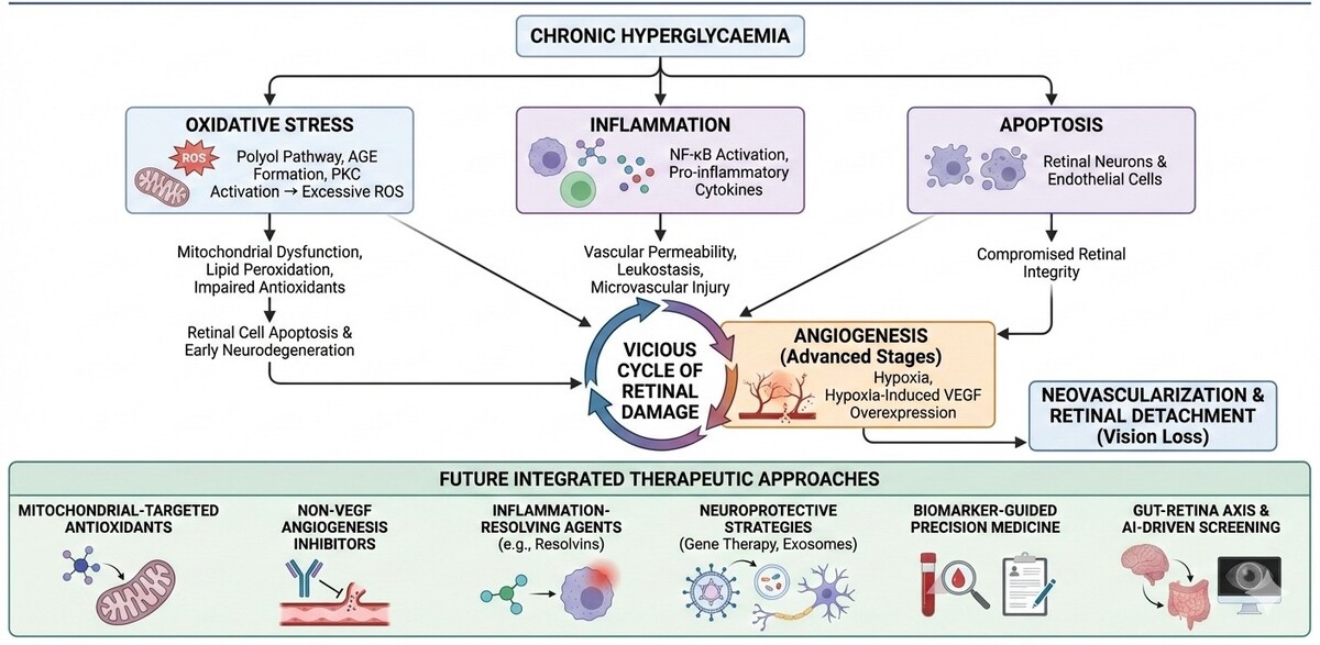
Diabetic retinopathy is a major cause of vision loss worldwide and arises from interconnected molecular mechanisms involving oxidative stress, inflammation, apoptosis, and angiogenesis. Chronic hyperglycaemia induces excessive reactive oxygen species production through pathways such as advanced glycation end-product formation and protein kinase C activation, leading to mitochondrial dysfunction, impaired antioxidant defences, and retinal cell apoptosis. In parallel, activation of inflammatory signalling cascades, including NF-κB–mediated cytokine release, promotes leukostasis, vascular permeability, and microvascular injury. Progressive neuronal and endothelial apoptosis further disrupts retinal integrity. At later stages, sustained hypoxia triggers VEGF overexpression and pathological angiogenesis. These processes reinforce one another, creating a self-amplifying pathogenic cycle. Effective disease control therefore requires integrated strategies that address multiple pathways simultaneously, including modulation of oxidative stress, suppression of chronic inflammation, preservation of retinal neuronal survival, and regulation of aberrant angiogenic signalling. Targeting shared upstream drivers is critical for slowing progression and limiting irreversible retinal damage.
REFERENCES (319)
1.
Kropp M, Golubnitschaja O, Mazurakova A, Koklesova L, Sargheini N, Vo T-TKS, et al. Diabetic retinopathy as the leading cause of blindness and early predictor of cascading complications—Risks and mitigation. EPMA Journal. 2023;14(1):21–42.
2.
Ptito M, Bleau M, Bouskila J. The retina: A window into the brain. MDPI; 2021. p. 3269.
3.
Abou Taha A, Dinesen S, Vergmann AS, Grauslund J. Present and future screening programs for diabetic retinopathy: a narrative review. International Journal of Retina and Vitreous. 2024;10(1):14.
4.
Sadikan MZ, Nasir NAA. Unravelling epidemiological trends and risk disparities in diabetic retinopathy. 2025.
5.
Dai Y, Chen H, Yu J, Cai J, Lu B, Dai M, et al. Global and regional trends in the incidence and prevalence of uterine fibroids and attributable risk factors at the national level from 2010 to 2019: A worldwide database study. Chinese Medical Journal. 2024:10.1097.
7.
Rastogi A, Rizvi TZ, Kanan D, editors. Diabetic Retinopathy-An Ensemble Approach. 2023 International Conference on Computer, Electrical & Communication Engineering (ICCECE); 2023: IEEE.
8.
Dutta S, Manideep B, Basha SM, Caytiles RD, Iyengar N. Classification of diabetic retinopathy images by using deep learning models. International Journal of Grid and Distributed Computing. 2018;11(1):89–106.
9.
Seewoodhary M. An overview of diabetic retinopathy and other ocular complications of diabetes mellitus. Eye. 2020.
10.
Mrowicka M, Mrowicki J, Majsterek I. Relationship between Biochemical Pathways and Non-Coding RNAs Involved in the Progression of Diabetic Retinopathy. Journal of Clinical Medicine. 2024;13(1):292.
11.
Wan T-T, Li X-F, Sun Y-M, Li Y-B, Su Y. Recent advances in understanding the biochemical and molecular mechanism of diabetic retinopathy. Biomedicine & pharmacotherapy. 2015;74(1):145–7.
12.
Lorenzi M. The polyol pathway as a mechanism for diabetic retinopathy: attractive, elusive, and resilient. Experimental diabetes research. 2007;2007(16):61038.
13.
Niimi N, Yako H, Takaku S, Chung SK, Sango K. Aldose Reductase and the Polyol Pathway in Schwann Cells: Old and New Problems. International Journal of Molecular Sciences. 2021;22(3):1031.
14.
Hotta N, Kawamura T, Umemura T. Are the polyol pathway and hyperuricemia partners in the development of non‐alcoholic fatty liver disease in diabetes? Journal of diabetes investigation. 2020;11(4):786–8.
15.
Haydinger CD, Oliver GF, Ashander LM, Smith JR. Oxidative stress and its regulation in diabetic retinopathy. Antioxidants. 2023;12(8):1649.
16.
Yan Lj. Redox imbalance stress in diabetes mellitus: Role of the polyol pathway. Animal models and experimental medicine. 2018;1(1):7–13.
17.
Mathebula SD. Polyol pathway: A possible mechanism of diabetes complications in the eye. African vision and eye health. 2015;74(1):5.
18.
Li W, Chen S, Mei Z, Zhao F, Xiang Y. Polymorphisms in sorbitol-aldose reductase (Polyol) pathway genes and their influence on risk of diabetic retinopathy among Han Chinese. Medical science monitor: international medical journal of experimental and clinical research. 2019;25(1):7073.
19.
Dagher Z, Park YS, Asnaghi V, Hoehn T, Gerhardinger C, Lorenzi M. Studies of rat and human retinas predict a role for the polyol pathway in human diabetic retinopathy. Diabetes. 2004;53(9):2404–11.
20.
Asnaghi V, Gerhardinger C, Hoehn T, Adeboje A, Lorenzi M. A role for the polyol pathway in the early neuroretinal apoptosis and glial changes induced by diabetes in the rat. Diabetes. 2003;52(2):506–11.
21.
Obrosova IG, Minchenko AG, Vasupuram R, White L, Abatan OI, Kumagai AK, et al. Aldose reductase inhibitor fidarestat prevents retinal oxidative stress and vascular endothelial growth factor overexpression in streptozotocin-diabetic rats. Diabetes. 2003;52(3):864–71.
22.
Iqbal Z, Morahan G, Arooj M, Sobolev AN, Hameed S. Synthesis of new arylsulfonylspiroimidazolidine-2ʹ, 4ʹ-diones and study of their effect on stimulation of insulin release from MIN6 cell line, inhibition of human aldose reductase, sorbitol accumulations in various tissues and oxidative stress. European journal of medicinal chemistry. 2019;168(1):154–75.
23.
Wojnar W, Zych M, Borymski S, Kaczmarczyk-Sedlak I. Chrysin reduces oxidative stress but does not affect polyol pathway in the lenses of type 1 diabetic rats. Antioxidants. 2020;9(2):160.
24.
Nishikawa T, Naruse K, Kobayashi Y, Miyajima S, Mizutani M, Kikuchi T, et al. Involvement of nitrosative stress in experimental periodontitis in diabetic rats. Journal of clinical periodontology. 2012;39(4):342–9.
25.
Garcia-Medina JJ, Rubio-Velazquez E, Foulquie-Moreno E, Casaroli-Marano RP, Pinazo-Duran MD, Zanon-Moreno V, et al. Update on the effects of antioxidants on diabetic retinopathy: In vitro experiments, animal studies and clinical trials. Antioxidants. 2020;9(6):561.
26.
Abdul Nasir NA, Agarwal R, Sheikh Abdul Kadir SH, Vasudevan S, Tripathy M, Iezhitsa I, et al. Reduction of oxidative-nitrosative stress underlies anticataract effect of topically applied tocotrienol in streptozotocin-induced diabetic rats. PloS one. 2017;12(3):e0174542.
27.
Aslan HE, Beydemir Ş. Phenolic compounds: the inhibition effect on polyol pathway enzymes. Chemico-biological interactions. 2017;266(1):47–55.
28.
Broadgate S, Kiire C, Halford S, Chong V. Diabetic macular oedema: under‐represented in the genetic analysis of diabetic retinopathy. Acta ophthalmologica. 2018;96(1):1–51.
29.
Tang W, Martin KA, Hwa J. Aldose reductase, oxidative stress, and diabetic mellitus. Frontiers in pharmacology. 2012;3(1):87.
30.
Stanton RC. Role of Glucose Metabolism and Mitochondrial Function in Diabetic Kidney Disease. Current Diabetes Reports. 2021;21(2):1–8.
31.
Han B, Wang L, Wei M, Rajani C, Yang H, Xie G. Fructose fuels tumor growth through the polyol pathway and GLUT 8 transporter. BioRxiv. 2020;2020(1):1–34.
32.
Amin R. Plagiat_Ramzi_Amin_The Relationships Between Sorbitol Dehydrogenase (SDH) Level and Diabetic Rethinopathy in Diabetes Melitus Type-2 Patients. 2017.
33.
Yumnamcha T, Guerra M, Singh LP, Ibrahim AS. Metabolic Dysregulation and Neurovascular Dysfunction in Diabetic Retinopathy. Antioxidants. 2020;9(12):1244.
34.
Taguchi K, Fukami K. RAGE signaling regulates the progression of diabetic complications. Frontiers in Pharmacology. 2023;14:1128872.
35.
Yadav UC, Srivastava SK, Ramana KV. Prevention of VEGF-induced growth and tube formation in human retinal endothelial cells by aldose reductase inhibition. Journal of diabetes and its complications. 2012;26(5):369–77.
36.
Ramana KV, Friedrich B, Bhatnagar A, Srivastava SK. Aldose reductase mediates cytotoxic signals of hyperglycemia and TNF‐α in human lens epithelial cells. The FASEB Journal. 2003;17(2):315–7.
37.
Chang K-C, Shieh B, Petrash JM. Role of aldose reductase in diabetes-induced retinal microglia activation. Chemico-biological interactions. 2019;302:46–52.
38.
Obrosova IG, Maksimchyk Y, Pacher P, Agardh E, Smith M-L, El-Remessy AB, et al. Evaluation of the aldose reductase inhibitor fidarestat on ischemia-reperfusion injury in rat retina. International journal of molecular medicine. 2010;26(1):135–42.
39.
Adki KM, Kulkarni YA. Paeonol attenuates retinopathy in streptozotocin-induced diabetes in rats by regulating the oxidative stress and polyol pathway. Frontiers in Pharmacology. 2022;13:891485.
40.
Petrash JM, Shieh B, Ammar DA, Pedler MG, Orlicky DJ. Diabetes-independent retinal phenotypes in an aldose reductase transgenic mouse model. Metabolites. 2021;11(7):450.
41.
Li W, Zhang Y, Shao N. Protective effect of glycine in streptozotocin-induced diabetic cataract through aldose reductase inhibitory activity. Biomedicine & Pharmacotherapy. 2019;114:108794.
42.
Reddy GB, Satyanarayana A, Balakrishna N, Ayyagari R, Padma M, Viswanath K, et al. Erythrocyte aldose reductase activity and sorbitol levels in diabetic retinopathy. Molecular vision. 2008;14:593.
43.
Preston GM, Calle RA. Elevated serum sorbitol and not fructose in type 2 diabetic patients. Biomarker insights. 2010;5:BMI. S4530.
44.
Kaur N, Vanita V. Association of aldose reductase gene (AKR1B1) polymorphism with diabetic retinopathy. Diabetes Research and Clinical Practice. 2016;121:41–8.
45.
Quan W, Li Y, Jiao Y, Xue C, Liu G, Wang Z, et al. Simultaneous generation of acrylamide, β-carboline heterocyclic amines and advanced glycation ends products in an aqueous Maillard reaction model system. Food Chemistry. 2020;332(1):127387.
46.
Khan H, Khanam A, Khan AA, Ahmad R, Husain A, Habib S, et al. The complex landscape of intracellular signalling in protein modification under hyperglycaemic stress leading to metabolic disorders. The Protein Journal. 2024:1–12.
47.
Wang Y, Yang X, Zhang Y, Hong L, Xie Z, Jiang W, et al. Single-cell RNA sequencing reveals roles of unique retinal microglia types in early diabetic retinopathy. Diabetology & Metabolic Syndrome. 2024;16(1):49.
48.
Akhter F, Chen D, Akhter A, Yan SF, Yan SS. Age-dependent accumulation of dicarbonyls and advanced glycation endproducts (AGEs) associates with mitochondrial stress. Free Radical Biology and Medicine. 2021;164(11):429–38.
49.
Dariya B, Nagaraju GP. Advanced glycation end products in diabetes, cancer and phytochemical therapy. Drug Discovery Today. 2020;25(9):1614–23.
50.
Hammes H-P. Diabetic retinopathy: hyperglycaemia, oxidative stress and beyond. Diabetologia. 2018;61(1):29–38.
51.
Fracasso BdM, Rangel JO, Machado AG, Curuja FS, Lopes A, Olsen V, et al. Characterization of advanced glycation end products and their receptor (RAGE) in an animal model of myocardial infarction. PloS one. 2019;14(1):e0209964.
52.
Stitt AW. AGEs and diabetic retinopathy. Investigative ophthalmology & visual science. 2010;51(10):4867–74.
53.
Stitt AW. The role of advanced glycation in the pathogenesis of diabetic retinopathy. Experimental and molecular pathology. 2003;75(1):95–108.
54.
Zgutka K, Tkacz M, Tomasiak P, Tarnowski M. A role for advanced glycation end products in molecular ageing. International Journal of Molecular Sciences. 2023;24(12):9881.
55.
Cho C-H, Yoo G, Kim M, Kurniawati UD, Choi I-W, Lee S-H. Dieckol, derived from the Edible Brown Algae Ecklonia cava, Attenuates Methylglyoxal-Associated Diabetic Nephropathy by Suppressing AGE–RAGE Interaction. Antioxidants. 2023;12(3):593.
56.
Tao D, Ni N, Zhang T, Li C, Sun Q, Wang L, et al. Accumulation of advanced glycation end products potentiate human retinal capillary endothelial cells mediated diabetic retinopathy. Molecular medicine reports. 2019;20(4):3719–27.
57.
Katagiri M, Shoji J, Inada N, Kato S, Kitano S, Uchigata Y. Evaluation of vitreous levels of advanced glycation end products and angiogenic factors as biomarkers for severity of diabetic retinopathy. International ophthalmology. 2018;38(2):607–15.
58.
Jud P, Sourij H. Therapeutic options to reduce advanced glycation end products in patients with diabetes mellitus: A review. Diabetes research and clinical practice. 2019;148(1):54–63.
59.
Milne R, Brownstein S. Advanced glycation end products and diabetic retinopathy. Amino Acids. 2013;44(6):1397–407.
60.
Singh VP, Bali A, Singh N, Jaggi AS. Advanced glycation end products and diabetic complications. The Korean journal of physiology & pharmacology: official journal of the Korean Physiological Society and the Korean Society of Pharmacology. 2014;18(1):1.
61.
Singh R, Barden A, Mori T, Beilin L. Advanced glycation end-products: a review. Diabetologia. 2001;44(2):129–46.
62.
Nakamura N, Hasegawa G, Obayashi H, Yamazaki M, Ogata M, Nakano K, et al. Increased concentration of pentosidine, an advanced glycation end product, and interleukin-6 in the vitreous of patients with proliferative diabetic retinopathy. Diabetes research and clinical practice. 2003;61(2):93–101.
63.
Zhao Z, Liu J, Shi B, He S, Yao X, Willcox MD. Advanced glycation end product (AGE) modified proteins in tears of diabetic patients. Molecular Vision. 2010;16(1):1576–84.
64.
Chen M, Curtis T, Stitt A. Advanced glycation end products and diabetic retinopathy. Current medicinal chemistry. 2013;20(26):3234–40.
65.
Pal R, Bhadada SK. AGEs accumulation with vascular complications, glycemic control and metabolic syndrome: A narrative review. Bone. 2023:116884.
66.
Nash A, Noh SY, Birch HL, de Leeuw NH. Lysine‐Arginine Advanced Glycation End‐Product Cross‐links and the Effect on Collagen Structure: A Molecular Dynamics Study. Proteins: Structure, Function, and Bioinformatics. 2020;89(1):1–10.
67.
Thangarajah H, Yao D, Chang EI, Shi Y, Jazayeri L, Vial IN, et al. The molecular basis for impaired hypoxia-induced VEGF expression in diabetic tissues. Proceedings of the National Academy of Sciences. 2009;106(32):13505–10.
68.
Barben M, Samardzija M, Grimm C. The role of hypoxia, hypoxia-inducible factor (HIF), and VEGF in retinal angiomatous proliferation. Retinal Degenerative Diseases. 2018;1074(1):177–83.
69.
Xu J, Chen L-J, Yu J, Wang H-J, Zhang F, Liu Q, et al. Involvement of advanced glycation end products in the pathogenesis of diabetic retinopathy. Cellular Physiology and Biochemistry. 2018;48(2):705–17.
70.
Hadzi-Petrushev N, Angelovski M, Mladenov M. Advanced Glycation End Products and Diabetes. Obesity, Diabetes and Inflammation: Molecular Mechanisms and Clinical Management: Springer; 2023. p. 99–127.
71.
Eilken HM, Diéguez-Hurtado R, Schmidt I, Nakayama M, Jeong H-W, Arf H, et al. Pericytes regulate VEGF-induced endothelial sprouting through VEGFR1. Nature communications. 2017;8(1):1–14.
72.
Raghu G, Akileshwari C, Reddy VS, Reddy GB. Attenuation of diabetic retinopathy in rats by ellagic acid through inhibition of AGE formation. Journal of food science and technology. 2017;54(8):2411–21.
73.
Kinuthia UM, Wolf A, Langmann T. Microglia and Inflammatory Responses in Diabetic Retinopathy. Frontiers in Immunology. 2020;11.
74.
Shekhtman A, Ramasamy R, Schmidt AM. Glycation & the RAGE axis: targeting signal transduction through DIAPH1. Expert review of proteomics. 2017;14(2):147–56.
75.
Lu Z, Fan B, Li Y, Zhang Y. RAGE plays key role in diabetic retinopathy: a review. BioMedical Engineering OnLine. 2023;22(1):128.
76.
Zhu J, Hu Z, Luo Y, Liu Y, Luo W, ShengLiang P, et al. Diabetic peripheral neuropathy: pathogenetic mechanisms and treatment. Frontiers in Endocrinology. 2024;14:1265372.
77.
Kandarakis SA, Piperi C, Topouzis F, Papavassiliou AG. Emerging role of advanced glycation-end products (AGEs) in the pathobiology of eye diseases. Progress in retinal and eye research. 2014;42(1):85–102.
78.
Wang X-L, Yu T, Yan Q-C, Wang W, Meng N, Li X-J, et al. AGEs promote oxidative stress and induce apoptosis in retinal pigmented epithelium cells RAGE-dependently. Journal of Molecular Neuroscience. 2015;56(2):449–60.
79.
Sheikpranbabu S, Haribalaganesh R, Gurunathan S. Pigment epithelium-derived factor inhibits advanced glycation end-products-induced cytotoxicity in retinal pericytes. Diabetes & metabolism. 2011;37(6):505–11.
80.
Pachydaki SI, Tari SR, Lee SE, Ma W, Tseng JJ, Sosunov AA, et al. Upregulation of RAGE and its ligands in proliferative retinal disease. Experimental eye research. 2006;82(5):807–15.
81.
Mahmud NM. The Effect of Cathepsin L Inhibitor on NF-κβ Signalling Pathway and Potential Modulation by Thymoquinone in Advanced Glycation End Products (Ages)-Exposed Retinal Pigment Epithelium Cells (RPE): The University of Liverpool (United Kingdom); 2019.
82.
Kim J, Kim C-S, Sohn E, Kim JS. Elevated N ε-(Carboxymethyl) lysine is associated with apoptosis of retinal pericytes in streptozotocin-induced diabetic rats. Ophthalmic Research. 2011;46(2):92–7.
83.
McVicar CM, Ward M, Colhoun LM, Guduric-Fuchs J, Bierhaus A, Fleming T, et al. Role of the receptor for advanced glycation endproducts (RAGE) in retinal vasodegenerative pathology during diabetes in mice. Diabetologia. 2015;58(5):1129–37.
84.
Berner A, Brouwers O, Pringle R, Klaassen I, Colhoun L, McVicar C, et al. Protection against methylglyoxal-derived AGEs by regulation of glyoxalase 1 prevents retinal neuroglial and vasodegenerative pathology. Diabetologia. 2012;55(3):845–54.
85.
Bolton WK, Cattran DC, Williams ME, Adler SG, Appel GB, Cartwright K, et al. Randomized trial of an inhibitor of formation of advanced glycation end products in diabetic nephropathy. American journal of nephrology. 2004;24(1):32–40.
86.
Freedman BI, Wuerth J-P, Cartwright K, Bain RP, Dippe S, Hershon K, et al. Design and baseline characteristics for the aminoguanidine Clinical Trial in Overt Type 2 Diabetic Nephropathy (ACTION II). Controlled clinical trials. 1999;20(5):493–510.
87.
Chun KH, Cho SJ, Lee JW, Seo JH, Kim KW, Lee SK. Protein kinase C‐δ interacts with and phosphorylates ARD1. Journal of Cellular Physiology. 2021;236(1):379–91.
88.
Yokoyama T, Suzuki R, Mizuguchi M. Crystal structure of death-associated protein kinase 1 in complex with the dietary compound resveratrol. International Union of Crystallography. 2021;8(1):131–8.
89.
Katan M, Cockcroft S. Phosphatidylinositol (4, 5) bisphosphate: diverse functions at the plasma membrane. Essays in Biochemistry. 2020;64(3):513–31.
90.
Irnaten M, Duff A, Clark A, O’Brien C. Intra-Cellular Calcium Signaling Pathways (PKC, RAS/RAF/MAPK, PI3K) in Lamina Cribrosa Cells in Glaucoma. Journal of Clinical Medicine. 2021;10(1):62.
91.
Sturgeon RM, Magoski NS. A closely associated phospholipase C regulates cation channel function through phosphoinositide hydrolysis. Journal of Neuroscience. 2018;38(35):7622–34.
92.
Mérida I, Arranz-Nicolás J, Rodríguez-Rodríguez C, Ávila-Flores A. Diacylglycerol kinase control of protein kinase C. Biochemical Journal. 2019;476(8):1205–19.
93.
Denis U, Lecomte M, Paget C, Ruggiero D, Wiernsperger N, Lagarde M. Advanced glycation end-products induce apoptosis of bovine retinal pericytes in culture: involvement of diacylglycerol/ceramide production and oxidative stress induction. Free Radical Biology and Medicine. 2002;33(2):236–47.
94.
Ighodaro OM. Molecular pathways associated with oxidative stress in diabetes mellitus. Biomedicine & Pharmacotherapy. 2018;108(1):656–62.
95.
Reisz JA, Wither MJ, Dzieciatkowska M, Nemkov T, Issaian A, Yoshida T, et al. Oxidative modifications of glyceraldehyde 3-phosphate dehydrogenase regulate metabolic reprogramming of stored red blood cells. Blood, The Journal of the American Society of Hematology. 2016;128(12):e32–e42.
96.
Hillion M, Imber M, Pedre B, Bernhardt J, Saleh M, Van Loi V, et al. The glyceraldehyde-3-phosphate dehydrogenase GapDH of Corynebacterium diphtheriae is redox-controlled by protein S-mycothiolation under oxidative stress. Scientific reports. 2017;7(1):1–14.
97.
Nogueira-Machado JA, Chaves MM. From hyperglycemia to AGE-RAGE interaction on the cell surface: a dangerous metabolic route for diabetic patients. Expert opinion on therapeutic targets. 2008;12(7):871–82.
98.
Kwiatek JM, Han G-S, Carman GM. Phosphatidate-mediated regulation of lipid synthesis at the nuclear/endoplasmic reticulum membrane. Biochimica et Biophysica Acta (BBA)-Molecular and Cell Biology of Lipids. 2020;1865(1):158434.
99.
Fu J, Yu MG, Li Q, Park K, King GL. Effects of Diabetes and Insulin Resistance on Endothelial Functions. Diabetes and Cardiovascular Disease: Springer; 2023. p. 45–80.
100.
Xie X, Chen Y, Liu J, Zhang W, Zhang X, Zha L, et al. High glucose induced endothelial cell reactive oxygen species via OGG1/PKC/NADPH oxidase pathway. Life Sciences. 2020;256(1):117886.
101.
Patil YK. A Review: Effect of Diabetes on Vascular Endothelium. Pharma News. 2021.
102.
Wu M-Y, Yiang G-T, Lai T-T, Li C-J. The oxidative stress and mitochondrial dysfunction during the pathogenesis of diabetic retinopathy. Oxidative medicine and cellular longevity. 2018;2018(1):1–12.
103.
Gliki G, Wheeler-Jones C, Zachary I. Vascular endothelial growth factor induces protein kinase C (PKC)-dependent Akt/PKB activation and phosphatidylinositol 3′-kinase-mediated PKCδ phosphorylation: role of PKC in angiogenesis. Cell biology international. 2002;26(9):751–9.
104.
Rask-Madsen C, King GL. Differential regulation of VEGF signaling by PKC-α and PKC-ε in endothelial cells. Arteriosclerosis, thrombosis, and vascular biology. 2008;28(5):919–24.
105.
Arden G. AM Joussen, TW Gardner, B. Kirchhof, SJ Ryan (eds): Retinal Vascular Disease. Springer; 2008.
106.
Joy SS, Siddiqui K. Molecular and pathophysiological mechanisms of diabetic retinopathy in relation to adhesion molecules. Current diabetes reviews. 2019;15(5):363–71.
107.
Group P-dS. The effect of ruboxistaurin on visual loss in patients with moderately severe to very severe nonproliferative diabetic retinopathy: initial results of the protein kinase C β inhibitor diabetic retinopathy study (PKC-DRS) multicenter randomized clinical trial. Diabetes. 2005;54(7):2188–97.
108.
Kim J-H, Kim JH, Jun H-O, Yu YS, Kim K-W. Inhibition of protein kinase C δ attenuates blood-retinal barrier breakdown in diabetic retinopathy. The American journal of pathology. 2010;176(3):1517–24.
109.
Amadio M, Bucolo C, Leggio G, Drago F, Govoni S, Pascale A. The PKCβ/HuR/VEGF pathway in diabetic retinopathy. Biochemical pharmacology. 2010;80(8):1230–7.
110.
Abdul Ghani NA, Abdul Nasir NA, Lambuk L, Sadikan MZ, Agarwal R, Ramli N. The effect of palm oil-derived tocotrienol-rich fraction in preserving normal retinal vascular diameter in streptozotocin-induced diabetic rats. Graefe's Archive for Clinical and Experimental Ophthalmology. 2023;261(6):1587–96.
111.
Giacco F, Brownlee M. Oxidative stress and diabetic complications. Circulation research. 2010;107(9):1058–70.
112.
Ekundayo BE, Obafemi TO, Adewale OB, Obafemi BA, Oyinloye BE, Ekundayo SK. Oxidative Stress, Endoplasmic Reticulum Stress and Apoptosis in the Pathology of Alzheimer’s Disease. Cell Biochemistry and Biophysics. 2024:1–21.
113.
Ma C, Xia F, Kelley SO. Mitochondrial Targeting of Probes and Therapeutics to the Powerhouse of the Cell. Bioconjugate Chemistry. 2020.
114.
M Santos J, Mohammad G, Zhong Q, A Kowluru R. Diabetic retinopathy, superoxide damage and antioxidants. Current pharmaceutical biotechnology. 2011;12(3):352–61.
115.
Dragoev SG. Lipid Peroxidation in Muscle Foods: Impact on Quality, Safety and Human Health. Foods. 2024;13(5):797.
116.
Pokrzywinski KL, Biel TG, Kryndushkin D, Rao VA. Therapeutic targeting of the mitochondria initiates excessive superoxide production and mitochondrial depolarization causing decreased mtDNA integrity. PLoS One. 2016;11(12):e0168283.
117.
Eshaq RS, Wright WS, Harris NR. Oxygen delivery, consumption, and conversion to reactive oxygen species in experimental models of diabetic retinopathy. Redox biology. 2014;2(1):661–6.
118.
Yaribeygi H, Mohammadi MT, Sahebkar A. PPAR-α agonist improves hyperglycemia-induced oxidative stress in pancreatic cells by potentiating antioxidant defense system. Drug research. 2018;68(06):355–60.
119.
Agarkov AA, Popova TN, Boltysheva YG. Influence of 10-(6-plastoquinonyl) decyltriphenylphosphonium on free-radical homeostasis in the heart and blood serum of rats with streptozotocin-induced hyperglycemia. World journal of diabetes. 2019;10(12):546.
120.
Ajala-Lawal R, Aliyu N, Ajiboye T. Betulinic acid improves insulin sensitivity, hyperglycemia, inflammation and oxidative stress in metabolic syndrome rats via PI3K/Akt pathways. Archives of physiology and biochemistry. 2020;126(2):107–15.
121.
Du Y, Miller CM, Kern T. Hyperglycemia increases mitochondrial superoxide in retina and retinal cells. Free Radical Biology and Medicine. 2003;35(11):1491–9.
122.
Zhou X, Ai S, Chen Z, Li C. Probucol promotes high glucose-induced proliferation and inhibits apoptosis by reducing reactive oxygen species generation in Müller cells. International ophthalmology. 2019;39(12):2833–42.
123.
Nishikawa T, Edelstein D, Du XL, Yamagishi S-i, Matsumura T, Kaneda Y, et al. Normalizing mitochondrial superoxide production blocks three pathways of hyperglycaemic damage. Nature. 2000;404(6779):787–90.
124.
Wang Y, Wang S, Zuo Z. Changes of TRIM16 expression in hippocampal neurons apoptosis of rats with cognitive dysfunction in type 2 diabetes mellitus. Chinese Journal of Biochemical Pharmaceutics. 2017;37(7):6–9.
125.
Kida T, Oku H, Osuka S, Horie T, Ikeda T. Hyperglycemia-induced VEGF and ROS production in retinal cells is inhibited by the mTOR inhibitor, rapamycin. Scientific reports. 2021;11(1):1–9.
126.
Taki K, Horie T, Kida T, Mimura M, Ikeda T, Oku H. Impairment of Autophagy Causes Superoxide Formation and Caspase Activation in 661 W Cells, a Cell Line for Cone Photoreceptors, under Hyperglycemic Conditions. International journal of molecular sciences. 2020;21(12):4240.
127.
Titialii K, Morris AC. Embryonic hyperglycemia is linked to a reduction in photoreceptor cells and increase in oxidative stress in the retina. Investigative Ophthalmology & Visual Science. 2019;60(9):3844–.
128.
Arumugam B, Palanisamy UD, Chua KH, Kuppusamy UR. Amelioration of hyperglycemia-induced oxidative damage in ARPE-19 cells by myricetin derivatives isolated from Syzygium malaccense. Journal of Functional Foods. 2020;67(1):103844.
129.
Shivarudrappa AH, Ponesakki G. Lutein reverses hyperglycemia-mediated blockage of Nrf2 translocation by modulating the activation of intracellular protein kinases in retinal pigment epithelial (ARPE-19) cells. Journal of cell communication and signaling. 2019;14(2):207–21.
130.
Asmat U, Abad K, Ismail K. Diabetes mellitus and oxidative stress—A concise review. Saudi Pharmaceutical Journal. 2016;24(5):547–53.
131.
Fathalipour M, Mahmoodzadeh A, Safa O, Mirkhani H. Puerarin as potential treatment in diabetic retinopathy. Journal of Herbmed Pharmacology. 2020;9(2):105–11.
132.
Adki KM, Kulkarni YA. Potential biomarkers in diabetic retinopathy. Current diabetes reviews. 2020;16(9):971–83.
133.
López-Contreras AK, Martínez-Ruiz MG, Olvera-Montaño C, Robles-Rivera RR, Arévalo-Simental DE, Castellanos-González JA, et al. Importance of the Use of Oxidative Stress Biomarkers and Inflammatory Profile in Aqueous and Vitreous Humor in Diabetic Retinopathy. Antioxidants. 2020;9(9):891.
134.
Qin Q, Lin N, Huang H, Zhang X, Cao X, Wang Y, et al. Ginsenoside Rg1 ameliorates cardiac oxidative stress and inflammation in streptozotocin-induced diabetic rats. Diabetes, Metabolic Syndrome and Obesity: Targets and Therapy. 2019;12(1):1091.
135.
Pall ML. The NO/ONOO-cycle as the central cause of heart failure. International journal of molecular sciences. 2013;14(11):22274–330.
136.
Han X-X, Jiang Y-P, Liu N, Wu J, Yang J-M, Li Y-X, et al. Protective effects of astragalin on spermatogenesis in streptozotocin-induced diabetes in male mice by improving antioxidant activity and inhibiting inflammation. Biomedicine & Pharmacotherapy. 2019;110(1):561–70.
137.
Bouzghaya S, Amri M, Homblé F. Improvement of diabetes symptoms and complications by an aqueous extract of Linum usitatissimum (L.) seeds in alloxan-induced diabetic mice. Journal of medicinal food. 2020;23(10):1077–82.
138.
Saio V, Syiem D, Sharma R, Pakyntein CL. Dichloromethane extract of Potentilla fulgens wall. Ex. Sims ameliorates alloxan-induced oxidative stress and inflammatory responses in mice. Clinical Phytoscience. 2021;7(1):1–12.
139.
Sadikan MZ, Abdul Nasir NA, Agarwal R, Mohd Ismail N. Protective effect of palm oil-derived tocotrienol-rich fraction against retinal neurodegenerative changes in rats with streptozotocin-induced diabetic retinopathy. Biomolecules. 2020;10(4):556.
140.
Sadikan MZ, Nasir NAA, Iezhitsa I, Agarwal R. Antioxidant and anti-apoptotic effects of tocotrienol-rich fraction against streptozotocin-induced diabetic retinopathy in rats. Biomedicine & Pharmacotherapy. 2022;153:113533.
141.
Panahi Y, Khalili N, Sahebi E, Namazi S, Karimian MS, Majeed M, et al. Antioxidant effects of curcuminoids in patients with type 2 diabetes mellitus: a randomized controlled trial. Inflammopharmacology. 2017;25(1):25–31.
142.
Sattarinezhad A, Roozbeh J, Yeganeh BS, Omrani G, Shams M. Resveratrol reduces albuminuria in diabetic nephropathy: A randomized double-blind placebo-controlled clinical trial. Diabetes & metabolism. 2019;45(1):53–9.
143.
Dworzański J, Strycharz-Dudziak M, Kliszczewska E, Kiełczykowska M, Dworzańska A, Drop B, et al. Glutathione peroxidase (GPx) and superoxide dismutase (SOD) activity in patients with diabetes mellitus type 2 infected with Epstein-Barr virus. Plos one. 2020;15(3):e0230374.
144.
Vijayan A, Chithra V, Sandhya C. The relationship of lipid peroxidation and antioxidant status to selected modifiable risk factors in coronary artery disease patients. International Journal of Cardiology Hypertension. 2021;8(1):100077.
145.
Li Q-r, Wang Z, Zhou W, Fan S-r, Ma R, Xue L, et al. Epalrestat protects against diabetic peripheral neuropathy by alleviating oxidative stress and inhibiting polyol pathway. Neural regeneration research. 2016;11(2):345.
146.
Siemianowicz K, Gminski J, Telega A, Wójcik A, Posielezna B, Grabowska-Bochenek R, et al. Blood antioxidant parameters in patients with diabetic retinopathy. International journal of molecular medicine. 2004;14(3):433–7.
147.
Ankamah E, Sebag J, Ng E, Nolan JM. Vitreous antioxidants, degeneration, and vitreo-retinopathy: Exploring the links. Antioxidants. 2020;9(1):7.
148.
Li C, Miao X, Li F, Wang S, Liu Q, Wang Y, et al. Oxidative stress-related mechanisms and antioxidant therapy in diabetic retinopathy. Oxidative Medicine and Cellular Longevity. 2017;2017(11):1–15.
149.
Wu Y, Tang L, Chen B. Oxidative stress: implications for the development of diabetic retinopathy and antioxidant therapeutic perspectives. Oxidative medicine and cellular longevity. 2014;2014(1):1–12.
150.
Da Silva S, Costa J, Pintado M, Ferreira D, Sarmento B. Antioxidants in the Prevention and Treatment of Diabetic Retinopathy A Review. Journal of Diabetes & Metabolism. 2010;1(3):1–10.
151.
Kowluru RA, Kennedy A. Therapeutic potential of anti-oxidants and diabetic retinopathy. Expert opinion on investigational drugs. 2001;10(9):1665–76.
152.
Tabatabaei-Malazy O, Ardeshirlarijani E, Namazi N, Nikfar S, Jalili RB, Larijani B. Dietary antioxidative supplements and diabetic retinopathy; a systematic review. Journal of Diabetes & Metabolic Disorders. 2019;18(2):705–16.
153.
Gao L, Mann GE. Vascular NAD (P) H oxidase activation in diabetes: a double-edged sword in redox signalling. Cardiovascular research. 2009;82(1):9–20.
154.
Augustine J, Troendle EP, Barabas P, McAleese CA, Friedel T, Stitt AW, et al. The role of lipoxidation in the pathogenesis of diabetic retinopathy. Frontiers in Endocrinology. 2020;11(1):1146.
155.
Shawki HA, Elzehery R, Shahin M, Abo-hashem EM, Youssef MM. Evaluation of some oxidative markers in diabetes and diabetic retinopathy. Diabetology International. 2021;12(1):108–17.
156.
Suryawanshi N, Bhutey A, Nagdeote A, Jadhav A, Manoorkar G. Study of lipid peroxide and lipid profile in diabetes mellitus. Indian journal of clinical Biochemistry. 2006;21(1):126.
157.
Rohrer B, Bandyopadhyay M, Beeson C. Reduced metabolic capacity in aged primary retinal pigment epithelium (RPE) is correlated with increased susceptibility to oxidative stress. Retinal Degenerative Diseases: Springer; 2016. p. 793–8.
158.
Şekeroğlu MR, Sahin H, Dülger H, Algün E. The effect of dietary treatment on erythrocyte lipid peroxidation, superoxide dismutase, glutathione peroxidase, and serum lipid peroxidation in patients with type 2 diabetes mellitus. Clinical biochemistry. 2000;33(8):669–74.
159.
Kaynar H, Meral M, Turhan H, Keles M, Celik G, Akcay F. Glutathione peroxidase, glutathione-S-transferase, catalase, xanthine oxidase, Cu–Zn superoxide dismutase activities, total glutathione, nitric oxide, and malondialdehyde levels in erythrocytes of patients with small cell and non-small cell lung cancer. Cancer letters. 2005;227(2):133–9.
160.
Yin H, Xu L, Porter NA. Free radical lipid peroxidation: mechanisms and analysis. Chemical reviews. 2011;111(10):5944–72.
161.
Kowluru RA. Diabetic retinopathy, metabolic memory and epigenetic modifications. Vision research. 2017;139(1):30–8.
162.
Natarajan R. Epigenetic Mechanisms in Diabetic Vascular Complications and Metabolic Memory: The 2020 Edwin Bierman Award Lecture. Diabetes. 2021;70(2):328–37.
163.
Testa R, Bonfigli AR, Prattichizzo F, La Sala L, De Nigris V, Ceriello A. The “metabolic memory” theory and the early treatment of hyperglycemia in prevention of diabetic complications. Nutrients. 2017;9(5):437.
164.
Engerman R, Bloodworth J, Nelson S. Relationship of microvascular disease in diabetes to metabolic control. Diabetes. 1977;26(8):760–9.
165.
Engerman RL, Kern TS. Progression of incipient diabetic retinopathy during good glycemic control. Diabetes. 1987;36(7):808–12.
166.
Ceriello A. The emerging challenge in diabetes: the “metabolic memory”. Vascular pharmacology. 2012;57(5-6):133–8.
167.
Nowotny K, Jung T, Höhn A, Weber D, Grune T. Advanced glycation end products and oxidative stress in type 2 diabetes mellitus. Biomolecules. 2015;5(1):194–222.
168.
Reddy MA, Zhang E, Natarajan R. Epigenetic mechanisms in diabetic complications and metabolic memory. Diabetologia. 2015;58(3):443–55.
169.
Costantino S, Paneni F, Cosentino F. Targeting chromatin remodeling to prevent cardiovascular disease in diabetes. Current pharmaceutical biotechnology. 2015;16(6):531–43.
170.
Mishra M, Zhong Q, Kowluru RA. Epigenetic modifications of Keap1 regulate its interaction with the protective factor Nrf2 in the development of diabetic retinopathy. Investigative ophthalmology & visual science. 2014;55(11):7256–65.
171.
Kowluru RA, Mohammad G. Epigenetics and mitochondrial stability in the metabolic memory phenomenon associated with continued progression of diabetic retinopathy. Scientific reports. 2020;10(1):1–13.
172.
Madsen–Bouterse SA, Mohammad G, Kanwar M, Kowluru RA. Role of mitochondrial DNA damage in the development of diabetic retinopathy, and the metabolic memory phenomenon associated with its progression. Antioxidants & redox signaling. 2010;13(6):797–805.
173.
Singh H, Singh R, Singh A, Singh H, Singh G, Kaur S, et al. Role of oxidative stress in diabetes-induced complications and their management with antioxidants. Archives of Physiology and Biochemistry. 2023:1–26.
174.
Zuo Z, Li N, Zhang Q, Liu Q, Qin H, Yao K. The role of non-coding RNAs in diabetic retinopathy: Mechanistic insights and therapeutic potential. Molecular Neurobiology. 2025;62(8):9829–60.
175.
Zhang Z, Huang Q, Zhao D, Lian F, Li X, Qi W. The impact of oxidative stress-induced mitochondrial dysfunction on diabetic microvascular complications. Frontiers in endocrinology. 2023;14:1112363.
176.
Xu Z, Wei Y, Gong J, Cho H, Park JK, Sung E-R, et al. NRF2 plays a protective role in diabetic retinopathy in mice. Diabetologia. 2014;57(1):204–13.
177.
Brzović-Šarić V, Landeka I, Šarić B, Barberić M, Andrijašević L, Cerovski B, et al. Levels of selected oxidative stress markers in the vitreous and serum of diabetic retinopathy patients. Molecular vision. 2015;21:649.
178.
Hartnett ME, Stratton RD, Browne RW, Rosner BA, Lanham RJ, Armstrong D. Serum markers of oxidative stress and severity of diabetic retinopathy. Diabetes care. 2000;23(2):234–40.
179.
López-Contreras AK, Arévalo-Simental DE, Pacheco-Moisés FP, Martínez-Ruíz MG, Olvera-Montaño C, Robles-Rivera RR, et al. Evaluation of Ocular and Systemic Oxidative Stress Markers in Patients with Diabetic Retinopathy. Life. 2024;14(12):1588.
180.
Rübsam A, Parikh S, Fort PE. Role of inflammation in diabetic retinopathy. International journal of molecular sciences. 2018;19(4):942.
181.
Tang J, Kern TS. Inflammation in diabetic retinopathy. Progress in retinal and eye research. 2011;30(5):343–58.
182.
Witkowski M, Witkowski M, Saffarzadeh M, Friebel J, Tabaraie T, Bao LT, et al. Vascular miR-181b controls tissue factor-dependent thrombogenicity and inflammation in type 2 diabetes. Cardiovascular diabetology. 2020;19(1):1–12.
183.
Charlton A, Garzarella J, Jandeleit-Dahm KA, Jha JC. Oxidative Stress and Inflammation in Renal and Cardiovascular Complications of Diabetes. Biology. 2021;10(1):18.
184.
Serhan CN. Resolution phase of inflammation: novel endogenous anti-inflammatory and proresolving lipid mediators and pathways. Annual Review of Immunology. 2007;25(1):101–37.
185.
Abdolmaleki F, Kovanen PT, Mardani R, Gheibi-Hayat SM, Bo S, Sahebkar A. Resolvins: emerging players in autoimmune and inflammatory diseases. Clinical reviews in allergy & immunology. 2020;58(1):82–91.
186.
Prasad M, Chen EW, Toh SA, Gascoigne NR. Autoimmune responses and inflammation in type 2 diabetes. Journal of leukocyte biology. 2020;107(5):739–48.
187.
Ortega ÁL. Oxidative Stress in Diabetic Retinopathy. Multidisciplinary Digital Publishing Institute; 2021.
188.
Goldfine AB, Fonseca V, Jablonski KA, Chen Y-DI, Tipton L, Staten MA, et al. Salicylate (salsalate) in patients with type 2 diabetes: a randomized trial. Annals of internal medicine. 2013;159(1):1–12.
189.
Goldfine AB, Fonseca V, Jablonski KA, Pyle L, Staten MA, Shoelson SE. The effects of salsalate on glycemic control in patients with type 2 diabetes: a randomized trial. Annals of internal medicine. 2010;152(6):346–57.
190.
Cvitkovic K, Sesar A, Sesar I, Pusic-Sesar A, Pejic R, Kelava T, et al., editors. Concentrations of selected cytokines and vascular endothelial growth factor in aqueous humor and serum of diabetic patients. Seminars in ophthalmology; 2020: Taylor & Francis.
191.
Boss JD, Singh PK, Pandya HK, Tosi J, Kim C, Tewari A, et al. Assessment of neurotrophins and inflammatory mediators in vitreous of patients with diabetic retinopathy. Investigative ophthalmology & visual science. 2017;58(12):5594–603.
192.
Gardner TW, Antonetti DA, Barber AJ, LaNoue KF, Levison SW, Group PSRR. Diabetic retinopathy: more than meets the eye. Survey of ophthalmology. 2002;47(1):S253–S62.
193.
Chalam KV, Grover S, Sambhav K, Balaiya S, Murthy RK. Aqueous interleukin-6 levels are superior to vascular endothelial growth factor in predicting therapeutic response to bevacizumab in age-related macular degeneration. Journal of ophthalmology. 2014;2014(9):502174.
194.
Giebel SJ, Menicucci G, McGuire PG, Das A. Matrix metalloproteinases in early diabetic retinopathy and their role in alteration of the blood–retinal barrier. Laboratory investigation. 2005;85(5):597–607.
195.
Vujosevic S, Micera A, Bini S, Berton M, Esposito G, Midena E. Aqueous humor biomarkers of Müller cell activation in diabetic eyes. Investigative ophthalmology & visual science. 2015;56(6):3913–8.
196.
Khalfaoui T, Lizard G, Ouertani-Meddeb A. Adhesion molecules (ICAM-1 and VCAM-1) and diabetic retinopathy in type 2 diabetes. Journal of molecular histology. 2008;39(2):243–9.
197.
Vujosevic S, Micera A, Bini S, Berton M, Esposito G, Midena E. Proteome analysis of retinal glia cells‐related inflammatory cytokines in the aqueous humour of diabetic patients. Acta Ophthalmologica. 2016;94(1):56–64.
198.
Min J-K, Kim Y-M, Kim SW, Kwon M-C, Kong Y-Y, Hwang IK, et al. TNF-related activation-induced cytokine enhances leukocyte adhesiveness: induction of ICAM-1 and VCAM-1 via TNF receptor-associated factor and protein kinase C-dependent NF-κB activation in endothelial cells. The Journal of Immunology. 2005;175(1):531–40.
199.
Nennig S, Schank J. The role of NFkB in drug addiction: beyond inflammation. Alcohol and Alcoholism. 2017;52(2):172–9.
200.
Nasir NAA, Sadikan MZ, Agarwal R. Modulation of NFκB signalling pathway by tocotrienol: a systematic review. Asia Pacific Journal of Clinical Nutrition. 2021;30(3):537–55.
201.
Thoma A, Lightfoot AP. NF-kB and inflammatory cytokine signalling: role in skeletal muscle atrophy. Muscle Atrophy. 2018;2018(1):267–79.
202.
Liang W-J, Yang H-W, Liu H-N, Qian W, Chen X-L. HMGB1 upregulates NF-kB by inhibiting IKB-α and associates with diabetic retinopathy. Life sciences. 2020;241(1):117146.
203.
Serasanambati M, Chilakapati SR. Function of nuclear factor kappa B (NF-kB) in human diseases-a review. South Indian Journal of Biological Sciences. 2016;2(4):368–87.
204.
Ahmad B, Rehman MU, Amin I, Ahmad SB, Farooq A, Muzamil S, et al. Zingerone (4-(4-hydroxy-3-methylphenyl) butan-2-one) protects against alloxan-induced diabetes via alleviation of oxidative stress and inflammation: probable role of NF-kB activation. Saudi pharmaceutical journal. 2018;26(8):1137–45.
205.
Kowluru RA, Zhong Q, Kanwar M. Metabolic memory and diabetic retinopathy: role of inflammatory mediators in retinal pericytes. Experimental eye research. 2010;90(5):617–23.
206.
Jayachandran M, Vinayagam R, Ambati RR, Xu B, Chung SSM. Guava leaf extract diminishes hyperglycemia and oxidative stress, prevents β-cell death, inhibits inflammation, and regulates NF-kB signaling pathway in STZ induced diabetic rats. BioMed research international. 2018;2018(5):1–14.
207.
Al-Kharashi AS. Role of oxidative stress, inflammation, hypoxia and angiogenesis in the development of diabetic retinopathy. Saudi journal of ophthalmology. 2018;32(4):318–23.
208.
Zhang G, Ghosh S. Toll-like receptor–mediated NF-κB activation: a phylogenetically conserved paradigm in innate immunity. The Journal of clinical investigation. 2001;107(1):13–9.
209.
Hao L-N, Wang M, Ma J-L, Yang T. Puerarin decreases apoptosis of retinal pigment epithelial cells in diabetic rats by reducing peroxynitrite level and iNOS expression. Sheng li xue bao:[Acta physiologica Sinica]. 2012;64(2):199–206.
210.
Leal EC, Manivannan A, Hosoya K-I, Terasaki T, Cunha-Vaz J, Ambrósio AF, et al. Inducible nitric oxide synthase isoform is a key mediator of leukostasis and blood-retinal barrier breakdown in diabetic retinopathy. Investigative ophthalmology & visual science. 2007;48(11):5257–65.
211.
Opatrilova R, Kubatka P, Caprnda M, Büsselberg D, Krasnik V, Vesely P, et al. Nitric oxide in the pathophysiology of retinopathy: evidences from preclinical and clinical researches. Acta ophthalmologica. 2018;96(3):222–31.
212.
Zheng L, Kern TS. Role of nitric oxide, superoxide, peroxynitrite and PARP in diabetic retinopathy. Front Biosci. 2009;14(10):3974–87.
213.
Crawford TN, Alfaro III DV, Kerrison JB, Jablon EP. Diabetic retinopathy and angiogenesis. Current diabetes reviews. 2009;5(1):8–13.
214.
Capitão M, Soares R. Angiogenesis and inflammation crosstalk in diabetic retinopathy. Journal of cellular biochemistry. 2016;117(11):2443–53.
215.
de Lemos L, Antas P, Ferreira IS, Santos IP, Felgueiras B, Gomes CM, et al. Modelling neurodegeneration and inflammation in early diabetic retinopathy using 3D human retinal organoids. In vitro models. 2024;3(1):33–48.
216.
Mugisho OO, Rupenthal ID, Squirrell DM, Bould SJ, Danesh-Meyer HV, Zhang J, et al. Intravitreal pro-inflammatory cytokines in non-obese diabetic mice: Modelling signs of diabetic retinopathy. PLoS one. 2018;13(8):e0202156.
217.
Scuderi S, D’amico AG, Federico C, Saccone S, Magro G, Bucolo C, et al. Different retinal expression patterns of IL-1α, IL-1β, and their receptors in a rat model of type 1 STZ-induced diabetes. Journal of Molecular Neuroscience. 2015;56(2):431–9.
218.
Portillo J-AC, Yu J-S, Vos S, Bapputty R, Lopez Corcino Y, Hubal A, et al. Disruption of retinal inflammation and the development of diabetic retinopathy in mice by a CD40-derived peptide or mutation of CD40 in Müller cells. Diabetologia. 2022;65(12):2157–71.
219.
Ucgun NI, Zeki-Fikret C, Yildirim Z. Inflammation and diabetic retinopathy. Molecular vision. 2020;26:718.
220.
Loporchio DF, Tam EK, Cho J, Chung J, Jun GR, Xia W, et al. Cytokine levels in human vitreous in proliferative diabetic retinopathy. Cells. 2021;10(5):1069.
221.
Jain M, Kasetty S, Khan S, Desai A. An insight to apoptosis. Journal of Research and Practice in Dentistry. 2014;2014(1):1–12.
222.
Nagata S, Tanaka M. Programmed cell death and the immune system. Nature Reviews Immunology. 2017;17(5):333–40.
223.
Ikwegbue PC, Masamba P, Oyinloye BE, Kappo AP. Roles of heat shock proteins in apoptosis, oxidative stress, human inflammatory diseases, and cancer. Pharmaceuticals. 2018;11(1):2.
224.
Lossi L. The concept of intrinsic versus extrinsic apoptosis. Biochemical Journal. 2022;479(3):357–84.
225.
Karadayian AG, Czerniczyniec A, Lores-Arnaiz S. Apoptosis due to after-effects of acute ethanol exposure in brain cortex: Intrinsic and extrinsic signaling pathways. Neuroscience. 2024.
226.
Min T, Lee S-H, Lee S. Angiogenesis and Apoptosis: Data Comparison of Similar Microenvironments in the Corpus Luteum and Tumor for Approaching the Novel Research Animal Model. 2024.
227.
Martinez-Ruiz G, Maldonado V, Ceballos-Cancino G, Grajeda JPR, Melendez-Zajgla J. Role of Smac/DIABLO in cancer progression. Journal of experimental & clinical cancer research. 2008;27(1):1–7.
228.
Jiang X, Wang X. Cytochrome C-mediated apoptosis. Annual review of biochemistry. 2004;73(1):87–106.
229.
Santucci R, Sinibaldi F, Cozza P, Polticelli F, Fiorucci L. Cytochrome c: an extreme multifunctional protein with a key role in cell fate. International journal of biological macromolecules. 2019;136(6):1237–46.
230.
Geto Z, Molla MD, Challa F, Belay Y, Getahun T. Mitochondrial dynamic dysfunction as a main triggering factor for inflammation associated chronic non-communicable diseases. Journal of inflammation research. 2020;13(1):97.
231.
Estaquier J, Arnoult D. Inhibiting Drp1-mediated mitochondrial fission selectively prevents the release of cytochrome c during apoptosis. Cell Death & Differentiation. 2007;14(6):1086–94.
232.
Monian P, Jiang X. Clearing the final hurdles to mitochondrial apoptosis: regulation post cytochrome C release. Experimental oncology. 2012;34(3):185–91.
233.
Gogvadze V, Orrenius S, Zhivotovsky B. Multiple pathways of cytochrome c release from mitochondria in apoptosis. Biochimica et Biophysica Acta (BBA)-Bioenergetics. 2006;1757(5-6):639–47.
234.
Franklin JL. Redox regulation of the intrinsic pathway in neuronal apoptosis. Antioxidants & redox signaling. 2011;14(8):1437–48.
235.
Yanumula A, Cusick JK. Biochemistry, Extrinsic Pathway of Apoptosis. StatPearls [Internet]. 2020.
236.
Mendilcioglu I, Karaveli S, Erdogan G, Simsek M, Taskin O, Ozekinci M. Apoptosis and expression of Bcl-2, Bax, p53, caspase-3, and Fas, Fas ligand in placentas complicated by preeclampsia. Clinical and experimental obstetrics & gynecology. 2011;38(1):38–42.
237.
Burlacu A. Regulation of apoptosis by Bcl‐2 family proteins. Journal of cellular and molecular medicine. 2003;7(3):249–57.
238.
Baba K, Minatoguchi S, Sano H, Kagawa T, Murata I, Takemura G, et al. Involvement of apoptosis in patients with diabetic nephropathy: A study on plasma soluble Fas levels and pathological findings. Nephrology. 2004;9(2):94–9.
239.
Ghule AE, Jadhav SS, Bodhankar SL. Trigonelline ameliorates diabetic hypertensive nephropathy by suppression of oxidative stress in kidney and reduction in renal cell apoptosis and fibrosis in streptozotocin induced neonatal diabetic (nSTZ) rats. International immunopharmacology. 2012;14(4):740–8.
240.
Edlich F. BCL-2 proteins and apoptosis: Recent insights and unknowns. Biochemical and Biophysical Research Communications. 2018;500(1):26–34.
241.
Andrés-Blasco I, Gallego-Martínez A, Machado X, Cruz-Espinosa J, Di Lauro S, Casaroli-Marano R, et al. Oxidative stress, inflammatory, angiogenic, and apoptotic molecules in proliferative diabetic retinopathy and diabetic macular edema patients. International journal of molecular sciences. 2023;24(9):8227.
242.
Sadikan MZ, Abdul Nasir NA, Lambuk L, Mohamud R, Reshidan NH, Low E, et al. Diabetic retinopathy: a comprehensive update on in vivo, in vitro and ex vivo experimental models. BMC ophthalmology. 2023;23(1):421.
243.
Roy S, Kim D, Sankaramoorthy A. Mitochondrial structural changes in the pathogenesis of diabetic retinopathy. Journal of clinical medicine. 2019;8(9):1363.
244.
El-Asrar AA, Meersschaert A, Dralands L, Missotten L, Geboes K. Inducible nitric oxide synthase and vascular endothelial growth factor are colocalized in the retinas of human subjects with diabetes. Eye. 2004;18(3):306–13.
245.
Adedara IA, Fasina OB, Ayeni MF, Ajayi OM, Farombi EO. Protocatechuic acid ameliorates neurobehavioral deficits via suppression of oxidative damage, inflammation, caspase-3 and acetylcholinesterase activities in diabetic rats. Food and chemical toxicology. 2019;125(1):170–81.
246.
Kern TS, Barber AJ. Retinal ganglion cells in diabetes. The Journal of physiology. 2008;586(18):4401–8.
247.
Park S-H, Park J-W, Park S-J, Kim K-Y, Chung J-W, Chun M-H, et al. Apoptotic death of photoreceptors in the streptozotocin-induced diabetic rat retina. Diabetologia. 2003;46(9):1260–8.
248.
Van Dijk HW, Kok PH, Garvin M, Sonka M, DeVries JH, Michels RP, et al. Selective loss of inner retinal layer thickness in type 1 diabetic patients with minimal diabetic retinopathy. Investigative ophthalmology & visual science. 2009;50(7):3404–9.
249.
Zhang SX, Wang JJ, Starr CR, Lee E-J, Park S, Zhylkibayev A, et al. The endoplasmic reticulum: Homeostasis and crosstalk in retinal health and disease. Progress in Retinal and Eye Research. 2023:101231.
250.
Barber AJ, Gardner TW, Abcouwer SF. The significance of vascular and neural apoptosis to the pathology of diabetic retinopathy. Investigative ophthalmology & visual science. 2011;52(2):1156–63.
251.
Yang Q, Huang Q, Hu Z, Tang X. Potential neuroprotective treatment of stroke: targeting excitotoxicity, oxidative stress, and inflammation. Frontiers in neuroscience. 2019;13(1):1036.
252.
Alomar SY, Barakat BM, Eldosoky M, Atef H, Mohamed AS, Elhawary R, et al. Protective effect of metformin on rat diabetic retinopathy involves suppression of toll-like receptor 4/nuclear factor-k B expression and glutamate excitotoxicity. International Immunopharmacology. 2021;90(1):107193.
253.
Li Q, Puro DG. Diabetes-induced dysfunction of the glutamate transporter in retinal Muller cells. Investigative ophthalmology & visual science. 2002;43(9):3109–16.
254.
Lieth E, LaNoue KF, Antonetti DA, Ratz M, Group PSRR. Diabetes reduces glutamate oxidation and glutamine synthesis in the retina. Experimental eye research. 2000;70(6):723–30.
255.
Rorbach-Dolata A, Kicinska A, Piwowar A. The Dephosphorylation of p70S6 (Thr 389) Kinase as a Marker of l-Glutamate-Induced Excitotoxicity Related to Diabetes Disturbances—an Unconventional In Vitro Model. Neurotoxicity research. 2020;37(3):628–39.
256.
Kritis AA, Stamoula EG, Paniskaki KA, Vavilis TD. Researching glutamate–induced cytotoxicity in different cell lines: a comparative/collective analysis/study. Frontiers in cellular neuroscience. 2015;9(1):91.
257.
Sadikan MZ, Nasir NAA, Iezhitsa I, Agarwal R. Open field mirror test as a tool for the assessment of visual functions in rats with streptozotocin-induced diabetes. Neuroscience Research Notes. 2021;4(3):11–20.
258.
Zeng H-y, Green WR, Tso MO. Microglial activation in human diabetic retinopathy. Archives of ophthalmology. 2008;126(2):227–32.
259.
Guo Y, Hong W, Wang X, Zhang P, Körner H, Tu J, et al. MicroRNAs in microglia: how do microRNAs affect activation, inflammation, polarization of microglia and mediate the interaction between microglia and glioma? Frontiers in molecular neuroscience. 2019;12(1):125.
260.
Abcouwer SF, Gardner TW. Diabetic retinopathy: loss of neuroretinal adaptation to the diabetic metabolic environment. Annals of the New York Academy of Sciences. 2014;1311(1):174.
261.
Simó R, Hernández C. Neurodegeneration in the diabetic eye: new insights and therapeutic perspectives. Trends in Endocrinology & Metabolism. 2014;25(1):23–33.
262.
Simo R, Hernandez C. Novel approaches for treating diabetic retinopathy based on recent pathogenic evidence. Progress in Retinal and eye Research. 2015;48:160–80.
263.
Stitt AW, Curtis TM, Chen M, Medina RJ, McKay GJ, Jenkins A, et al. The progress in understanding and treatment of diabetic retinopathy. Progress in retinal and eye research. 2016;51(1):156–86.
264.
Krady JK, Basu A, Allen CM, Xu Y, LaNoue KF, Gardner TW, et al. Minocycline reduces proinflammatory cytokine expression, microglial activation, and caspase-3 activation in a rodent model of diabetic retinopathy. Diabetes. 2005;54(5):1559–65.
265.
Zhang T, Mei X, Ouyang H, Lu B, Yu Z, Wang Z, et al. Natural flavonoid galangin alleviates microglia-trigged blood–retinal barrier dysfunction during the development of diabetic retinopathy. The Journal of nutritional biochemistry. 2019;65(1):1–14.
266.
Arroba AI, Alcalde-Estevez E, García-Ramírez M, Cazzoni D, de la Villa P, Sánchez-Fernández EM, et al. Modulation of microglia polarization dynamics during diabetic retinopathy in db/db mice. Biochimica et Biophysica Acta (BBA)-Molecular Basis of Disease. 2016;1862(9):1663–74.
267.
Altmann C, Schmidt MH. The role of microglia in diabetic retinopathy: inflammation, microvasculature defects and neurodegeneration. International journal of molecular sciences. 2018;19(1):110.
268.
Hu Y, Xu Q, Li H, Meng Z, Hao M, Ma X, et al. Dapagliflozin reduces apoptosis of diabetic retina and human retinal microvascular endothelial cells through ERK1/2/cPLA2/AA/ROS pathway independent of hypoglycemic. Frontiers in Pharmacology. 2022;13:827896.
269.
Martin PM, Roon P, Van Ells TK, Ganapathy V, Smith SB. Death of retinal neurons in streptozotocin-induced diabetic mice. Investigative ophthalmology & visual science. 2004;45(9):3330–6.
270.
Sadikan MZ, Abdul Nasir NA, Bakar NS, Iezhitsa I, Agarwal R. Tocotrienol-rich fraction reduces retinal inflammation and angiogenesis in rats with streptozotocin-induced diabetes. BMC Complementary Medicine and Therapies. 2023;23(1):179.
271.
Yang J-H, Guo Z, Zhang T, Meng XX, Sun T, Wu J. STZ treatment induced apoptosis of retinal cells and effect of up-regulation of calcitonin gene related peptide in rats. Journal of Diabetes and its Complications. 2013;27(6):531–7.
272.
Goh Y, Sadikan MZ, Jaiprakash H, Nasir NAA, Agarwal R, Iezhitsa I, et al. Tocotrienol-rich fraction (TRF) protects against retinal cell apoptosis and preserves visual behavior in rats with streptozotocin-induced diabetic retinopathy. BMC Complementary Medicine and Therapies. 2024;24(1):322.
273.
Suarez S, McCollum GW, Jayagopal A, Penn JS. High glucose-induced retinal pericyte apoptosis depends on association of GAPDH and Siah1. Journal of Biological Chemistry. 2015;290(47):28311–20.
275.
Goradel NH, Mohammadi N, Haghi‐Aminjan H, Farhood B, Negahdari B, Sahebkar A. Regulation of tumor angiogenesis by microRNAs: State of the art. Journal of cellular physiology. 2019;234(2):1099–110.
276.
Rao N, Lee YF, Ge R. Novel endogenous angiogenesis inhibitors and their therapeutic potential. Acta Pharmacologica Sinica. 2015;36(10):1177–90.
277.
Shao D, He S, Ye Z, Zhu X, Sun W, Fu W, et al. Identification of potential molecular targets associated with proliferative diabetic retinopathy. BMC ophthalmology. 2020;20(1):1–10.
278.
Cao D, Hou M, Guan Y-s, Jiang M, Yang Y, Gou H-f. Expression of HIF-1alpha and VEGF in colorectal cancer: association with clinical outcomes and prognostic implications. BMC cancer. 2009;9(1):1–9.
279.
Li H-Y, Yuan Y, Fu Y-H, Wang Y, Gao X-Y. Hypoxia-inducible factor-1α: A promising therapeutic target for vasculopathy in diabetic retinopathy. Pharmacological research. 2020;159(3):104924.
280.
Qian H, Hu J, Zhang Q. Effects of Hydroxylation at Proline 567 in HIF-1α on the Binding to pVHL. Biophysical Journal. 2018;114(3):407a.
281.
Gunton JE. Hypoxia-inducible factors and diabetes. The Journal of Clinical Investigation. 2020;130(10):1–12.
282.
Loukovaara S, Koivunen P, Inglés M, Escobar J, Vento M, Andersson S. Elevated protein carbonyl and HIF‐1α levels in eyes with proliferative diabetic retinopathy. Acta ophthalmologica. 2014;92(4):323–7.
283.
Xie W, Zhou P, Qu M, Dai Z, Zhang X, Zhang C, et al. Ginsenoside Re attenuates high glucose-induced RF/6A injury via regulating PI3K/AKT inhibited HIF-1α/VEGF signaling pathway. Frontiers in Pharmacology. 2020;11(1):695.
284.
Meng D, Mei A, Liu J, Kang X, Shi X, Qian R, et al. NADPH oxidase 4 mediates insulin-stimulated HIF-1α and VEGF expression, and angiogenesis in vitro. PLoS One. 2012;7(10):e48393.
285.
Zhao Y, Singh RP. The role of anti-vascular endothelial growth factor (anti-VEGF) in the management of proliferative diabetic retinopathy. Drugs in context. 2018;7(1):212532.
286.
Gupta N, Mansoor S, Sharma A, Sapkal A, Sheth J, Falatoonzadeh P, et al. Diabetic retinopathy and VEGF. The open ophthalmology journal. 2013;7(1):4.
287.
Gehlbach P, Demetriades AM, Yamamoto S, Deering T, Xiao WH, Duh EJ, et al. Periocular gene transfer of sFlt-1 suppresses ocular neovascularization and vascular endothelial growth factor-induced breakdown of the blood-retinal barrier. Human gene therapy. 2003;14(2):129–41.
288.
Bainbridge J, Mistry A, De Alwis M, Paleolog E, Baker A, Thrasher A, et al. Inhibition of retinal neovascularisation by gene transfer of soluble VEGF receptor sFlt-1. Gene therapy. 2002;9(5):320–6.
289.
Simons M, Gordon E, Claesson-Welsh L. Mechanisms and regulation of endothelial VEGF receptor signalling. Nature reviews Molecular cell biology. 2016;17(10):611.
290.
Miller JW, Le Couter J, Strauss EC, Ferrara N. Vascular endothelial growth factor a in intraocular vascular disease. Ophthalmology. 2013;120(1):106–14.
291.
Koch S, Claesson-Welsh L. Signal transduction by vascular endothelial growth factor receptors. Cold Spring Harbor perspectives in medicine. 2012;2(7):a006502.
292.
Campochiaro PA, Aiello LP, Rosenfeld PJ. Anti–vascular endothelial growth factor agents in the treatment of retinal disease: from bench to bedside. Ophthalmology. 2016;123(10):S78–S88.
293.
Heloterä H, Alitalo K. The VEGF family, the inside story. Cell. 2007;130(4):591–2.
294.
Murakami M, Iwai S, Hiratsuka S, Yamauchi M, Nakamura K, Iwakura Y, et al. Signaling of vascular endothelial growth factor receptor-1 tyrosine kinase promotes rheumatoid arthritis through activation of monocytes/macrophages. Blood. 2006;108(6):1849–56.
295.
Mesquita J, Castro-de-Sousa JP, Vaz-Pereira S, Neves A, Passarinha LA, Tomaz CT. Vascular endothelial growth factors and placenta growth factor in retinal vasculopathies: current research and future perspectives. Cytokine & growth factor reviews. 2018;39(1):102–15.
296.
Demircan N, Safran B, Soylu M, Ozcan A, Sizmaz S. Determination of vitreous interleukin-1 (IL-1) and tumour necrosis factor (TNF) levels in proliferative diabetic retinopathy. Eye. 2006;20(12):1366–9.
297.
Rusnak S, Vrzalova J, Sobotova M, Hecova L, Ricarova R, Topolcan O. The measurement of intraocular biomarkers in various stages of proliferative diabetic retinopathy using multiplex xMAP technology. Journal of ophthalmology. 2015;2015(1):1–6.
298.
Drankowska J, Kos M, Kościuk A, Marzęda P, Boguszewska-Czubara A, Tylus M, et al. MMP targeting in the battle for vision: Recent developments and future prospects in the treatment of diabetic retinopathy. Life sciences. 2019;229(1):149–56.
299.
Kowluru RA, Zhong Q, Santos JM. Matrix metalloproteinases in diabetic retinopathy: potential role of MMP-9. Expert opinion on investigational drugs. 2012;21(6):797–805.
300.
Wang Q, Zhang X, Wang K, Zhu L, Qiu B, Chen X, et al. An in vitro model of diabetic retinal vascular endothelial dysfunction and neuroretinal degeneration. Journal of diabetes research. 2021;2021(1):9765119.
301.
Gong C-Y, Lu B, Hu Q-W, Ji L-L. Streptozotocin induced diabetic retinopathy in rat and the expression of vascular endothelial growth factor and its receptor. International journal of ophthalmology. 2013;6(5):573.
302.
Wells JA, Glassman AR, Ayala AR, Jampol LM, Bressler NM, Bressler SB, et al. Aflibercept, bevacizumab, or ranibizumab for diabetic macular edema: two-year results from a comparative effectiveness randomized clinical trial. Ophthalmology. 2016;123(6):1351–9.
303.
Gross JG, Glassman AR, Jampol LM, Inusah S, Aiello LP, Antoszyk AN, et al. Panretinal photocoagulation vs intravitreous ranibizumab for proliferative diabetic retinopathy: a randomized clinical trial. Jama. 2015;314(20):2137–46.
304.
Liu J, Yao J, Li X, Song Y, Wang X, Li Y, et al. Pathogenic role of lncRNA-MALAT1 in endothelial cell dysfunction in diabetes mellitus. Cell death & disease. 2014;5(10):e1506–e.
305.
Radhakrishnan R, Kowluru RA. Long noncoding RNA MALAT1 and regulation of the antioxidant defense system in diabetic retinopathy. Diabetes. 2021;70(1):227–39.
306.
Chen Z, Yang J, Gao Y, Jiang S, Li Z, Wang Y, et al. LncRNA MALAT1 aggravates the retinal angiogenesis via miR-320a/HIF-1α axis in diabetic retinopathy. Experimental Eye Research. 2022;218:108984.
307.
Li Q, Pang L, Yang W, Liu X, Su G, Dong Y. Long non-coding RNA of myocardial infarction associated transcript (LncRNA-MIAT) promotes diabetic retinopathy by upregulating transforming growth factor-β1 (TGF-β1) signaling. Medical science monitor: international medical journal of experimental and clinical research. 2018;24:9497.
308.
Tong P, Peng Q-H, Gu L-M, Xie W-W, Li W-J. LncRNA-MEG3 alleviates high glucose induced inflammation and apoptosis of retina epithelial cells via regulating miR-34a/SIRT1 axis. Experimental and Molecular Pathology. 2019;107:102–9.
309.
Shao J, Zhang Y, Fan G, Xin Y, Yao Y. Transcriptome analysis identified a novel 3-LncRNA regulatory network of transthyretin attenuating glucose induced hRECs dysfunction in diabetic retinopathy. BMC Medical Genomics. 2019;12(1):134.
310.
Sadikan MZ, Oo KT, Anasamy T, Shuen FTY, Sahoo S. Understanding Pharmacological Treatments for Ocular Diseases with Focus on Mechanisms and Adverse Effects. Vascular and Endovascular Review. 2025;8(3):154–70.
311.
Lambuk L, Suhaimi NAA, Sadikan MZ, Jafri AJA, Ahmad S, Nasir NAA, et al. Nanoparticles for the treatment of glaucoma-associated neuroinflammation. Eye and Vision. 2022;9(1):26.
312.
Nordin NA, Sadikan MZ, Lambuk L, Hashim S, Airuddin S, Mohd Nasir N-A, et al. Liposomal topical drug administration surpasses alternative methods in glaucoma therapeutics: a novel paradigm for enhanced treatment. Journal of Pharmacy and Pharmacology. 2025;77(4):475–91.
313.
Lambuk L, Ahmad S, Sadikan MZ, Nordin NA, Kadir R, Nasir NAA, et al. Targeting differential roles of tumor necrosis factor receptors as a therapeutic strategy for glaucoma. Frontiers in Immunology. 2022;13:857812.
314.
Sadikan MZ, Abdul Nasir NA. Diabetic retinopathy: emerging concepts of current and potential therapy. Naunyn-Schmiedeberg's Archives of Pharmacology. 2023;396(12):3395–406.
315.
Goldstein J, Weitzman D, Lemerond M, Jones A. Determinants for scalable adoption of autonomous AI in the detection of diabetic eye disease in diverse practice types: key best practices learned through collection of real-world data. Frontiers in Digital Health. 2023;5:1004130.
316.
Li Z, Qian K. Application and Challenges of Artificial Intelligence in Diabetic Retinopathy Screening: A Comprehensive Analysis. Spectrum of Research. 2025;5(2).
317.
Kumar Y, Sivasubramanian N. Artificial intelligence in healthcare: Transforming services in low-resource settings—Evidence from Bihar, India. Artificial Intelligence in Health. 2025:025420089.
318.
Kaštelan S, Konjevoda S, Sarić A, Urlić I, Lovrić I, Čanović S, et al. Resveratrol as a novel therapeutic approach for diabetic retinopathy: molecular mechanisms, clinical potential, and future challenges. Molecules. 2025;30(15):3262.
319.
Niu J, Liu Y, Peng H, Chen J, Chen L. Early-stage diabetic retinopathy: gut-metabolic triggers, immune-neurodegeneration, and interventions. Graefe's Archive for Clinical and Experimental Ophthalmology. 2025;263(10):2723–36.
Share
RELATED ARTICLE
| eISSN: | 1896-9151 |
| ISSN: | 1734-1922 |

We process personal data collected when visiting the website. The function of obtaining information about users and their behavior is carried out by voluntarily entered information in forms and saving cookies in end devices. Data, including cookies, are used to provide services, improve the user experience and to analyze the traffic in accordance with the Privacy policy. Data are also collected and processed by Google Analytics tool (more).
You can change cookies settings in your browser. Restricted use of cookies in the browser configuration may affect some functionalities of the website.
You can change cookies settings in your browser. Restricted use of cookies in the browser configuration may affect some functionalities of the website.
